Abstract
Matrix-assisted laser desorption/ionization time-of-flight (MALDI-TOF) mass spectrometry (MS) has been recently used to identify disease markers by directly profiling and quantifying the pep-tide/proteins in biological samples under differ-ent physiological or experimdiffer-ental conditions. The information of reproducibility of such quantitative profiling method has not been available. It is important to evaluate and reduce error from tech-nical variation. In this study, an unbiased signal acquisition strategy was used to evaluate the effects of three sample-matrix spotting methods and two matrix chemicals, ␣-cyano-4-hydroxycin-namic acid (CHCA) and sinapinic acid, on the
reproducibility of the peptide/protein signal intensities. The sandwich spotting method using 0.1% nitrocellulose coating film and CHCA gave the best quantitative results for the standard pep-tides and proteins with mass <66.5 kDa. The nor-malized signal intensities of the standard peptides and proteins were directly proportional to their concentrations with intra-assay (within-day) co-efficient of variations (CVs) ranging from 6.5% to 17%. When analyzing serum peptides <6000 m/z, the interassay (between-days) CVs of all the eval-uated peptide peaks were <15%. These data indi-cate that with the right MS analysis conditions, MALDI-TOF MS appears to be a feasible tool for directly profiling and quantifying the peptide/ proteins in biological samples.
259
Original Article
Technical Evaluation of MALDI-TOF Mass
Spectrometry for Quantitative Proteomic Profiling
Matrix Formulation and Application
Ronald T. K. Pang,
1Philip J. Johnson,
3Charles M. L. Chan,
2Ebenezer K. C. Kong,
2Anthony T. C. Chan,
2Joseph J. Y. Sung,
1and Terence C. W. Poon
1,*
Departments of 1Medicine and Therapeutics and 2Clinical Oncology, the Sir Y. K. Pao Center for Cancer,
The Chinese University of Hong Kong, Shatin, Hong Kong SAR, the People’s Republic of China; and
3Cancer Research UK Institute for Cancer Studies, The University of Birmingham, Edgbaston,
Birmingham, England
Clinical Proteomics Journal
Copyright © Humana Press Inc.
All rights of any nature whatsoever are reserved. ISSN 1542-6416/04/01:259–270/$25.00
*Author to whom all correspondence and reprint requests should be addressed:
Prof. Terence C. W. Poon, Department of Medicine and Therapeutics, Chinese University of Hong Kong, Prince of Wales Hospital, Shatin, Hong Kong.
Key Words:Matrix-assisted laser desorption/ion-ization time-of-flight mass spectrometry; quantita-tive proteomics; nitrocellulose; reproducibility; variation.
Introduction
Quantitative proteomics is concerned with the identification of biological and physiolog-ical changes in protein expression patterns between biological samples under different physiological, pathological, or experimental conditions. Two-dimensional polyacrylamide gel electrophoresis (2D PAGE) is the most commonly used technology to separate indi-vidual proteins, followed by image analysis to obtain the quantity of individual protein spots. Subsequently, the protein identity of a spot of interest can be obtained by peptide mass finger-printing with the use of matrix-assisted laser desorption/ionization time-of-flight (MALDI-TOF) mass spectrometry (MS) (1).
Recently, there has been increasing interest in using gel-free approaches, such as surface-enhanced laser desorption/ionization (SELDI)-TOF ProteinChip technology (2–4) and direct application of MALDI-TOF MS (5–8), to profil-ing and quantification of individual peptides/ proteins in biological samples. MALDI-TOF MS was applied to direct profiling and imag-ing of peptides and proteins from mammalian cells and tissue sections (5,6). Liquid-phase isoelectric focusing electrophoresis has been used to reduce signal suppression effects in the MALDI-TOF MS analysis of serum pro-teins (7). This approach was used to identify potential lung cancer markers by comparing the serum peptide/protein profiles between lung cancer patients and individuals without cancer (8).
MALDI-TOF MS has been being regarded as a semiquantitative technology, and the quantitative aspects have not been well exam-ined. It has been shown that when a mixture of very pure peptides/proteins is analyzed, the relative ion yields of different peptide and
protein molecules can vary significantly from preparation to preparation (9). As quantitative proteomics identifies disease markers by mea-suring relative differences between biological samples under different physiological or ex-perimental conditions, it is important to evaluate the signal linearity and error caused by technical variation and to increase the precision.
In this study, using an unbiased automatic mass spectrum acquisition protocol, we exam-ined how different sample-matrix spotting methods and matrix chemicals affect the reproducibility of peptide/protein ion signals when MALDI-TOF MS was used for direct quantitative measurement of mixtures of pep-tides and proteins. Finally, using serum as an example, we attempted to apply our opti-mized methodology in analyzing the peptide profile of a complex peptide-protein mixture and examined the interassay variation.
Materials and Methods
Nitrocellulose Preparation
A 2% (w/v, 20 g/L) nitrocellulose (NC) solution was prepared by dissolving Hybond C NC membrane (Amersham Biosciences, Piscataway, NJ) in pure acetone and subse-quently diluting it with same volume of iso-propanol to form the 1% NC stock solution. Different concentrations of NC solution (0.01%, 0.05%, 0.1%) were then prepared by diluting with 1:1 (v/v) acetone-isopropanol solution.
Preparation of Matrix Chemical Solution
and Standard Peptide/Protein Mixture
An ␣-cyano-4-hydroxycinnamic acid (CHCA; Sigma, St. Louis, MO) matrix solution was prepared by dissolving 1 mg of CHCA in 250
Volume 1, 2004 ________________________________________________________________ Clinical Proteomics
(1 g/L) was prepared by dissolving SA in 30% ACN and 0.2% TFA. Peptide and protein stan-dards were prepared from the Sequazyme peptide mass standard kit (Applied Biosys-tems, Foster City, CA). Components of the standard mixture are listed in Table 1.
Sample-Matrix Spotting Methods
The application of sample and matrix chemical on a 100-shallow-well gold MALDI sample stage (Applied Biosystems) was per-formed on open bench at 24°C and approx 70% humidity. Three types of sample-matrix spotting methods were used, as summarized below:
Direct Mixing Method
One microliter of sample was mixed with 1 L of matrix chemical. Two microliters of the
sample-matrix mixture was then deposited onto a shallow well (3.14 mm2) and air-dried.
Overlaying Method
One microliter of sample was directly spot-ted onto a shallow well and air-dried. Then 1
L of matrix chemical was overlaid on the sample afterward and air-dried.
NC Sandwich Method
One microliter of NC solution (0.01%, 0.05%, 0.1%, 0.25%, 0.5%, or 1.0%) was first spotted onto a shallow well with a positive displacement pipette and air-dried. Then 1 L of matrix chemical was spotted and air-dried, followed by addition of 1 L of sample and air-drying. Finally, another 1 L of matrix chemical was spotted over the sample and air-dried.
Unbiased Automatic MALDI-TOF MS
Mass Spectrum Acquisition
All MALDI-TOF MS mass spectra were acquired on a Voyager DE-PRO Biospectrom-etry Workstation (Applied Biosystems) in pos-itive linear mode using a nitrogen laser (337 nm). The acceleration voltage was 25 kV. The low- (600–10,000 Dalton) and high- (10–200 kDa) molecular-weight proteins were detected by different settings to achieve highest resolu-tions. The laser power, grid voltage, and guide wire voltage were optimized for different sample-matrix spotting methods, different matrix chemicals, and different mass ranges.
To overcome the problems of variations of MS signals from different sample-matrix cocrystals, we collected each sample mass spectra automatically from 104 different posi-tions on the sample-matrix spot in a spiral dis-tribution pattern. For each position, MS data were collected from 35 laser shots. The final mass spectrum of a sample was the average of the data (total of 3640 laser shots) from the 104 different positions. This automatic MS
Evaluation of MALDI-TOF MS for Quantitative Proteomic Profiling ________________________________ 261
Table 1
Peptide and Protein Standards Used in the Present Study
Theoretical
Peptide/protein average Concentration standard mass, MH⫹ (pmol/L)
Des-Arg-bradykinin 905.05 1.00
Angiotensin I 1297.51 3.30
Glu-fibrinopeptide B 1571.61 1.30 ACTH (clip 1–17) 2094.46 2.00 ACTH (clip 18–39) 2466.72 1.50 ACTH (clip 7–38) 3660.19 3.00 Insulin (bovine) 5734.59 4.50 Thioredoxin
(E. coli) 11674.48 5.50
Apomyoglobin
(horse) 16952.56 8.00
Bovine serum
albumin 66431.00 2.40
IgG (mouse) 148500.00 6.00
acquisition method aimed to avoid biased selection of a “good” sample-matrix cocrystal for mass spectrum collection, which might not reflect accurately the peptide/protein content of a sample.
Normalization of Peptide/Protein
Signal Intensity
The raw spectra were processed with the Voyager Data explorer software (version 4.0.0, Applied Biosystems). The whole isotope envelope was used to calculate the signal intensity of a peptide/protein. After baseline subtraction, the signal intensity of each pep-tide/protein was normalized by presenting either as a percentage of total peak area, or as a relative peak intensity with the use of the following equations.
Percentage of total peak area
Peak area of the interested peak
⫽ ⫻100%
Sum of areas of all peaks
Relative peak intensity
Peak area of the interested peak
⫽ ⫻100%
Peak area of an internal reference peptide
Measurement of Signal Variations
and Linearity
The peptide/protein standards and their quantities (at low picomole levels) used in the evaluation of intra-assay error are listed in Table 1. For each combination of sample-matrix spotting method and sample-matrix chemical, 10 identical sample-matrix spots were pre-pared and subjected to MS analysis indepen-dently for the examination of intra-assay (within-day) coefficient of variation (CV) for each standard peptide/protein. For determi-nation of interassay (between-days) CV, a con-trol sample was analyzed in duplicate, and the MS analysis was performed on five different
days for a total of five times. For assessing the linearity of the peptide/protein signals, the amount of a peptide/protein of interest was varied while fixing the concentration of the internal reference peptide.
Quantitative Profiling of Serum Peptides
One microliter of peptide standards at low picomole levels (Table 1) were spiked into 20
L serum as an internal reference. The serum was diluted and acidified with 60 L of 1% TFA solution. The mixture was then sub-jected to ultrafiltration with a 30-kDa-cutoff Microcon centrifugal filter device (Millipore, Billerica, MA) according to the manufacturer’s instructions. Two microliters of the filtrate was analyzed with the use of the NC sandwich method and CHCA matrix. The signals within the range of 900–6000 m/z were collected with the use of the unbiased automated acquisition method. When examining the interassay vari-ation, the control serum sample was processed and applied onto the source plate on the day of measurement.
Results
Effect of Sample-Matrix Spotting
Methods and Matrix Chemicals
on Reproducibility of
Peptide/Protein Signals
T
able 2
Summary of Coef
ficient of V
ariations (CV
, %) of Dif
fer
ent Sample-Matrix Spotting Methods in Detecting
Low-Molecular W
eight Pr
oteins (Peptides)
CV (%) of r
elative peak intensity
CV (%) of per
centage of total peak ar
ea
(angiotensin as internal r
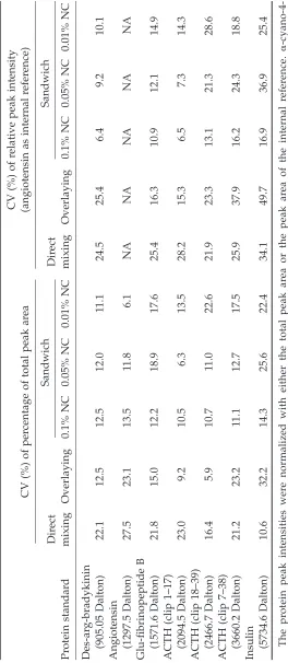
efer ence) Dir ect Sandwich Dir ect Sandwich Pr otein standar d mixing Overlaying 0.1% NC 0.05% NC 0.01% NC mixing Overlaying 0.1% NC 0.05% NC 0.01% NC Des-ar g-bradykinin (905.05 Dalton) 22.1 12.5 12.5 12.0 11.1 24.5 25.4 6.4 9.2 10.1
Angiotensin (1297.5 Dalton)
27.5 23.1 13.5 11.8 6.1 NA NA NA NA NA
Glu-fibrinopeptide B (1571.6 Dalton)
21.8 15.0 12.2 18.9 17.6 25.4 16.3 10.9 12.1 14.9
ACTH (clip 1–17) (2094.5 Dalton)
23.0 9.2 10.5 6.3 13.5 28.2 15.3 6.5 7.3 14.3
ACTH (clip 18–39) (2466.7 Dalton)
16.4 5.9 10.7 11.0 22.6 21.9 23.3 13.1 21.3 28.6
ACTH (clip 7–38) (3660.2 Dalton)
21.2 23.2 11.1 12.7 17.5 25.9 37.9 16.2 24.3 18.8
Insulin (5734.6 Dalton)
10.6 32.2 14.3 25.6 22.4 34.1 49.7 16.9 36.9 25.4 The pr
otein peak intensities wer
e normalized with either the total peak ar
ea or the peak ar
ea of the internal r
efer
ence.
␣
-cyano-4-hydr
oxycinnamic acid was used as the matrix chemical. NA, Not applicable;
ACTH, adr
enocorticotr
opic hormone; NC, nitr
ocellulose
methods, the sandwich method with the use of 0.1% NC provided the best reproducibility of the peptide ion signals. The peak intensity could be normalized with either the total peak area or with the peak area of an internal reference peptide. These two normalization procedures resulted in intra-assay CVs in the range of 6.5 to 17%.
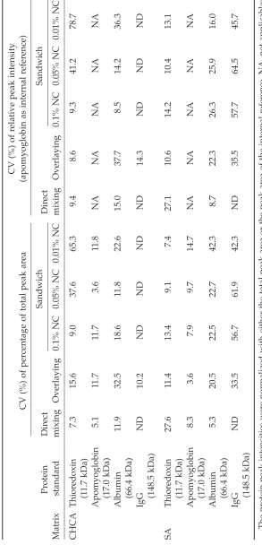
For profiling proteins in the range of 10–150 kDa, the effects of the three sample-matrix spotting methods and two matrix chemicals were examined. The results are summarized in Table 3. When only thioredoxin (11.67 kDa), apomyoglobin (16.85 kDa), and bovine serum albumin (BSA; 66.43 kDa) were considered, the sandwich method with the use of 0.1% NC and CHCA matrix was still a reliable method, resulting in the low CV values for various pro-teins. When the signal intensity was normal-ized with an internal reference, the CV for of measurement of BSA was only 8.5%.
When SA was used as the matrix chemical, all of the proteins within the detectable range, including mouse IgG, were ionized and de-tected. However, in general, the results were less reproducible (relatively higher CVs) when compared with those obtained with the use of CHCA. Because the sandwich method (0.1% NC and CHCA) gave the best quantitative results for profiling peptides/proteins of mass <66,5 kDa, this approach was used and referred to as NC-MALDI-TOF MS in the following experiments.
Assay Linearity of the
Nitrocellulose-MALDI TOF MS
for Peptides <6000 Dalton
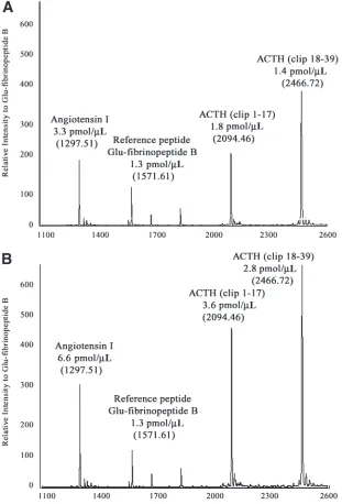
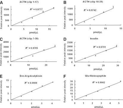
The linearity of the NC-MALDI-TOF MS method in quantitative profiling of a mixture of peptides between 900 and 6000 Dalton was examined. Adrenocorticotropic hormone (ACTH; clip 18–39) or glu-fibrinopeptide B was used as the internal reference peptide, and was added to the samples at a fixed concentration. Figure 1 shows typical mass spectra of the
quantitative analysis of the peptides when using glu-fibrinopeptide B as the internal ref-erence. Using glu-fibrinopeptide B (1571.61 m/z) as the internal reference peptide, the rel-ative peak intensities of ACTH clip 1–17 (2094.46 m/z), ACTH clip 18–39 (2466.72 m/z), ACTH clip 7–38 (3660.19), and insulin (5734.59 m/z) increased with their concentra-tions (Fig. 2A–D). Using ACTH clip 18–39 as the internal reference peptide, similar correla-tions were observed when measuring the intensities of des-arg-bradykinin (905.1 m/z) and glu-fibrinopeptide B (Fig. 2E,F). Linear equation was used in the curve fitting. All R2
values were ≥0.97. The results demonstrate that the relative peak intensity is directly pro-portional to the peptide concentration regard-less of the mass of a peptide.
Accuracy of Mass Measurement
The accuracy and precision of the NC-MALDI-TOF MS method in obtaining the pep-tide mass values were examined. The results are summarized in Table 4. For the assessed mass range between 900 and 6000 Dalton, per-centage errors for all of the tested standard peptides were <0.05%. The CVs were all <0.03%. These results suggest that introduc-tion of a NC layer does not significantly affect the mass accuracy of MALDI-TOF MS.
Application in Quantitative Profiling
of Serum Peptides
T
able 3
Summary of Coef
ficient of V
ariations (CV
, %) of Dif
fer
ent Sample-Matrix Spotting Methods in Detecting
High-Molecular
-W
eight Pr
oteins
CV (%) of r
elative peak intensity
CV (%) of per
centage of total peak ar
ea
(apomyoglobin as internal r
efer ence) Pr otein Dir ect Sandwich Dir ect Sandwich Matrix standar d mixing Overlaying 0.1% NC 0.05% NC 0.01% NC mixing Overlaying 0.1% NC 0.05% NC 0.01% NC CHCA Thior edoxin 7.3 15.6 9.0 37.6 65.3 9.4 8.6 9.3 41.2 78.7 (1 1.7 kDa) Apomyoglobin 5.1 11.7 11.7 3.6 11.8 NA NA NA NA NA (17.0 kDa) Albumin 1 1.9 32.5 18.6 11.8 22.6 15.0 37.7 8.5 14.2 36.3 (66.4 kDa) IgG ND 10.2 ND ND ND ND 14.3 ND ND ND (148.5 kDa) SA Thior edoxin 27.6 11.4 13.4 9.1 7.4 27.1 10.6 14.2 10.4 13.1 (1 1.7 kDa) Apomyoglobin 8.3 3.6 7.9 9.7 14.7 NA NA NA NA NA (17.0 kDa) Albumin 5.3 20.5 22.5 22.7 42.3 8.7 22.3 26.3 25.9 16.0 (66.4 kDa) IgG ND 33.5 56.7 61.9 42.3 ND 35.5 57.7 64.5 45.7 (148.5 kDa) The pr
otein peak intensities wer
e normalized with either the total peak ar
ea or the peak ar
ea of the internal r
efer
ence. NA, no
t applicable;
ND, not detectable; CHCA,
␣
-cyano-4-hydr
oxycinnamic acid; SA, sinapinic acid; NC, nitr
weight proteins before MS analysis. After removing the proteins >30 kDa, peptide peaks were observed in the mass spectra of the serum samples (Fig. 3). One of the serum samples was used to examine the interassay
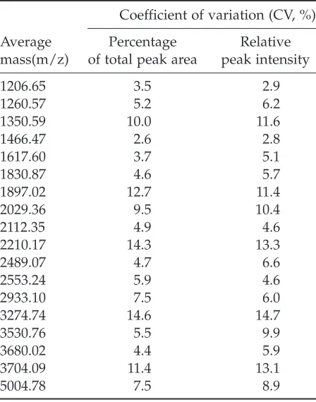
errors. The interassay errors of 18 repre-sentative peptide peaks (sample A) were examined and are summarized in Table 5. All of the interassay CVs were between 3.5 and 15%.
Volume 1, 2004 ________________________________________________________________ Clinical Proteomics
Discussion
Understanding the precision and linearity is crucial to all quantitative studies, including quantitative proteomic experiments. In exper-iments aiming to identify significant differ-ences between two sample groups, the statistical power depends on the CVs of the quantitative measurements. For assays with higher CVs, a larger sample size is needed to achieve the same degree of statistical power
Evaluation of MALDI-TOF MS for Quantitative Proteomic Profiling ________________________________ 267
(10). In this study, we have demonstrated that different sample-matrix spotting methods and matrix chemicals do affect the reproducibility of the intensity of peptide/protein ion signals. The quantitative capability of MALDI-TOF MS can be easily improved with the use of a thin film of NC coating for sample-matrix cocrystallization. The normalized signal inten-sities of the peptides/proteins were directly proportional to their concentrations with
intra-assay and interassay CVs <17%. The reproducibility of the NC-MALDI-TOF MS method is comparable to technical variations (20–30% CV) of the 2D PAGE technique (11)
Fig. 3. Representative mass spectra of serum samples from four individuals with the peptide range of 900–6000 m/z.The peptide standard adrenocorticotropic hormone 1–17 clip (2094.46 m/z) was used as the internal ref-erence. The interassay errors for sample A are listed in Table 5.
Table 4
Summary of the Accuracy and Precision of Measurement of the Masses for the Peptide Standards by the NC MALDI-TOF MS Method
Mean of masses from Theoretical average five independent
Peptide mass, MH+ assays, MH+ Percentage error CV (%)
Des-arg-bradykinin 905.05 904.68 0.041 0.025
Angiotensin I 1297.51 1297.18 0.025 0.024
Glu-fibrinopeptide B 1571.61 1571.35 0.017 0.026
ACTH (clip 1–17) 2094.46 2094.32 0.007 0.025
ACTH (clip 18–39) 2466.72 2466.68 0.002 0.025
ACTH (clip 7–38) 3660.19 3660.26 –0.002 0.025
Insulin 5734.59 5734.95 –0.006 0.025
The masses shown were obtained from five independent assays. NC, nitrocellulose; MALDI-TOF MS, matrix-assisted laser desorption/ionization time-of-flight mass spectrometry analysis; CV, coefficient of variations; ACTH, adrenocorticotropic hormone.
and those (CVs <15%) of SELDI ProteinChip technology (12).
Volume 1, 2004 ________________________________________________________________ Clinical Proteomics
denser network and provides more crystalliza-tion cores for the formacrystalliza-tion of a layer of homogeneous small sample-matrix cocrystals, resulting in more homogeneous relative pep-tide ion signals. NC concentrations of 0.01–3% were tested in initial experiments. We found that low NC concentrations (0.01 and 0.05%) were ineffective in the formation of a layer of homogenous NC film (data not shown). When the NC concentration was too high (>1%), the signal intensities of the peptide/protein peaks fall significantly (data not shown). It is possible that peptides/proteins are adsorbed to the NC fiber by physicochemical forces such as hydrophobic interactions, hydrogen bonding, and electrostatic interactions when too much NC is coated on the MALDI sample stage.
It is important to emphasize that in this study we have only evaluated the effect of matrix formulation and application on the reproducibility of MALDI-TOF MS in quanti-tative proteomic profiling. In future studies, other matrix factors, such as drying condi-tions, should be examined.
In conclusion, with the right MS analysis conditions, MALDI-TOF MS appears to be a feasible tool for directly profiling and quanti-fying the peptides/proteins in biological sam-ples with intra- and interassay CVs <17%.
Acknowledgments
The work was partially supported by the Strategic Research Area Grant from the Chinese University of Hong Kong. Dr. Ronald Pang was supported by the Postdoctoral Fellowship Scheme from the Chinese Univer-sity of Hong Kong.
References
1. Poon, T.C.W. and Johnson, P.J. (2001). Pro-teome analysis and its impact on the discov-ery of serological tumor markers. Clin. Chim. Acta 313:231–239.
2. Petricoin, E.F., Ardekani, A.M., Hitt, B.A., Levine, P.J., Fusaro, V.A., Steinberg, S.M., et al. Evaluation of MALDI-TOF MS for Quantitative Proteomic Profiling ________________________________ 269
Table 5
Interassay Errors of Quantitative Measurements of 18 Representative Peptides Identified in
Serum Sample A
Coefficient of variation (CV, %)
Average Percentage Relative
mass(m/z) of total peak area peak intensity
1206.65 3.5 2.9
1260.57 5.2 6.2
1350.59 10.0 11.6
1466.47 2.6 2.8
1617.60 3.7 5.1
1830.87 4.6 5.7
1897.02 12.7 11.4
2029.36 9.5 10.4
2112.35 4.9 4.6
2210.17 14.3 13.3
2489.07 4.7 6.6
2553.24 5.9 4.6
2933.10 7.5 6.0
3274.74 14.6 14.7
3530.76 5.5 9.9
3680.02 4.4 5.9
3704.09 11.4 13.1
5004.78 7.5 8.9
The signal intensities were normalized with the total peak area or with the peak area of the internal reference, adrenocorticotropic hormone (clip 1–17).
(2002). Use of proteomic patterns in serum to identify ovarian cancer. Lancet 359:572–577. 3. Poon, T.C.W., Yip, T.T., Chan, A.T.C., Yip, C.,
Yip, V., Mok, T.S., et al. (2003). Comprehensive proteomic profiling identifies serum pro-teomic signatures for detection of hepatocel-lular carcinoma and its subtypes. Clin. Chem. 49:752–760.
4. Poon, T.C.W., Chan, K.C.A., Ng, P.C., Chiu, R.W., Ang, I.L., Tong, Y.K., et al. (2004). Serial analysis of plasma proteomic signatures in pediatric patients with severe acute respira-tory syndrome and correlation with viral load. Clin. Chem. 50:1452–1455.
5. Stoeckli, M., Chaurand, P., Hallahan, D.E., and Caprioli, R.M. (2001). Imaging mass spec-trometry: a new technology for the analysis of protein expression in mammalian tissues. Nat. Med. 7:493–496.
6. Chaurand, P. and Caprioli, R.M. (2002). Direct profiling and imaging of peptides and pro-teins from mammalian cells and tissue sec-tions by mass spectrometry. Electrophoresis 23:3125–3135.
7. Wang, M.Z., Howard, B., Campa, M.J., Patz, E.F. Jr., and Fitzgerald, M.C. (2003). Analysis of human serum proteins by liquid phase isoelectric focusing and matrix-assisted laser desorption/ionization-mass spectrometry. Pro-teomics 3:1661–1666.
8. Howard, B.A., Wang, M.Z., Campa, M.J., Corro, C., Fitzgerald, M.C., and Patz, E.F. Jr. (2003). Identification and validation of a poten-tial lung cancer serum biomarker detected by matrix-assisted laser desorption/ionization-time of flight spectra analysis. Proteomics 3:1720–1724.
9. Beavis, R.C. and Chait, B.T. (1996). Matrix-assisted laser desorption ionization mass-spectrometry of proteins. Methods Enzymol. 270:519–551.
10. Lachin, J. (1981). Introduction to sample size determination and power analysis for clinical trials. Control Clin. Trials 2:93–113.
11. Molloy, M.P., Brzezinski, E.E., Hang, J., McDowell, M.T., and VanBogelen, R.A. (2003). Overcoming technical variation and biological variation in quantitative proteomics. Pro-teomics 3:1912–1919.
12. Paweletz, C.P., Gillispie, J.W., Ornstein, D.K., Simone, N.L., Brown, M.R., Cole, K.A., et al. (2000). Rapid protein display profiling of cancer progression directly from human tissue using a protein biochip. Drug Develop Res. 49:34–42.