S H O R T G E N O M E R E P O R T
Open Access
Non-contiguous finished genome sequence and
description of
Clostridium saudii
sp. nov
Emmanouil Angelakis
1, Fehmida Bibi
2, Dhamodharan Ramasamy
1, Esam I Azhar
2,3, Asif A Jiman-Fatani
4,
Sally M Aboushoushah
2, Jean-Christophe Lagier
1, Catherine Robert
1, Aurelia Caputo
1, Muhammad Yasir
2,
Pierre-Edouard Fournier
1and Didier Raoult
1,2*Abstract
Clostridium saudiistrain JCCTsp. nov. is the type strain ofC. saudiisp. nov., a new species within the genus
Clostridia. This strain, whose genome is described here, was isolated from a fecal sample collected from an obese 24-year-old (body mass index 52 kg/m2) man living in Jeddah, Saudi Arabia.C. saudiiis a Gram-positive, anaerobic bacillus. Here we describe the features of this organism, together with the complete genome sequence and annotation. The 3,653,762 bp long genome contains 3,452 protein-coding and 53 RNA genes, including 4 rRNA genes.
Keywords:Clostridium saudii, Genome, Culturomics, Taxono-genomics
Introduction
Clostridium saudii strain JCCT (=CSUR P697 = DSM
27835) is the type strain ofC. saudii sp. nov. This bac-terium is a Gram-positive, anaerobic, spore-forming in-dole negative bacillus that was isolated from the stool sample of an obese 24 year-old Saudi individual, as a part of a culturomics study as previously reported [1-3].
The current prokaryote species classification method, known as polyphasic taxonomy, is based on a combin-ation of genomic and phenotypic properties [4]. The usual parameters used to delineate a bacterial species include 16S rDNA sequence identity and phylogeny [2,3], genomic G + C content diversity and DNA–DNA hybridization (DDH) [4,5]. Nevertheless, some limita-tions appear, notably because the cutoff values vary dramatically between species and genera [6]. The intro-duction of high-throughput sequencing techniques has made genomic data for many bacterial species available [7]. To date, more than 4,000 bacterial genomes have been published and approximately 15,000 genomes pro-ject are anticipated to be completed in a near future [5]. We recently proposed a new method (taxono-genomics),
which integrates genomic information in the taxonomic framework, combining phenotypic characteristics, including MALDI-TOF MS spectra, and genomic analysis [8-38].
The genus Clostridium was first created in 1880 [39] and consists of obligate anaerobic rod-shaped bacilli able to produce endospores [39]. To date, more than 200 species have been described (http://www.bacterio.cict.fr/c/ clostridium.html). Members of the genus Clostridium are mostly environmental bacteria or associated with the com-mensal digestive flora of mammals. However,C. botulinum,
C. difficileandC. tetaniare major human pathogens [39].
Classification and features
A stool sample was collected from an obese 24-year-old male Saudi volunteer patient from Jeddah. The patient gave an informed and signed consent, and the agreement of the local Ethical Committee of the King Abdulaziz University, King Fahd medical Research Centre, Saudi Arabia, and of the local ethics committee of the IFR48 (Marseille, France) were obtained under agreement number 014-CEGMR-2-ETH-P and 09–022 respectively. The fecal specimen was preserved at−80°C after collection and sent to Marseille. C. saudii strain JCCT (Table 1) was iso-lated in July 2013 by anaerobic cultivation on 5% sheep blood-enriched Columbia agar (BioMerieux, Marcy l’Etoile, France) after a 5-day preincubation on blood culture bottle with rumen fluid. This strain exhibited a 98.3% nucleotide sequence similarity withClostridium * Correspondence:[email protected]
1Unité de Recherche sur les Maladies Infectieuses et Tropicales Emergentes,
UMR CNRS, Institut Hospitalo-Universitaire Méditerranée-Infection, Faculté de médecine, Aix-Marseille Université, Marseille, France
2
Special Infectious Agents Unit, King Fahd Medical Research Center, King Abdulaziz University, Jeddah, Saudi Arabia
Full list of author information is available at the end of the article
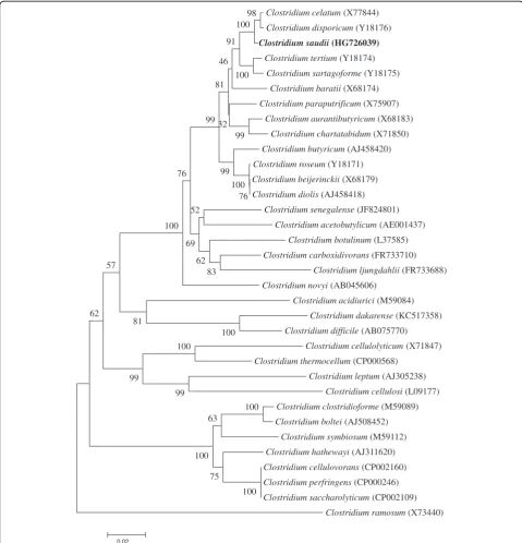
disporicum (Y18176) (Figure 1). This value was lower than the 98.7% 16S rRNA gene sequence threshold recommended by Stackebrandt and Ebers to delineate a new species without carrying out DNA-DNA hybridization [3] and was in the 78.4 to 98.9% range of 16S rRNA identity values observed among 41Clostridiumspecies with validly published names [40].
For the growth ofC. saudiiwe tested four temperatures (25, 30, 37, 45°C); growth occurred between 25 and 37°C, however optimal growth occurred at 37°C, 24 hours after inoculation. No growth occurred at 45°C. Colonies were translucent on 5% sheep blood-enriched Columbia agar (BioMerieux). Colonies on blood-enriched Columbia agar were about 0.2 to 0.3 mm in diameter and translucent light
grey. Growth of the strain was tested in the same agar under anaerobic and microaerophilic conditions using GENbag anaer and GENbag microaer systems, respect-ively (BioMerieux), and in aerobic conditions, with or without 5% CO2. Growth was observed only under anaer-obic conditions and no growth occurred under aeranaer-obic or microaerophilic conditions. Gram staining showed Gram-positive rods able to form spores (Figure 1) and the motility test was positive. Cells grown on agar ex-hibit a mean diameter of 1 μm and a mean length of 1.22μm in electron microscopy (Figure 2, Figure 3).
C. saudii did not have catalase or oxidase activity
(Table 2). On an API Rapid ID 32A strip (BioMerieux),
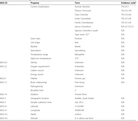
C. saudii presented positive reactions for α-galactosidase, Table 1 Classification and general features ofClostridium saudiistrain JCCT
MIGS ID Property Term Evidence codea
Current classification DomainBacteria TAS [41]
PhylumFirmicutes TAS [42-44]
ClassClostridia TAS [45,46]
OrderClostridiales TAS [47,48]
FamilyClostridiaceae TAS [47,49]
GenusClostridium IDA [47,50,51]
SpeciesClostridium saudii IDA
Type strain JCCT IDA
Gram stain Positive IDA
Cell shape Rod IDA
Motility Motile IDA
Sporulation Sporulating IDA
Temperature range Mesophile IDA
Optimum temperature 37°C IDA
MIGS-6.3 Salinity Unknown IDA
MIGS-22 Oxygen requirement Anaerobic IDA
Carbon source Unknown IDA
Energy source Unknown IDA
MIGS-6 Habitat Human gut IDA
MIGS-15 Biotic relationship Free living IDA
Pathogenicity Unknown
Biosafety level 2
MIGS-14 Isolation Human feces
MIGS-4 Geographic location Jeddah, Saudi Arabia IDA
MIGS-5 Sample collection time July 2013 IDA
MIGS-4.1 Latitude 21.422487 IDA
MIGS-4.1 Longitude 39.856184 IDA
MIGS-4.3 Depth surface IDA
MIGS-4.4 Altitude 0 m above sea level IDA
β-galactosidase,β-galactosidase-6-phosphatase,α-glucosidase, β-glucosidase, α-arabinosidase, N-acetyl-β-glucosaminidase, alkaline phosphatase, arginine arylamidase, pyroglutamic acid arylamidase, tyrosine arylamidase, alanine arylamidase, glycine arylamidase and histidine arylamidase. Negative reactions were obtained for urease, arginine dihydrolase,β -glucuronidase, fermentation of mannose and raffinose,
glutamic acid decarboxylase, α-fucosidase, nitrate reduc-tion, indole producreduc-tion, proline arylamidase, leucyl glycine arylamidase, phenylalanine arylamidase, leucine arylami-dase, glutamyl glutamic acid arylamidase and serine arylamidase.C. saudiiwas asaccharolytic on an API 50CH strip (Biomerieux). C. saudii is susceptible imipenem, trimethoprim-sulfamethoxazole, metronidazole, doxycycline,
Clostridium celatum (X77844)
Clostridium disporicum (Y18176) Clostridium saudii (HG726039)
Clostridium tertium (Y18174)
Clostridium sartagoforme (Y18175)
Clostridium baratii (X68174)
Clostridium paraputrificum (X75907)
Clostridium aurantibutyricum (X68183)
Clostridium chartatabidum (X71850)
Clostridium butyricum (AJ458420)
Clostridium roseum (Y18171)
Clostridium beijerinckii (X68179)
Clostridium diolis (AJ458418)
Clostridium senegalense (JF824801)
Clostridium acetobutylicum (AE001437)
Clostridium botulinum (L37585)
Clostridium carboxidivorans (FR733710)
Clostridium ljungdahlii (FR733688)
Clostridium novyi (AB045606)
Clostridium acidiurici (M59084)
Clostridium dakarense (KC517358)
Clostridium difficile (AB075770)
Clostridium cellulolyticum (X71847)
Clostridium thermocellum (CP000568)
Clostridium leptum (AJ305238)
Clostridium cellulosi (L09177)
Clostridium clostridioforme (M59089)
Clostridium boltei (AJ508452)
Clostridium symbiosum (M59112)
Clostridium hathewayi (AJ311620)
Clostridium cellulovorans (CP002160)
Clostridium perfringens (CP000246)
Clostridium saccharolyticum (CP002109)
Clostridium ramosum(X73440) 100
100 75
63
100 100 100
99 99
81 62
57
83 62 52
69 100
76
76 100 99
99 98 100
100
99 91
46
32 81
0.02
Figure 1A consensus phylogenetic tree highlighting the position ofClostridium saudiistrain JCCTrelative to other type strains within
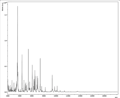
rifampicin, vancomycin and amoxicillin-clavulanate and resistant to amoxicillin, ciprofloxacine, erythromycin and gentamicin. The differential phenotypic characteristics with otherClostridiumspecies are summarized in Table 2. Matrix-assisted laser-desorption/ionization time-of-flight (MALDI-TOF) MS protein analysis was carried out as pre-viously described [53]. Briefly, a pipette tip was used to pick one isolated bacterial colony from a culture agar plate and spread it as a thin film on a MTP 384 MALDI-TOF target plate (Bruker Daltonics, Leipzig, Germany). Twelve distinct deposits from twelve isolated colonies were performed for
C. saudii JCCT. Each smear was overlaid with 2 μL of
matrix solution (saturated solution of alpha-cyano-4-hydro-xycinnamic acid) in 50% acetonitrile, 2.5% tri-fluoracetic acid, and allowed to dry for 5 minutes. Measurements were performed with a Microflex spectrometer (Bruker). Spectra were recorded in the positive linear mode for the mass range of 2,000 to 20,000 Da (parameter settings: ion source 1 (ISI), 20 kV; IS2, 18.5 kV; lens, 7 kV). A spectrum was obtained after 675 shots with variable laser power. The time of acquisition was between 30 seconds and 1 minute per spot. The twelve JCCT spectra were imported into the MALDI BioTyper software (version 2.0, Bruker) and ana-lyzed by standard pattern matching (with default param-eter settings) against the main spectra of 3,769 bacteria, including 228 spectra from 96 Clostridium species. The method of identification included the m/z from 3,000 to 15,000 Da. For every spectrum, a maximum of 100 peaks were compared with spectra in database. The resulting score enabled the identification of tested species, or not: a score≥2 with a validly published species enabled identifi-cation at the species level, a score≥1.7 but < 2 enabled identification at the genus level, and a score < 1.7 did not enable any identification. No significant MALDI-TOF score was obtained for strain JCCT against the Bruker database, suggesting that our isolate was not a member of a known species. We added the spectrum from strain JCCT to our database (Figure 4). Finally, the gel view showed the spectral differences with other members of the genusClostridium(Figure 5).
Genome sequencing information
Genome project history
The organism was selected for sequencing on the basis of its phylogenetic position and 16S rRNA similarity to members of the genusClostridium, and is part of a study of the human digestive flora aiming at isolating all bac-terial species in human feces [1]. It was the 101st gen-ome of a Clostridium species and the first genome of
C. saudii sp. nov. The GenBank accession number is
HG726039 and consists of 104 contigs. Table 2 shows the project information and its association with MIGS version 2.0 compliance [54].
Growth conditions and DNA isolation
C. saudii sp. nov., strain JCCT (=CSUR P697 = DSM
27835) was grown anaerobically on 5% sheep blood-enriched Columbia agar (BioMerieux) at 37°C.
Bac-teria grown on three Petri dishes were harvested and
resuspended in 4×100μL of TE buffer. Then, 200μL of this suspension was diluted in 1 ml TE buffer for lysis treatment that included a 30 minute incubation with 2.5 μg/μL lysozyme at 37°C, followed by an overnight incubation with 20 μg/μL proteinase K at 37°C. Extracted DNA was then purified using 3 suc-cessive phenol-chloroform extractions and ethanol
Figure 2Gram stain ofClostridium saudiistrain JCCT.
Figure 3Transmission electron micrograph ofC. saudiistrain JCCT, taken using a Morgani 268D (Philips) at an operating
precipitation at −20°C overnight. After centrifugation, the DNA was resuspended in 160 μL TE buffer. The yield and concentration was measured by the Quant-it Picogreen kit (Invitrogen) on the Genios-Tecan fluorometer.
Genome sequencing and assembly
Genomic DNA of C. saudii was sequenced on a MiSeq instrument (Illumina Inc, San Diego, CA, USA) with 2 applications: paired end and mate pair. The paired end and the mate pair strategies were barcoded in order to be mixed respectively with 14 other genomic projects prepared with the Nextera XT DNA sample prep kit (Illumina) and 11 other projects with the Nextera Mate Pair sample prep kit (Illumina). The gDNA was quanti-fied by a Qubit assay with the high sensitivity kit (Life technologies, Carlsbad, CA, USA) at 36.6 ng/μl and
dilution was performed such that 1 ng of each genome was used to prepare the paired end library. The “ tag-mentation”step fragmented and tagged the DNA with a mean size of 1.5 kb. Then limited cycle PCR amplifica-tion (12 cycles) completed the tag adapters and in-troduced dual-index barcodes. After purification on AMPure XP beads (Beckman Coulter Inc, Fullerton, CA, USA), the libraries were then normalized on specific beads according to the Nextera XT protocol (Illumina). Normalized libraries were pooled into a single library for sequencing on the MiSeq. The pooled single strand li-brary was loaded onto the reagent cartridge and then onto the instrument along with the flow cell. Automated cluster generation and paired end sequencing with dual index reads were performed in a single 39-hours run with a 2x250 bp read length. Total information of 5.3 Gb was obtained from a 574 K/mm2cluster density with Table 2 Differential characteristics ofClostridium saudiiJCCT,C. beijerinckiistrain NCIMB 8052,C. disporicumNCIB 12424,C. carboxidivoransstrain P7,C. senegalensestrain JC122,C. dakarensestrain FF1 andC. difficilestrain B1
Properties C. saudii C. beijerinckii C. disporicum C. carboxidivorans C. senegalense C. dakarense C. difficile
Cell diameter (μm) 1.0 1.7 1.5 1.5 1.1 1.2 3.0
Oxygen requirement Strictly Strictly Strictly Strictly Strictly Strictly Strictly
anaerobic anaerobic anaerobic anaerobic anaerobic anaerobic anaerobic
Gram stain Positive Variable Positive Positive Positive Positive Variable
Motility Motile Motile Na Motile Motile Motile Motile
Endospore formation + + Na + + + +
Indole - Na - - - + Na
Production of
Alkaline phosphatase - Na Na Na - + Na
Catalase - - - Na
Oxidase - Na Na - - - Na
Nitrate reductase - - Na - - -
-Urease - - Na - - - Na
β-galactosidase - Na Na Na - - Na
N-acetyl-glucosamine - Na Na Na + Na
Acid from
L-Arabinose - + Na + Na -
-Ribose - - + + Na -
-Mannose - + + + Na - +
Mannitol - + + + Na - +
Sucrose - + + + Na - +
D-glucose - + + + Na + Na
D-fructose - + + + Na - +
D-maltose - + + + Na +
-D-lactose - + + + Na -
-G + C content (%) 28 28 29 31 26.8 27.98 28
Habitat Human gut Human gut Rat gut Environment Human gut Human gut Human gut
95.4% (11,188,000) of the clusters passing quality control filters. Within this run, the index representation for C. saudiiwas determined to be 6.9%. The 710,425 reads were filtered according to the read qualities.
The mate pair library was prepared with 1 μg of gen-omic DNA using the Nextera mate pair Illumina guide. The genomic DNA sample was simultaneously fragmen-ted and tagged with a mate pair junction adapter. The profile of the fragmentation was validated on an Agilent 2100 BioAnalyzer (Agilent Technologies Inc, Santa Clara, CA, USA) with a DNA 7500 labchip. The DNA fragments ranged in size from 1.4 kb up to 10 kb with a mean size of 5 kb. No size selection was performed and 600 ng of tagmented fragments were circularized. The circularized DNA was mechanically sheared to small fragments with a mean size of 625 bp on the Covaris
device S2 in microtubes (Covaris, Woburn, MA, USA). The library profile was visualized on a High Sensitivity Bioanalyzer LabChip (Agilent Technologies Inc, Santa Clara, CA, USA). The libraries were normalized at 2 nM and pooled. After a denaturation step and dilution at 10 pM, the pool of libraries was loaded onto the reagent cartridge and then onto the instrument along with the flow cell. Automated cluster generation and sequencing run were performed in a single 42-hours run with a 2×250 bp read length.
Total information of 3.2 Gb was obtained from a 690 K/mm2 cluster density with 95.4% (13,264,000) of the clusters passing quality control filters. Within this run, the index representation for C. saudii was deter-mined to be 8.2%. The 1,037,710 reads were filtered according to the read qualities.
Genome annotation
Open Reading Frames (ORFs) were predicted using Prodigal [55] with default parameters. However, the pre-dicted ORFs were excluded if they spanned a sequencing gap region. The predicted bacterial protein sequences were searched against the GenBank [56] and Clusters of Orthologous Groups (COG) databases using BLASTP. The tRNAs and rRNAs were predicted using the tRNAScanSE [57] and RNAmmer [58] tools, respectively. Lipoprotein signal peptides and numbers of transmembrane helices
were predicted using SignalP [59] and TMHMM [60], re-spectively. Mobile genetic elements were predicted using PHAST [61] and RAST [62]. ORFans were identified if their BLASTPE-value was lower than 1e-03 for alignment length greater than 80 amino acids. If alignment lengths were smaller than 80 amino acids, we used an E-value of 1e-05. Such parameter thresholds have already been used in previous works to define ORFans. Artemis [63] and DNA Plotter [64] were used for data management and visualization of genomic features, respectively. Mauve Clostridium tertium
1048_NCTC 541 BOG
Clostridium saudii
Clostridium sartagoforme
100617_58 PNU
Clostridium paraputrificum
110706_B3 LUMC
Clostridium disporicum
5521T DSM
Clostridium carboxidivorans
DSM 15243T DSM
Clostridium botulinum
1028_NCTC 7272_BOG
Clostridium beijerinckii
1072_ATCC 25752T BOG
Clostridium baratii
1084_ATCC 25782 BOG
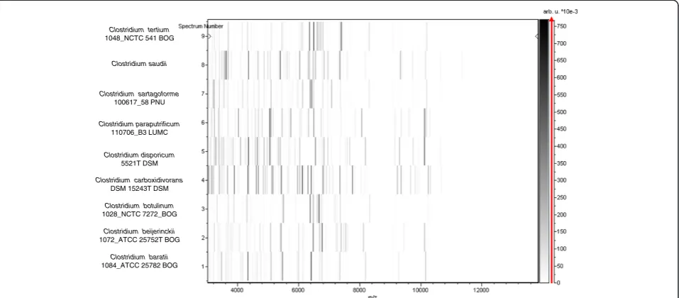
Figure 5Gel view comparing spectra fromClostridium saudiistrain JCCT,Clostridium tertium,Clostridium sartagoforme,Clostridium
baratii,Clostridium beijerinckii,Clostridium botulinum,Clostridium carboxidivoransandClostridium paraputrificum.The gel view presents the raw spectra of loaded spectrum files as a pseudo-electrophoretic gel. The x-axis records the m/z value. The left y-axis displays the running spectrum number originating from subsequent spectra loading. The peak intensity is expressed by a grey scale scheme code. The grey scale bar on the right y-axis indicates the relation between the shade of grey a peak is displayed with and the peak intensity in arbitrary units. Species are listed on the left.
Table 3 Genomic comparison ofC. saudiiand 9 other members ofClostridiumspecies†
C. sma C.bej C. bot C. car C. cel C. dak C. dif C. par C. per C. sen
C. sma 5,786 1,479 1,181 1,034 1,779 1,100 1,037 1,554 1,351 1,137
C. bej 72.92 4,911 1,438 1,132 1,017 1,069 1,003 1,539 1,312 1,129
C. bot 71.34 73.00 5,719 1,533 1,275 1,101 1,099 1,046 1,337 1,210
C. car 71.11 71.66 73.13 4,184 1,426 1,334 1,182 1,162 1,294 1,252
C. cel 81.95 71.20 71.34 71.10 4,066 1,302 1,081 1,111 1,144 1,378
C. dak 70.13 70.38 71.06 71.46 74.04 4,778 1,149 1,119 1,076 1,137
C. dif 69.57 69.70 69.56 69.02 69.80 72.54 3,553 1,015 1,303 1,066
C. par 73.94 73.96 69.23 68.54 69.23 70.30 69.34 3,244 1,018 961
C. per 73.21 73.32 79.95 72.01 71.94 69.47 77.70 69.09 4,485 957
C. sen 71.94 72.07 71.53 71.10 73.11 72.16 70.40 71.58 69.58 4,663
†The numbers of orthologous protein shared between genomes (above diagonal), average percentage similarity of nucleotides corresponding to orthologous
protein shared between genomes (below diagonal) and the numbers of proteins per genome (bold).
alignment tool (version 2.3.1) was used for multiple gen-omic sequence alignment [65]. To estimate the Average Genome Identity of Orthologous Sequences (AGIOS) [7] at the genome level between C. saudii and another 9 mem-bers of the Clostridiumgenus (Table 3), orthologous pro-teins were detected using the Proteinortho [66] and we compared genomes two by two and determined the mean percentage of nucleotide sequence identity among ortholo-gous ORFs using BLASTn.
Genome properties
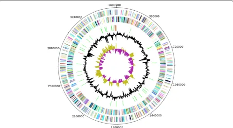
The genome is 3,653,762 bp long (one chromosome, no plasmid) with a GC content of 27.9% (Figure 6 and Table 4). Of the 3,509 predicted chromosomal genes, 3,452 were protecoding genes and 57 were RNAs in-cluding 49 tRNAs and 8 rRNAs (5S = 6, 23S = 1, 16S = 1). A total of 2144 genes (61.10%) were assigned a putative function. One hundred and twenty eight genes were iden-tified as ORFans (3.65%) and the remaining genes were annotated as hypothetical proteins. The properties and statistics of the genome are summarized in Tables 4 and 5.
Figure 6Graphical circular map of the chromosome.From outside to the center: Genes on the forward strand (colored by COG categories), genes on the reverse strand (colored by COG categories), RNA genes (tRNAs green, rRNAs red), GC content, and GC skew.
Table 4 Nucleotide content and gene count levels of the genome
Attribute Value % of totala
Genome size (bp) 3,653,762
DNA G + C content (bp) 1,019,399 27.9
DNA coding region (bp) 3,057,234 83.67
Total genes 3509 100
RNA genes 57 1.62
Protein-coding genes 3452 98.37
Genes with function prediction 2144 61.10
Genes assigned to COGs 2514 71.64
Genes with peptide signals 135 3.85
Genes with transmembrane helices 887 25.27
a
The total is based on either the size of the genome in base pairs or the total number of protein coding genes in the annotated genome.
Table 5 Project information
MIGS ID Property Term
MIGS-31 Finishing quality High-quality draft
MIGS-28 Libraries used One paired-end 454 3-kb library
MIGS-29 Sequencing platforms MiSeq Illumina
MIGS-31.2
Fold coverage 85.77×
MIGS-30 Assemblers Newbler version 2.5.3
MIGS-32 Gene calling method Prodigal
GenBank ID CBYM00000000
GenBank Date of Release
February 12, 2014
The distribution of genes into COGs functional categories is presented in Table 6.
Genome comparison of C. saudii with 9 otherClostridium
genomes
We compared the genome of C. saudii strain JCCT with those of C. beijerinckiistrain NCIMB 8052,C. botulinum
strain ATCC 3502,C. carboxidivoransstrain P7,C. celatum
strain DSM 1785,C. dakarensestrain FF1,C. difficiles train
B1, C. perfringens strain AGR 2156, C. paraputrificum
strain ATCC 13124 and C. senegalense strain JC122 (Tables 6 and 7). The draft genome sequence ofC. saudii
strain JCCT is smaller than those of C. beijerinckii, C. botulinum, C. carboxidivorans, C. dakarense, C. difficile,
and C. senegalense (3.9, 4.41, 3.73, 4.46 and 3.89 Mb
respectively), but larger than those of C. celatum, C.
paraputrificum and C. perfringens (3.55, 3.56 and
3.26 Mb, respectively). The G + C content ofC. saudii
is lower than those of C. beijerinckii,C. botulinum, C. carboxidivorans,C. dakarense,C. difficile,C. perfringens
and C. paraputrificum (29.0, 28.2, 29.7, 27.98, 28.4,
29.6 and 28.4%, respectively) but greater than those
of C. celatum and C. senegalense (26.8 and 27.7
re-spectively). The gene content of C. saudii (3462) is smaller to those ofC. beijerinckii,C. botulinum,C. dif-ficile,C. carboxidivorans,C. paraputrificum,C.
dakar-ense andC. senegalense (5020, 3590, 3934, 4174, 3568,
3843, and 3704 respectively) but larger that ofC.
per-fringens and C. celatum (2876 and 3453 respectively).
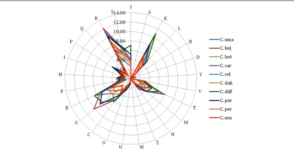
The distribution of genes into COG categories was al-most similar in all the 10 compared genomes except the unique presence of cytoskeleton associated pro-teins inC. difficile(Figure 7).
In addition,C. saudii shared 1479, 1181, 1034, 1779, 1100, 1037, 1554, 1351, and 1137 orthologous genes with C. beijerinckii, C. botulinum, C. carboxidivorans,
C. celatum, C. dakarense,C. difficile, C. perfringens, C.
paraputrificumandC. senegalense, respectively. Among
compared genomes AGIOS values ranged from 68.54 between C. carboxidivorans and C. paraputrificum to 79.95% betweenC. botulinumandC. perfringens. When
C. saudiiwas compared to other species, AGIOS values
ranged from 69.57 with C. difficile to 81.95% with C. celatum(Table 7).
Table 6 Number of genes associated with the 25 general COG functional categories
Code Value % agea Description
J 154 4.46 Translation
A 0 0 RNA processing and modification
K 296 8.57 Transcription
L 138 4 Replication, recombination and repair
B 1 0.03 Chromatin structure and dynamics
D 24 0.7 Cell cycle control, mitosis and meiosis
Y 0 0 Nuclear structure
V 73 2.11 Defense mechanisms
T 156 4.52 Signal transduction mechanisms
M 116 3.36 Cell wall/membrane biogenesis
N 62 1.8 Cell motility
Z 0 0 Cytoskeleton
W 0 0 Extracellular structures
U 48 1.4 Intracellular trafficking and secretion
O 66 1.91 Posttranslational modification, protein turnover, chaperones
C 153 4.43 Energy production and conversion
G 237 6.86 Carbohydrate transport and metabolism
E 328 9.5 Amino acid transport and metabolism
F 56 1.62 Nucleotide transport and metabolism
H 92 2.66 Coenzyme transport and metabolism
I 85 2.46 Lipid transport and metabolism
P 164 4.75 Inorganic ion transport and metabolism
Q 53 1.53 Secondary metabolites biosynthesis, transport and catabolism
R 346 10.02 General function prediction only
S 195 5.65 Function unknown
- 948 27.46 Not in COGs
a
The total is based on the total number of protein coding genes in the annotated genome.
Table 7 Genomic comparison ofC. saudiiand 9 other members ofClostridiumspecies†
Species Strain Genome accession number
Genome size (Mb)
G + C content
C. saudii JCC In progress 3.65 27.9
C. beijerinckii NCIMB 8052
NC_009617 6.0 29.0
C. botulinum ATCC 3502
NC_009495 3.9 28.2
C.
carboxidivorans
P7 NZ_ADEK00000000 4.41 29.7
C. celatum DSM 1785
AMEZ01000000 3.55 27.7
C. dakarense FF1 CBTZ010000000 3.73 27.98
C. difficile B1 NC_017179 4.46 28.4
C.
paraputrificum
AGR2156 AUJC01000000 3.56 29.6
C. perfringens ATCC 13124
NC_008261 3.26 28.4
C. senegalense JC122 CAEV01000001 3.89 26.8
†Species name, Strain, GenBank accession number, Genome size and GC
Conclusion
On the basis of phenotypic, phylogenetic and genomic analyses, we formally propose the creation ofClostridium saudiisp. nov. that contains the strain JCCT. This bacter-ial strain was isolated in Marseille, France.
Description ofClostridium saudiisp. nov
Clostridium saudii(sa.u'di.i N.L. gen. n. saudii, of Saudi Arabia, for the country where the strain originates). Iso-lated from an obese Saudi patient sample. Transparent colonies were 0.2 to 0.3 mm in diameter on blood-enriched agar.C. saudiiis a Gram-positive, obligate anaer-obic, endospore-forming bacterium with a mean diameter of 1μm. Optimal growth was observed at 37°C.C. saudii
is catalase and oxidase negative. Alpha-galactosidase, β -galactosidase,β-galactosidase-6-phosphatase,α-glucosidase, β-glucosidase,α-arabinosidase, N-acetyl-β-glucosaminidase, alkaline phosphatase, arginine arylamidase, pyroglutamic acid arylamidase, tyrosine arylamidase, alanine arylami-dase, glycine arylamidase and histidine arylamidase were positive. Urease, arginine dihydrolase, β-glucuronidase, fermentation of mannose and raffinose, glutamic acid de-carboxylase,α-fucosidase, nitrate reduction, indole produc-tion, proline arylamidase, leucyl glycine arylamidase, phenylalanine arylamidase, leucine arylamidase, gluta-myl glutamic acid arylamidase and serine arylamidase were negative. Asaccharolytic.C. saudiiis susceptible to imipenem, trimethoprim-sulfamethoxazole, metronida-zole, doxycycline, rifampicin, vancomycin and
amoxicillin-clavulanate and resistant to amoxicillin, ciprofloxacine, erythromycin and gentamicin.
The G + C content of the genome is 28%. The 16S rRNA and genome sequences are deposited in GenBank under accession numbers HG726039 and CBYM00 000000, respectively. The type strain is JCCT (=CSUR P697 = DSM 27835).
Competing interests
The authors declare that they have no competing interests.
Authors’contributions
EA: wrote the manuscript and analysed the data; FB: analysed the data; DhR: analysed the genome; EIA: organized the study in Saudi Arabia; AJF: collected samples in Saudi Arabia; SA: collected samples in Saudi Arabia; JCL: cultured the samples and analyzed microbiological data; CR: performed the sequencing analysis; AC: analyzed sequences; MH: collected data in Saudi Arabia; PEF: organized the study and wrote the manuscript; DR: organized the study.
Acknowledgements
This work was funded by the Deanship of Scientific Research (DSR), King Abdulaziz University, under grant No. (1-141/1433 HiCi). The authors, therefore, acknowledge technical and financial support of KAU. The authors thank the Xegen Company (www.xegen.fr) for automating the genomic annotation process.
Author details
1
Unité de Recherche sur les Maladies Infectieuses et Tropicales Emergentes, UMR CNRS, Institut Hospitalo-Universitaire Méditerranée-Infection, Faculté de médecine, Aix-Marseille Université, Marseille, France.2Special Infectious Agents Unit, King Fahd Medical Research Center, King Abdulaziz University, Jeddah, Saudi Arabia.3Department of Medical Laboratory Technology, Faculty of Applied Medical Sciences, King Abdulaziz University, Jeddah, Saudi Arabia.4Department of Medical Microbiology and Parasitology, Faculty of Medicine, King Abdulaziz University, Jeddah, Saudi Arabia.
Received: 12 June 2014 Accepted: 16 June 2014 Published: 8 December 2014
References
1. Lagier JC, Armougom F, Million M, Hugon P, Pagnier I, Robert C, Bittar F, Fournous G, Gimenez G, Maraninchi M, Trape JF, Koonin EV, La Scola B, Raoult D.Microbial culturomics: paradigm shift in the human gut microbiome study.Clin Microbiol Infect.2012;18:1185–93. PubMed. 2. Tindall BJ, Rosselló-Móra R, Busse HJ, Ludwig W, Kämpfer P.Notes on the
characterization of prokaryote strains for taxonomic purposes.Int J Syst Evol Microbiol.2010;60:249–66. PubMed http://dx.doi.org/10.1099/ijs.0.016949-0. 3. Stackebrandt E, Ebers J.Taxonomic parameters revisited: tarnished gold
standards.Microbiol Today.2006;33:152–5.
4. Wayne LG, Brenner DJ, Colwell PR, Grimont PAD, Kandler O, Krichevsky MI, Moore LH, Moore WEC, Murray RGE, Stackebrandt E, Starr MP, Truper HG.
Report of thead hoccommittee on reconciliation of approaches to bacterial systematics.Int J Syst Bacteriol.1987;37:463–4. http://dx.doi.org/ 10.1099/00207713-37-4-463.
5. Rossello-Mora R.DNA-DNA Reassociation Methods Applied to Microbial Taxonomy and Their Critical Evaluation.In: Stackebrandt E, editor. Molecular Identification, Systematics, and population Structure of Prokaryotes. Berlin: Springer; 2006: p. 23–50.
6. Welker M, Moore ER.Applications of whole-cell matrix-assisted laser-desorption/ionization time-of-flight mass spectrometry in systematic microbiology.Syst Appl Microbiol.2011;34:2–11. PubMed http://dx.doi.org/ 10.1016/j.syapm.2010.11.013.
7. Ramasamy D, Mishra AK, Lagier JC, Padhmanabhan R, Rossi-Tamisier M, Sentausa E, Raoult D, Fournier PE.A polyphasic strategy incorporating genomic data for the taxonomic description of new bacterial species.
Int J Syst Evol Microbiol.2014;64:384–91. PubMed http://dx.doi.org/10.1099/ ijs.0.057091-0.
8. Kokcha S, Mishra AK, Lagier JC, Million M, Leroy Q, Raoult D, Fournier PE.
Non-contiguous finished genome sequence and description ofBacillus timonensissp. nov.Stand Genomic Sci.2012;6:346–55. PubMed http://dx. doi.org/10.4056/sigs.2776064.
9. Lagier JC, El Karkouri K, Nguyen TT, Armougom F, Raoult D, Fournier PE.
Non-contiguous finished genome sequence and description of
Anaerococcus senegalensissp. nov.Stand Genomic Sci.2012;6:116–25. PubMed http://dx.doi.org/10.4056/sigs.2415480.
10. Mishra AK, Gimenez G, Lagier JC, Robert C, Raoult D, Fournier PE. Non-contiguous finished genome sequence and description ofAlistipes senegalensissp. nov.Stand Genomic Sci.2012;6:304–14. http://dx.doi.org/ 10.4056/sigs.2625821.
11. Lagier JC, Armougom F, Mishra AK, Ngyuen TT, Raoult D, Fournier PE.
Non-contiguous finished genome sequence and description ofAlistipes timonensissp. nov.Stand Genomic Sci.2012;6:315–24. PubMed http://dx. doi.org/10.4056/sigs.2685971.
12. Mishra AK, Lagier JC, Robert C, Raoult D, Fournier PE.Non-contiguous finished genome sequence and description ofClostridium senegalense
sp. nov.Stand Genomic Sci.2012;6:386–95. PubMed.
13. Mishra AK, Lagier JC, Robert C, Raoult D, Fournier PE.Non-contiguous finished genome sequence and description ofPeptoniphilus timonensissp. nov.Stand Genomic Sci.2012;7:1–11. PubMed http://dx.doi.org/10.4056/sigs.2956294. 14. Mishra AK, Lagier JC, Rivet R, Raoult D, Fournier PE.Non-contiguous
finished genome sequence and description ofPaenibacillus senegalensis
sp. nov.Stand Genomic Sci.2012;7:70–81. PubMed http://dx.doi.org/ 10.4056/sigs.3056450.
15. Lagier JC, Gimenez G, Robert C, Raoult D, Fournier PE.Non-contiguous finished genome sequence and description ofHerbaspirillum massiliense
sp. nov.Stand Genomic Sci.2012;7:200–9. PubMed.
16. Kokcha S, Ramasamy D, Lagier JC, Robert C, Raoult D, Fournier PE. Non-contiguous finished genome sequence and description ofBrevibacterium senegalensesp. nov.Stand Genomic Sci.2012;7:233–45. PubMed http://dx. doi.org/10.4056/sigs.3256677.
17. Ramasamy D, Kokcha S, Lagier JC, N’Guyen TT, Raoult D, Fournier PE.
Non-contiguous finished genome sequence and description of
Aeromicrobiummassilensesp. nov.Stand Genomic Sci.2012;7:246–57. PubMed http://dx.doi.org/10.4056/sigs.3306717.
18. Lagier JC, Ramasamy D, Rivet R, Raoult D, Fournier PE.Non-contiguous finished genome sequence and description ofCellulomonas massiliensis
sp. nov.Stand Genomic Sci.2012;7:258–70. PubMed http://dx.doi.org/ 10.4056/sigs.3316719.
19. Lagier JC, Karkouri K, Rivet R, Couderc C, Raoult D, Fournier PE.
Non contiguous-finished genome sequence and description of
Senegalemassilia anaerobiagen. nov., sp. nov.Stand Genomic Sci. 2013;7:343–56. PubMed http://dx.doi.org/10.4056/sigs.3246665.
20. Mishra AK, Hugon P, Nguyen TT, Robert C, Couderc C, Raoult D, Fournier PE.
Non contiguous-finished genome sequence and description of
Peptoniphilus obesisp. nov.Stand Genomic Sci.2013;7:357–69. PubMed http://dx.doi.org/10.4056/sigs.32766871.
21. Mishra AK, Lagier JC, Nguyen TT, Raoult D, Fournier PE.Non contiguous-finished genome sequence and description ofPeptoniphilus senegalensis
sp. nov.Stand Genomic Sci.2013;7:357–69. PubMed http://dx.doi.org/ 10.4056/sigs.32766871.
22. Lagier JC, Karkouri K, Mishra AK, Robert C, Raoult D, Fournier PE.Non contiguous-finished genome sequence and description ofEnterobacter massiliensissp. nov.Stand Genomic Sci.2013;7:399–412. PubMed http://dx. doi.org/10.4056/sigs.3396830.
23. Hugon P, Ramasamy D, Rivet R, Raoult D, Fournier PE.Non contiguous-finished genome sequence and description ofAlistipes obesisp. nov.Stand Genomic Sci.2013;7:427–39. PubMed http://dx.doi.org/10.4056/sigs.3336746. 24. Hugon P, Mishra AK, Nguyen TT, Raoult D, Fournier PE.Non-contiguous finished
genome sequence and description ofBrevibacillus massiliensissp. nov.Stand Genomic Sci.2013;8:1–14. PubMed http://dx.doi.org/10.4056/sigs.3466975. 25. Mishra AK, Hugon P, Nguyen TT, Raoult D, Fournier PE.Non
contiguous-finished genome sequence and description ofEnorma massiliensisgen. nov., sp. nov., a new member of the familyCoriobacteriaceae.Stand Genomic Sci.2013;8:290–305. PubMed http://dx.doi.org/10.4056/ sigs.3426906.
26. Ramasamy D, Lagier JC, Gorlas A, Raoult D, Fournier PE.Non contiguous-finished genome sequence and description ofBacillus
massiliosenegalensissp. nov.Stand Genomic Sci.2013;8:264–78. PubMed http://dx.doi.org/10.4056/sigs.3496989.
27. Ramasamy D, Lagier JC, Nguyen TT, Raoult D, Fournier PE.Non contiguous-finished genome sequence and description ofDielma fastidiosagen. nov., sp. nov., a new member of the familyErysipelotrichaceae.Stand Genomic Sci.2013;8:336–51. PubMed http://dx.doi.org/10.4056/sigs.3567059. 28. Mishra AK, Pfleiderer A, Lagier JC, Robert C, Raoult D, Fournier PE.Non
contiguous-finished genome sequence and description ofBacillus massilioanorexiussp. nov.Stand Genomic Sci.2013;8:465–79. PubMed http://dx.doi.org/10.4056/sigs.4087826.
29. Hugon P, Ramasamy D, Robert C, Couderc C, Raoult D, Fournier PE. Non-contiguous finished genome sequence and description ofKallipyga massiliensisgen. nov., sp. nov., a new member of the familyClostridiales Incertae Sedis XI.Stand Genomic Sci.2013;8:500–15. PubMed http://dx.doi. org/10.4056/sigs.4047997.
30. Padhmanabhan R, Lagier JC, Dangui NPM, Michelle C, Couderc C, Raoult D, Fournier PE.Non-contiguous finished genome sequence and description ofMegasphaera massiliensis.Stand Genomic Sci.2013;8:525–38. PubMed http://dx.doi.org/10.4056/sigs.4077819.
31. Mishra AK, Edouard S, Dangui NPM, Lagier JC, Caputo A, Blanc-Tailleur C, Ravaux I, Raoult D, Fournier PE.Non-contiguous finished genome sequence and description ofNosocomiicoccus massiliensissp. nov.Stand Genomic Sci. 2013;9:205–19. PubMed http://dx.doi.org/10.4056/sigs.4378121.
32. Mishra AK, Lagier JC, Robert C, Raoult D, Fournier PE.Genome sequence and description ofTimonella senegalensisgen. nov., sp. nov., a new member of the suborderMicrococcineae.Stand Genomic Sci.2013;
8:318–35. PubMed http://dx.doi.org/10.4056/sigs.3476977.
33. Keita MB, Diene SM, Robert C, Raoult D, Fournier PE.Non contiguous-finished genome sequence and description ofBacillus massiliogorillaesp. nov.
Stand Genomic Sci.2013;9:93–105. PubMed http://dx.doi.org/10.4056/ sigs.4388124.
34. Mediannikov O, El Karkouri K, Robert C, Fournier PE, Raoult D.Non contiguous-finished genome sequence and description ofBartonella florenciaesp. nov.Stand Genomic Sci.2013;9:185–96. PubMed http://dx. doi.org/10.4056/sigs.4358060.
36. Mishra AK, Hugon P, Robert C, Raoult D, Fournier PE.Non contiguous-finished genome sequence and description ofPeptoniphilus grossensis
sp. nov.Stand Genomic Sci.2012;7:320–30. PubMed.
37. Mediannikov O, El Karkouri K, Diatta G, Robert C, Fournier PE, Raoult D.
Non contiguous-finished genome sequence and description of
Bartonella senegalensissp. nov.Stand Genomic Sci.2013;8:279–89. PubMed http://dx.doi.org/10.4056/sigs.3807472.
38. Roux V, Million M, Robert C, Magne A, Raoult D.Non-contiguous finished genome sequence and description ofOceanobacillus massiliensissp. nov.
Stand Genomic Sci.2013;9:370-84 doi: 10.4056/sigs.4267953.
39. Wells CL, Wilkins TD.Clostridia:Spore forming AnaerobicBacilli.In:Medical Microbiology. 4th ed. Baron S et al., editor. Galveston (TX): University of Texas Medical Branch at Galveston; 1996.
40. 16S Yourself database. http://www.mediterranee-infection.com/article.php? larub=152&titre=16s-yourself.
41. Woese CR, Kandler O, Wheelis ML.Towards a natural system of organisms: proposal for the domainsArchaea, Bacteria, andEucarya.Proc Natl Acad Sci USA.1990;87:4576–9. PubMed http://dx.doi.org/10.1073/pnas.87.12.4576. 42. Gibbons NE, Murray RGE.Proposals concerning the higher taxa of
bacteria.Int J Syst Bacteriol.1978;28:1–6. http://dx.doi.org/10.1099/ 00207713-28-1-1.
43. Murray RGE.The Higher Taxa, or, a Place for Everything…?In: Holt JG, editor.Bergey’s Manual of Systematic Bacteriology. First Edition, Volume 1st ed. Baltimore: The Williams and Wilkins Co; 1984: p. 31–4.
44. Garrity GM, Holt JG.The Road Map to the Manual.In: Garrity GM, Boone DR, Castenholz RW, editors.Bergey’s Manual of Systematic Bacteriology, Volume 1. 2nd ed. New York: Springer; 2001: p. 119–69.
45. List of new names and new combinations previously effectively, but not validly, published. List no. 132.Int J Syst Evol Microbiol.2010;60:469–72. http://dx.doi.org/10.1099/ijs.0.022855-0.
46. Rainey FA, Class II.Clostridiaclass nov.In: De Vos P, Garrity G, Jones D, Krieg NR, Ludwig W, Rainey FA, Schleifer KH, Whitman WB, editors.Bergey’s Manual of Systematic Bacteriology. Second Edition, Volume 3rd ed. New York: Springer; 2009: p. 736.
47. Skerman VBD, Sneath PHA.Approved list of bacterial names.Int J Syst Bact.1980;30:225–420. http://dx.doi.org/10.1099/00207713-30-1-225. 48. Prevot AR.Dictionnaire des Bactéries Pathogens. In: Hauduroy P, Ehringer G,
Guillot G, Magrou J, Prevot AR, Rosset D, Urbain A, editors. Paris, France: Masson; 1953: p. 1–692.
49. Pribram E.Klassification der Schizomyceten. Klassifikation der Schizomyceten (Bakterien). Leipzig: Franz Deuticke; 1933: p. 1–143.
50. Prazmowski A.“Untersuchung Über die Entwickelungsgeschichte und Fermentwirking Einiger Bakterien-Arten”, Ph.D. Dissertation. Germany: University of Leipzig; 1880: p. 366–71.
51. Smith LDS, Hobbs G.Genus III.ClostridiumPrazmowski 1880, 23.In: Buchanan RE, Gibbons NE, editors.Bergey’s Manual of Determinative Bacteriology. Eighthth ed. Baltimore: The Williams and Wilkins Co; 1974: p. 551–72.
52. Ashburner M, Ball CA, Blake JA, Botstein D, Butler H, Cherry JM, Raoult D.Gene ontology: tool for the unification of biology. The gene ontology consortium.
Nat Genet.2000;25:25–9. PubMed http://dx.doi.org/10.1038/75556. 53. Seng P, Drancourt M, Gouriet F, La SB, Fournier PE, Rolain JM, Raoult D.
Ongoing revolution in bacteriology: routine identification of bacteria by matrix-assisted laser desorption ionization time-of-flight mass spectrometry.
Clin Infect Dis.2009;49:543–51. PubMed http://dx.doi.org/10.1086/600885. 54. Field D, Garrity G, Gray T, Morrison N, Selengut J, Sterk P, Tatusova T,
Thomson N, Allen MJ, Angiuoli SV, Ashburner M, Axelrod N, Baldauf S, Ballard S, Boore J, Cochrane G, Cole J, Dawyndt P, De Vos P, DePamphilis C, Edwards R, Faruque N, Feldman R, Gilbert J, Gilna P, Glöckner FO, Goldstein P, Guralnick R, Haft D, Hancock D, et al.The minimum information about a genome sequence (MIGS) specification.Nat Biotechnol.2008;26:541–7. PubMed http://dx.doi.org/10.1038/nbt1360.
55. Prodigal. http://prodigal.ornl.gov/.
56. Benson DA, Karsch-Mizrachi I, Clark K, Lipman DJ, Ostell J, Sayers EW.
GenBank.Nucleic Acids Res.2012;40:D48–53. PubMed http://dx.doi.org/ 10.1093/nar/gkr1202.
57. Lowe TM, Eddy SR.tRNAscan-SE: a program for improved detection of transfer RNA genes in genomic sequence.Nucleic Acids Res.1997;
25:955–64. PubMed http://dx.doi.org/10.1093/nar/25.5.0955.
58. Lagesen K, Hallin P, Rodland EA, Staerfeldt HH, Rognes T, Ussery DW.RNAmmer: consistent and rapid annotation of ribosomal RNA genes.Nucleic Acids Res. 2007;35:3100–8. PubMed http://dx.doi.org/10.1093/nar/gkm160.
59. Bendtsen JD, Nielsen H, von Heijne G, Brunak S.Improved prediction of signal peptides: signalP 3.0.J Mol Biol.2004;340:783–95. PubMed http://dx.doi.org/10.1016/j.jmb.2004.05.028.
60. Krogh A, Larsson B, von Heijne G, Sonnhammer EL.Predicting transmembrane protein topology with a hidden Markov model: application to complete genomes.J Mol Biol.2001;305:567–80. PubMed http://dx.doi.org/10.1006/jmbi.2000.4315.
61. Zhou Y, Liang Y, Lynch KH, Dennis JJ, Wishart DS.PHAST: a fast phage search tool.Nucleic Acids Res.2011;39:3W347–52.
62. Aziz RK, Bartels D, Best AA, DeJongh M, Disz T, Edwards RA, Formsma K, Gerdes S, Glass EM, Kubal M, Meyer F, Olsen GJ, Olson R, Osterman AL, Overbeek RA, McNeil LK, Paarmann D, Paczian T, Parrello B, Pusch GD, Reich C, Stevens R, Vassieva O, Vonstein V, Wilke A, Zagnitko O.The RAST server: rapid annotations using subsystems technology.BMC Genomics.2008;9:75. PubMed http://dx.doi.org/10.1186/1471-2164-9-75.
63. Rutherford K, Parkhill J, Crook J, Horsnell T, Rice P, Rajandream MA, Barrell B.
Artemis: sequence visualization and annotation.Bioinformatics.2000;
16:944–5. PubMed http://dx.doi.org/10.1093/bioinformatics/16.10.944. 64. Carver T, Thomson N, Bleasby A, Berriman M, Parkhill J.DNAPlotter:
circular and linear interactive genome visualization.Bioinformatics.2009;
25:119–20. PubMed http://dx.doi.org/10.1093/bioinformatics/btn578. 65. Darling AC, Mau B, Blattner FR, Perna NT.Mauve: multiple alignment of
conserved genomic sequence with rearrangements.Genome Res.2004;
14:1394–403. PubMed http://dx.doi.org/10.1101/gr.2289704. 66. Lechner M, Findeib S, Steiner L, Marz M, Stadler PF, Prohaska SJ.
Proteinortho: detection of (Co-)orthologs in large-scale analysis.BMC Bioinformatics.2011;12:124. PubMed http://dx.doi.org/10.1186/1471-2105-12-124.
doi:10.1186/1944-3277-9-8
Cite this article as:Angelakiset al.:Non-contiguous finished genome sequence and description ofClostridium saudiisp. nov.Standards in Genomic Sciences20149:8.
Submit your next manuscript to BioMed Central and take full advantage of:
• Convenient online submission
• Thorough peer review
• No space constraints or color figure charges
• Immediate publication on acceptance
• Inclusion in PubMed, CAS, Scopus and Google Scholar
• Research which is freely available for redistribution