2019
Angiogram negative subarachnoid hemorrhage in the setting of sexual
Angiogram negative subarachnoid hemorrhage in the setting of sexual
intercourse and chronic cannabis use
intercourse and chronic cannabis use
Dakota T. May and Dominika Lozowska Author Affiliations
Author Affiliations
Dakota May MS (Marshall University Joan C. Edwards School of Medicine, Huntington, West Virginia)
The authors have no financial disclosures to declare and no conflicts of interest to report.
Corresponding Author Corresponding Author
Dakota May MS
Marshall University Joan C. Edwards School of Medicine Huntington, West Virginia
Email: may110@marshall.edu
Follow this and additional works at: https://mds.marshall.edu/mjm
Part of the Neurology Commons
Recommended Citation
May, Dakota T. and Lozowska, Dominika (2019) "Angiogram negative subarachnoid hemorrhage in the setting of sexual intercourse and chronic cannabis use," Marshall Journal of Medicine: Vol. 5: Iss. 2, Article 4.
DOI: 10.33470/2379-9536.1211
Available at: https://mds.marshall.edu/mjm/vol5/iss2/4 DOI: 10.33470/2379-9536.1211
Abstract
Etiology of unprovoked subarachnoid hemorrhage (SAH) is predominantly from cerebral aneurysm rupture and manifests classically as a thunderclap headache. Orgasmic cephalgia may herald SAH given that 4-12% of SAH sufferers were found to have engaged in prior sexual activity.1
Precipitating causes of SAH leading to aneurysmal rupture may be the rise in blood pressure caused by physical activity. A CT angiogram (CTA) is used to reveal a source of the bleed but
occasionally this is normal, and is labelled angiogram- negative SAH or non-aneurysmal SAH. In those cases digital subtraction imaging (DSA, conventional angiogram) is needed for verification. Herein we discuss an instance of angiogram negative SAH which occurred after sexual activity in a young male with a chronic cannabis habit.
Keywords
Subarachnoid Hemorrhage, Non-Aneurysmal SAH, Marijuana use, Cannabis habit, Post-Coital Headache
Case Report
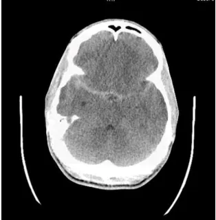
A 26-year-old African American male with no significant past medical history presented with complaints of a severe occipital headache that started during sexual intercourse and was non-radiating. His mother passed away from stroke in her thirties and father’s medical history was unknown. His siblings had no medical problems. He had used his usual dose of marijuana daily for the last 3 years and smoked 3-4 cigarettes per day. He was allergic to iodine and was not on any medications. He had no intake of alcohol or any toxin exposures. Patient described the headache as throbbing and graded it as 8/10 in intensity. He denied nausea, vomiting, visual disturbance, chest pain, palpitations, numbness, tingling, weakness or gait difficulties. He was afebrile, heart rate was 58, blood pressure 117/68. His neurologic exam was normal, including cranial nerves, strength reflexes, coordination, sensation, gait and station. CT head (Figure 1) showed an acute subarachnoid hemorrhage within basilar cisterns and right temporal lobe along the sylvian fissure with no
hydrocephalus. Neurology recommended BP goal <140/90 and admission to ICU for further management. He was started on nimodipine 60mg q4h for 21 days (7 of which he spent as
inpatient). Laboratory testing showed a normal white blood cell count, electrolytes, liver functions, and urine drug screen showed presence of cannabinoids. Patient’s headache went down to 3/10
intensity in the first 12 hours and after that he was asymptomatic. CTA head (Figure 2) showed no evidence of arteriovenous malformation (AVM) or arterio-venous (AV) fistula or intracranial aneurysm. DSA showed that there was mild hypervascularity in the right temporal region, possibly from an AVM. Dural venous sinuses were patent throughout. No aneurysm was identified. MRI brain without contrast (Figure 3) showed no clear source for the observed subarachnoid hemorrhage and the brain appeared normal. Transcranial dopplers performed daily over 7 days were negative for any significant vasospasm. CTA (Figure 4) after 1 week from onset showed resolution of the SAH and possible dissection flaps involving the cavernous portions of bilateral internal carotid arteries which ultimately were determined to be bony artifact.
Figure 1. CT head showing the SAH upon presentation.
Figure 3. A) MRI brain SWAN sequence the day after presentation.
Figure 3 B) MRI T2 Flair sequence
Discussion
Angiogram negative SAH (or non-aneurysmal SAH) accounts for 15-20% of all SAH2 and is believed to be caused by: venous drainage anomalies or venous sinus thrombosis; spinal or cerebral dural or parenchymal vascular malformation or tumors in the brain, spine, or subarachnoid space; infections; blood dyscrasias, complications of anticoagulants and fluoxetine, trauma, and small perforating vessel rupture.2 Our patient may have had a small vessel rupture that was not picked up on any of the three angiograms that were performed.
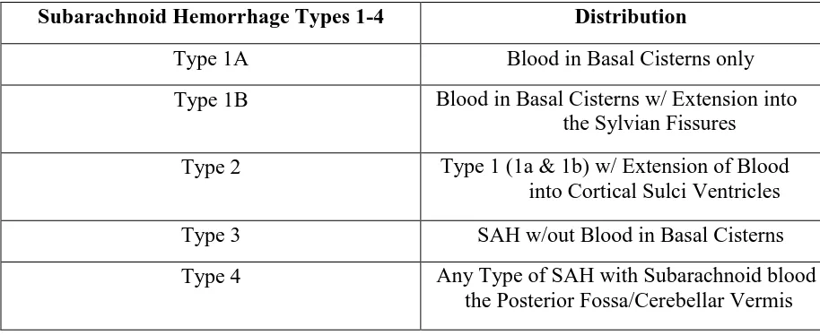
As per MacKinnon et al, the incidence of spontaneous SAH worldwide is approximately 9 per 100,000 persons peaking in the sixth decade.3 According to Nayak et al., in their classification system (Table 1), Type 1a and 1b SAH which are localized to basal cisterns and sylvian fissure only, have benign outcomes, based on their study of 129 type 1 SAH patients.2 Despite the likelihood that his prognosis would likely be benign, our patient was not discharged but admitted to observation in the hospital because the initial CT imaging raised the possibility of an underlying AVM and also because most cases of vasospasm occur on days 3-7. Once the MRI brain disproved any AVM, DSA was employed to not miss any aneurysm which may not have been noticed, as sometimes CTA misses a small aneurysm due to various reasons such as small aneurysm lumen size, technical factors like poor contrast timing, and large blood burden.4 Foreman et al reported finding that 4 out of 16 patients retrospectively studied with post-coital SAH had no brain pathology.4
Table 1.Illustration of the SAH classification categories from Nayak et al.1
Subarachnoid Hemorrhage Types 1-4 Distribution
Type 1A Blood in Basal Cisterns only
Type 1B Blood in Basal Cisterns w/ Extension into
the Sylvian Fissures
Type 2 Type 1 (1a & 1b) w/ Extension of Blood
into Cortical Sulci Ventricles
Type 3 SAH w/out Blood in Basal Cisterns
Type 4 Any Type of SAH with Subarachnoid blood in
the Posterior Fossa/Cerebellar Vermis
Non-aneurysmal SAH during sexual activity may be due to high blood pressure affecting spiking of mean arterial pressure (MAP) and impacting cerebral autoregulation negatively. Also, intercourse leads to elevated heart rate causing a hyperdynamic state.3 Oral erectile dysfunction treatment popular nowadays is with phosphodiesterase-5 inhibitors and these have been reported to be linked to both aneurysmal and non-aneurysmal SAH, especially during their illicit use;5,6,7,8 our patient, however, denied using these medications.
It would be helpful to have information about further relatives of patient in order to determine if there was any possibility of Ehlers-Danlos (EDS) vascular type in the family, however, he was lost to follow up. EDS makes one prone to dissection and possibly hemorrhage. The angiogram did not indicate any trace of Call – Fleming syndrome either, also known as
reversible vasoconstriction syndrome (RCVS), or of cerebral vasculitis which may cause acute headache.
Patient’s cannabis habit may have been a risk factor because vasoactive substance use may contribute to a subarachnoid hemorrhage. Rumalla et al reported that recreational marijuana use is independently associated with an increased likelihood of SAH.9 The incidence of a SAH among cannabis users was 10.67 times greater than expected when adjusted for age, 13.20 times greater than expected when adjusted for gender, and 8.71 times greater than expected when adjusted for race.9 This fact is not widely known, and further studies are needed.
The main psychoactive component of cannabis (tetrahydrocannbinol) effects mitochondria and oxidative stress, while cannabis cessation showed resolution of multifocal intracranial stenosis in 21% of young patients in a series with stroke.10 Transcranial doppler (TCD) studies have shown elevated cerebrovascular resistance in cannabis users, which may increase
susceptibililty to delayed cerebral ischemia, along with elevated systolic velocities.10 Curiously our patient did not have concerning TCD results. He did not have any striking structural
differences on vascular imaging (Figure 2, Figure 4) so there may still be an unknown
Rumalla et al saw a risk of an 18% increase for SAH correlating to cannabis, which is not insignificant, caution should be exercised when using vasoactive substances in the setting of activities that already act to increase heart rate and blood pressure naturally, as does intercourse. So perhaps when taking social history physicians could consider counselling marijuana users to take care, or at least be informed about their risk for rare but potentially grave neurologic
consequences, the mechanism of which is not fully understood. With legalization of medical and in some states recreational marijuana, it is useful to present this case for discussion. Certain corollaries exist; for example, valsalva manouvers have occasionally been linked to aneurysmal rupture and straining when constipated may potentially increase intracranial pressure, but there are many factors at work as to why sometimes a serious complication results. Similarly, it is conceivable that blood redistribution and cerebrovascular resistance may play a role in causing SAH when vasovagal substances like marijuana are used but there may be other understudied or unknown predictors.
The rationale for these conclusions is that while this patient had no long lasting repercussions, it was still a serious encounter that could have had dire consequences. He was an otherwise normal young male with the only risk factor of stroke being smoking. It could have turned out quite differently for another patient in a similar situation who had more vascular risk factors, or at another time for this same patient if he combined another vasovagal drug with the marijuana. Essentially, further study is needed on this subject, preferably as a case series. Cannabis has had a long history of use in the world, so it may be useful to keep track of similar cases of non-aneurysmal post-coital SAH coinciding with marijuana use, if they can be amassed, although they may be rather under-reported due to the sensitive and divisive nature of the topic.
References
1. Singh D, Jan A. Subarachnoid haemorrhage with orgasmic cephalgia. BMJ Case Rep. 2013;1–2.
2. Nayak S, Kunz AB, Kieslinger K, Ladurner G, Killer M. Classification of non-aneurysmal subarachnoid haemorrhage : CT correlation to the clinical outcome. Clin Radiol.
2010;65(8):623–8.
3. Mackinnon AD, Clifton AG, Rich PM. Acute subarachnoid haemorrhage: Is a negative CT angiogram enough? Clin Radiol. 2013;68(3):232–8.
4. Foreman PM, Griessenauer CJ, Selim MH, Searls DEC, Safdar A, Kasper EM, et al. Sexual activity as a trigger for intracranial hemorrhage. Acta Neurochirurgica. 2016;189–95.
5. Kaneria MV, Pagar S, Samant H, Yeole S, Patil S. Subarachnoid haemorrhage: possibly caused by the illegitimate use of sildenafil citrate. Journal Assoc Physicians India. 2006;56:809-811.
6. Byoun HS, Lee YJ, Yi HJ. Subarachnoid hemorrhage and intracerebral hematoma due to sildenafil ingestion in a young adult. Journal of Korean Neurosurgery Society. 201;47(3):210-212.
7. DeGiorgio F, Arena V, Arena E, Lodise M, Valerio L, d’Aloja E, Chiarott M. Subarachnoid hemorrhage during sexual activity after sildenafil intake: an accidental association. American Journal Forensic Med Pathology. 2011;23(4)310-311.
8. Antar V, Sutpideler N, Baran O, Bitirak G. Subarachnoid and intracerebral hemorrhage after alcohol ingestion and illicit use of sildenafil. Turkish Neurosurgery. 2015;25(3):485- 7.
9. Rumalla K, Reddy AY, Mittal MK. Association of recreational marijuana use with aneurysmal subarachnoid hemorrhage. J Stroke Cerebrovasc Dis. 2016;25(2):452–60.