*Corresponding author: Sumer S Choudhary
Department of Pulmonary/Respiratory Medicine, NKP Salve Institute of Medical Sciences And Research centre & Lata Mangeshkar ISSN: 0976-3031
Research Article
STUDY OF PATIENTS WITH RESTRICTIVE SPIROMETRY HAVING SIGNIFICANT
BRONCHODILATOR RESPONSE
*Sumer S Choudhary., Tayade B.O., Rini Abraham., Vishal More.,
Anchit Bhatnagar and Sonal Arsude
Department of Pulmonary/Respiratory Medicine, NKP Salve Institute of Medical Sciences And Research
centre & Lata Mangeshkar Hospital, Hingna Digdoh Hills Nagpur Maharashtra India 440019
DOI: http://dx.doi.org/10.24327/ijrsr.2017.0806.0354
ARTICLE INFO ABSTRACT
Introduction: Obstructive airway disease is characterized by reversibility on bronchodilator therapy measured by pulmonary function tests; however this is not often seen is restrictive diseases. We studied the clinical significance of bronchodilator reversibility in patients of restrictive pattern of spirometry. Materials and Methods: 30 patients with restrictive spirometry having significant bronchodilators response, were included in our study. Restiction was defined as decreased FVC and FEV1, with normal FEVI/FVC and a bronchodilator response as improvement of 12% and 200ml in FEV1 and FVC. Patients demographics, clinical history, treatment history, X-Ray characteristics, spirometry, diffusing lung capacity, and lung volumes measurements on Body Plethysmography were recorded.Results: The mean age was 48.60 + 14.06 years, majority of the patients were male 70 % and 22.23 + 3 .59 was females BMI slightly higher than males .40% were smokers, with shortness of breath the most common symptom, followed by cough, wheeze and chest pain..Asthma was the most common diagnosed medical condition and most of them were on bronchodialtors. The mean post bronchodilator FEV1% and FVC% was 66.88 + 24.28 and 70.95 + 24.99, with a reversibility of 12.91%.The FEV1/FVC% was 96.80+16.95. The mean TLC was normal whereas the RV, TLC, RV/TLC was increased.Conclusions: It can be concluded that post bronchodilator reversibility in patients of restrictive spirometry may be because of decrease elastic recoil resulting in early airway closure leading to air trapping and low FVC. Even though the numbers of such patients are low if symptomatic they would be benefited with bronchodilator therapy.
INTRODUCTION
Aims and Objectives
1. To evaluate patients clinical characteristics having restrictive spirometry with significant post bronchodilator response.
2. To study the reasons for significant response to bronchodilators in patients of restrictive spirometry. 3. To co-relate bronchodilator responsive restrictive
diseases with their radiographic and lung volume findings.
4. To study diffusion lung capacity (DLCO) and body plethysmography in patients of restrictive diseases having significant bronchodilator response.
Pulmonary function testing (PFT) is important investigatory tool for pulmonologists.PFT is a basic investigatory tool for
evaluation of pulmonary symptoms. It is used for long term management as well as for evaluation of potential disability. It is also used for epidemiological and research purposes[1]. Spirometry helps to differentiate between obstructive, restrictive and mixed disease.
It is a routine practice to use short acting bronchodialators while performing Pulmonary Function test (PFT) to assess the post bronchodilator reversibility. The patient’s response to the bronchodilator therapy is a physiological reaction due to the involvement of airway epithelium, nerves, smooth muscles and mediators.[2] In certain respiratory diseases like asthma and chronic obstructive airway diseases, PFT has a diagnostic, theurapeutic and prognostic importance. Bronchodialtors helps to measure the reversibility of obstruction and a good/significant response is recorded in Forced Expiratory
Available Online at http://www.recentscientific.com
International Journal of
Recent Scientific
Research
International Journal of Recent Scientific Research
Vol. 8, Issue, 6, pp. 17438-17442, June, 2017
Copyright © Sumer S Choudhary et al, 2017, this is an open-access article distributed under the terms of the Creative Commons Attribution License, which permits unrestricted use, distribution and reproduction in any medium, provided the original work is properly cited.
DOI: 10.24327/IJRSR CODEN: IJRSFP (USA)
Article History:
Received 15th March, 2017 Received in revised form 25th April, 2017
Accepted 23rd May, 2017 Published online 28th June, 2017
Key Words:
Volume in 1 second (FEV1) and Forced Vital Capacity (FVC) or both.
Patients having purely restrictive pattern on spirometry, with a significant response to bronchodilator as defined earlier have not been thoroughly evaluated. This bronchodilator responsiveness in patient subsets of restrictive disease is because of underlying airway disease or some other reason was studied.
MATERIALS AND METHODS
Study Design
Prospective cross sectional study. Inclusion Criteria
1. Patients aged 18 years and above
2. Patients and cares willing to participate. 3. Patients presenting with respiratory complaints of
shortness of breath, cough, wheezing, and chest pain. 4. Patients basic initial spirometry of restrictive patt
5. Patients with restrictive spirometry having significant post bronchodilator response willing
pulmonary lung volumes measurement on body
plethysmogrph. Exclusion Criteria
1. Patients age below 18 years. 2. Patients and carers not willing to give consent for
participation.
Patients with significant post bronchodilator response on spirometry attending Department of Pulmonary/Respiratory Medicine OPD satisfying the inclusion and exclusion criteria participated in study. Total of 121 patients of restrictive spirometry were initially screened of which 90 patients agreed for post bronchodilator test of which 55 patients had significant reversibility on post bronchodialation, however only 30 patients consented for Body Plethysmography and DLCO and were included in study population.
After written informed consent, data information was collected as per the specialized proforma., prepared for the study.
ATS guidelines were followed to define normal and restrictive pattern on pulmonary function test. Normal FEV1/FVC w values greater than 95% of the predicted for age, gender and height [1, 3]and restriction as decreased FVC and FEV1, with normal FEV1/FVC. Patients were subjected to pre and post bronchodilator test. Significant response was considered if post bronchodilator improvement of minimum 12% and 200 ml
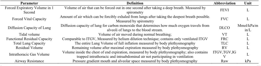
Table 1 Spirometry, Diffusion & Lung Volumes Measurements and their meaning with units
Parameter
Forced Expiratory Volume in 1 Second
Volume of air that can be forced out
Forced Vital Capacity Amount of air which can be forcibly exhaled from lungs after taking the deepest breath possible.
Diffusion Capacity of Lung Diffusion capacity of lung for carbon monoxide that determines how much oxygen travels from
Tidal volume
Functional Residual Capacity Comparable to ITGV,
Total Lung Capacity The entire Lung Volume of full inflation measured by body plethysmography
Residual Volume Remaining volume after maximal expiration measured by
Intrathoracic Gas Volume Volume inside the chest of end expiration, measured by body plethysmography;
trapped intrathoracic and intraabdominal air not participating in ventilation
Airway Resistance Pressure gradient mouth and alveolar space measured by body plethysmography
Forced Vital Capacity (FVC)
Patients having purely restrictive pattern on spirometry, with a bronchodilator as defined earlier have not been thoroughly evaluated. This bronchodilator responsiveness estrictive disease is because of underlying airway disease or some other reason was studied.
Patients aged 18 years and above Patients and cares willing to participate. Patients presenting with respiratory complaints of
shortness of breath, cough, wheezing, and chest pain.
Patients basic initial spirometry of restrictive pattern Patients with restrictive spirometry having significant
post bronchodilator response willing for DLCO and pulmonary lung volumes measurement on body
plethysmogrph.
Patients age below 18 years. Patients and carers not willing to give consent for
Patients with significant post bronchodilator response on spirometry attending Department of Pulmonary/Respiratory Medicine OPD satisfying the inclusion and exclusion criteria participated in study. Total of 121 patients of restrictive rometry were initially screened of which 90 patients agreed for post bronchodilator test of which 55 patients had significant reversibility on post bronchodialation, however only 30 patients consented for Body Plethysmography and DLCO and
After written informed consent, data information was collected , prepared for the study. ATS guidelines were followed to define normal and restrictive pattern on pulmonary function test. Normal FEV1/FVC was values greater than 95% of the predicted for age, gender and
and restriction as decreased FVC and FEV1, with Patients were subjected to pre and post bronchodilator test. Significant response was considered if post odilator improvement of minimum 12% and 200 ml was
recorded in FEV1 and or FVC
Thoracic society/European Respiratory Society in their guidelines. [2]. Improvements of less than 8% on
bronchodilator spirometry because of measurement variability. All those patients with restrictive spirometry pattern and having significant response to bronchhodialtor, and were willing for lung volume measurements were recruited for our study. DLCO and alveolar volume (VA) were measured using single breath method, lung volumes, airway resistance and specific conductance were also measured [4] on Geratherm Body Plethysmiograph. Patients clinical characteristics which included demographics including
symptoms, smoking history, co morbidities, and medication
used were recorded. Chest X-rays findings were noted. The different parameters along with their definitions were
followed as shown above in table 1.
To summarize following were recorded.
1. Patients Clinical charact
included demographics, clinical symptoms, smoking history, co-morbidities, and treatment history.
2. Chest X-rays findings were recorded.. 3. DLCO was measured by single breath method. 4. Lung volumes were measured using whole
plethysmography. 5. Total airway resistance was measured. Criteria for acceptability of lung volumes on body box:
1. Maneuver shows a closed loop without drift. 2. Tracing does not go off the screen.
3. Breathing is at 0.5-1 Hz 4. Tangents should be within 10%
5. At least 3 TGV values should agree within 5% and the mean value reported.
Spirometry, Diffusion & Lung Volumes Measurements and their meaning with units
Definition
Volume of air that can be forced out in one second after taking a deep breath. Measured by spirometry
Amount of air which can be forcibly exhaled from lungs after taking the deepest breath possible. Measured by spirometry
Diffusion capacity of lung for carbon monoxide that determines how much oxygen travels from alveoli of lungs to the blood stream.
Volume of air moved during normal breathing;
o ITGV, Measured by helium dilution technique; contains only ventilated ITGV The entire Lung Volume of full inflation measured by body plethysmography Remaining volume after maximal expiration measured by body plethysmography Volume inside the chest of end expiration, measured by body plethysmography; also contains
trapped intrathoracic and intraabdominal air not participating in ventilation Pressure gradient mouth and alveolar space measured by body plethysmography
Graph 1 Loop Tracings on Body Plerthysmograph
shortness of breath, cough, wheezing, and chest pain. ern
FEV1 and or FVC as defined by American Thoracic society/European Respiratory Society in their Improvements of less than 8% on post may because of measurement variability. All those patients with restrictive spirometry pattern and having significant response to bronchhodialtor, and were willing for lung volume measurements were recruited for our study. CO and alveolar volume (VA) were measured using single breath method, lung volumes, airway resistance and specific conductance were also measured [4] on Geratherm Body Patients clinical characteristics which included demographics including body mass index (BMI), symptoms, smoking history, co morbidities, and medication
rays findings were noted. The different parameters along with their definitions were
followed as shown above in table 1.
To summarize following were recorded.
Patients Clinical characteristics were recorded that included demographics, clinical symptoms, smoking
morbidities, and treatment history.
rays findings were recorded.. DLCO was measured by single breath method. Lung volumes were measured using whole-body
plethysmography. Total airway resistance was measured.
Criteria for acceptability of lung volumes on body box: Maneuver shows a closed loop without drift. Tracing does not go off the screen.
1 Hz hould be within 10%
At least 3 TGV values should agree within 5% and the mean value reported.
Spirometry, Diffusion & Lung Volumes Measurements and their meaning with units
Abbreviation Unit
in one second after taking a deep breath. Measured by
FEVI L
Amount of air which can be forcibly exhaled from lungs after taking the deepest breath possible.
FVC L
Diffusion capacity of lung for carbon monoxide that determines how much oxygen travels from DLCO Mmol/kPa/m
in/L
VT L
Measured by helium dilution technique; contains only ventilated ITGV FRC L
The entire Lung Volume of full inflation measured by body plethysmography TLC L
body plethysmography RV L
also contains trapped intrathoracic and intraabdominal air not participating in ventilation
ITGV,TGV,IG
V L
Pressure gradient mouth and alveolar space measured by body plethysmography Raw kPa
International Journal of Recent Scientific Research Vol. 8, Issue, 6, pp. 17438-17442, June, 2017
The graph 1 showing the normal effort and inadequate effort tracing on bodyplethysmography.
Ethical Issues
1. Ethical Committee approval. 2. Informed consent. 3. Confidentiality.
RESULTS
Total 30 patients were included in our study and following characteristics and lung volumes were recorded.
Majority of patients were male 21. The mean age of males 51.62 + 13.79 years and was greater than females. The BMI of females 22.23 + 3.59 kg/m2 which was slightly higher than males however were within normal limits. More than half of patients were smokers. They had mean pack years of 22.09 +5.41. (Table2)
All of them were symptomatic with SOB being the commonest symptom. The most common co morbid disease was asthma. Most of the patients were on steroids and on chest X-ray COPD was the most common finding (Table 3).
All of them showed restrictive impairment with mean post bronchodilator FEV1% 66.88+ 24.28 and FVC % 70.95+ 24.99 with normal FEV1/FVC 98.80+ 16.95.(Table 4)
DLCO was suggestive of moderate disease 52.27 + 12.59, TLC was normal whereas RV, RV/TLC, TGV/TLC and Raw tot was increased.(Table 5)
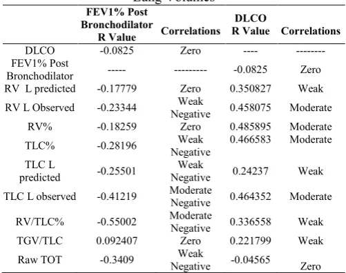
The above table no.6 showing the correlation of FEV1 and DLCO with different lung volume variables.FEV1 had a negative association with RV,TLC, Raw tot whereas DLCO showed weak to moderate association with RV,TLC, RV/ TLC,TGV/TLC and no association with FEV1 and Raw tot.
Photo 1 Body Plethysmography and DLCO
Table 2 Patients Clinical Characteristics
Distribution of Patients according to Sex n(%)
Male 21(70%)
Female 9(30%)
Total 30(100%)
Age in accordance to Gender Distribution
Male (n=21) 51.62 + 13.79*
Female (n=9) 41.56 + 12.72
Total (n=30) 48.60 +14.06
BMI in accordance to Gender Distribution
Male (n=21) 21.12 +3.05*
Female (n=9) 22.23 +3.59
Total (n=30) 21.50 +3.24
Distribution of Smokers, Ex Smokers and
Non-Smokers n(%)
Smokers 12(40%)
Ex Smokers 7(23.33%)
Non Smokers 11(36.67%)
Smokers Pack Years n
12 22.09 +5.41
n- Frequency,*Mean + Standard Deviation
Table 3 Symptoms, Chest X-Ray, Medical and Treatment History
Symptom n %
Shortness of Breath 22 73.33
Cough 18 60
Wheezing 8 26.67
Chest Pain 5 16.67
Chest X-Ray Findings
COPD 7 23.33
Kyphosis/Scoliosis 6 20
Pleuroparenchymal Fibrosis 3 10
Reticulonodular Shadows 2 6.67
Parenchymal Fibrosis 2 6.67
Pleural Fibrosis 1 3.33
Diagnosed Medical History
Asthma 8 26.67
COPD 6 20
Autoimmune Disease 3 10
Interstitial Lung Disease 2 6.67
Medications
Bronchodilator 16 53.33
Inhaled Steroids 11 36.67
Systemic Steroids 4 13.33
Home Oxygen Therapy 4 13.33
Immunosuppressive Therapy 2 6.67
Table 4 Baseline Pulmonary Function Tests
FEV1
Pre FEV1 In L 1.50 + 0.63*
Pre FEVI in % 53.99 + 17.78
Post FEV1 in L 1.90 + 0.66
Post FEVI in % 66.88 + 24.28
FVC
Pre FVC in L 2.17 + 0.70
Pre FVC in % 66.5 + 16.96
Post FVC in L 2.43 + 0.73
Post FVC in % 70.95 + 24.99
FEV1/FVC
FEV1/FVC L 76.47 + 14.56
FEV1/FVC % 96.80 + 16.95
*Mean + Standard Deviation
Table 5 Lung Volume Measurements
DLCO 52.27+12.59*
TLC in L 4.98+1.18
TLC% 99.8+59.50
RV in L 3.27+3.87
RV% 150.77+121.18
RV/TLC% 145.03+40.95
TGV/TLC in L 71.03+15.09
TGV/TLC % 131.90+22.35
Raw total 0.70+0.88
Raw total% 272.97+245.17
DISCUSSION
Our study was aimed to understand clinical significance and the reasons in subset of patients having restrictive pattern on spirometry with significant post bronchodilator response defined as decreased FVC, FEV1 with normal FEV1/FVC. The following observations were recorded:
In the present study most of them were males 70%, the mean age was 48.60 +14.06 years, males having higher mean age then females. The BMI of the study group was 21.50 +3.24 kg/m2 with females having slightly higher BMI however not significantly increased.40% of patients were smokers with pack years of 22.09 +5.41.
Most of patients in present study have low FEV1 and FVC with normal FEV1/FVC and normal TLC which is a non specific pattern. This was similar to the study group of patients of Hyatt
et al.[5]
The above recording may be because the patients are unable to exhale completely to empty their lungs to RV.[2]Early airway closure and gas trapping due to airflow limitation results in incomplete emptying of the lungsalong with hyperinflation.[6] In these patients the lung volumes would be misinterpreted as restrictive disease although the pathology is obstructive.
The measured DLCO was reduced in the majority of patients with the mean of 52.27 + 12.59 Mmol/kPa/min/L. This is usually seen in restrictive lung disease; however certain obstructive airway diseases like emphysema may have similar results.[7] DLCO/VA often normalizes in ILD, may be elevated in asthma, and it is however decreased in emphysema, ILD and many forms of chronic lung disease.
TLC is usually used as a marker of effects of obstructive airways diseases and a key index to confirm presence of a restrictive ventilator defect. The measured TLC in our study was of 99.8% + 59.50 or 4.98L+ 1.18.TLC is increased in asthma due to airway narrowing with air trapping and emphysema because of loss of elastic recoil.
Individual patient case reports of bronchodilator responsive restrictive disease described previously in literature had both restrictive spirometry and low TLC.They had concluded that contraction of alveolar ducts results in closure of terminal lung units and the reasons for low lung volumes.[8]
TLC was within normal range, only few patients had low TLC. There was no reduction in FEVI/FVC% 96.80 + 16.95, Most of the patients having spirometry interpretation of moderate restriction had normal TLC, excluding significant restriction in these subjects [9]. The clue to the presence of the obstructive defect was the response to bronchodilators.
The vital capacity may be decreased in both restrictive and obstructive disease hence not a specific parameter to determine restrictive defects, and has to be confirmed by low TLC. Similar to our study <60% of patients of Aaron et al. with restrictive pattern on spirometry, restrictive disease was confirmed on lung volume measurements.[10]
Residual volume usually accounts for 25% of TLC; this is a marker of gas trapping which reflects the effects of obstructive or restrictive diseases on lung volumes. Air trapping, results in increased RV and RV/TLC. In our study the mean RV was 150.77% +121.18 or 3.27L+3.87.Residual volume was increased in all of the patients, which decreases in restrictive disease. Residual volume may increase in asthma because of air trapping and emphysema because of loss of elastic recoil. It is reduced in pulmonary fibrosis due to increase elastic recoil. RV/TLC ratio reflects poor gas mixing and hence gas trapping. In fact, the average RV/TLC% was elevated 145.03+40.95 %.[11]
More than half of the patients had elevated RV, TLC and TGV which may be due to pseudo restriction. In one of the study of 413 patients done by Miller and Palecki having diagnosis of asthma found most of the patients had restrictive pattern on spirometry and was associated with increased BMI.[12] Pseudo restriction with air trapping was seen in only 7 patients. This is in contrast to the present study in which half of the patients had an elevated residual volume.
In our study the mean TGV/TLC was 131.90 % + 22.35 or 71.03L + 15.09.Patients with obstructive lung disease airway closure occurs at abnormally high lung volume resulting in increased TLC, RV and FRC and in diseases where the lung compliance decrease like diffuse intestinal diseases there would be decrease in TLC, RV and FRC, because of increase stiffness of the lungs and recoil of the lungs to a smaller resting volumes.
In our study the mean Airway Resistance was 272.97% +245.17 or 0 .70+ 0.87.The normal threshold abnormality of total airway resistance in adults is 0.3kPa/ (L/S). However we were not able to measure sReff i.e. specific effective airway resistance which, measures the larger central airways and sRtot specific total airway resistance which measures the smaller peripheral airways. The elevated airway resistance is a typical finding of obstructive lung disease
Most of the patients had low FEV1 and FVC with normal FEV1/FVC and TLC, indicating a non specific type of PFT similar to as described by Hyatt et al.5 Mayo clinic reported 10 % of the patients having similar pattern on spirometry. In 2004 12 patients were described to have similar pattern and was Table 6 Correlation between FEVI % and DLCO with
Lung Volumes
FEV1% Post Bronchodilator
R Value Correlations
DLCO
R Value Correlations
DLCO -0.0825 Zero ---- ---
FEV1% Post
Bronchodilator --- --- -0.0825 Zero
RV L predicted -0.17779 Zero 0.350827 Weak
RV L Observed -0.23344 Weak
Negative 0.458075 Moderate
RV% -0.18259 Zero 0.485895 Moderate
TLC% -0.28196 Weak
Negative
0.466583 Moderate
TLC L
predicted -0.25501
Weak
Negative 0.24237 Weak
TLC L observed -0.41219 Moderate Negative 0.464352 Moderate
RV/TLC% -0.55002 Moderate
Negative 0.336558 Weak
TGV/TLC 0.092407 Zero 0.221799 Weak
Raw TOT -0.3409 Weak
Negative -0.04565 Zero
International Journal of Recent Scientific Research Vol. 8, Issue, 6, pp. 17438-17442, June, 2017
thought to be of small airway disease.[13] In one of the follow up study by mayo clinic a significant response to bronchodilator was seen in 11 % of patients with nonspecific pattern.
We tried to corelate FEV1 and DLCO with different lung volume variables.FEV1 had a negative association with RV, TLC, Raw tot whereas DLCO showed weak to moderate association with RV,TLC,RV/TLC,TGV/TLC and no association with FEV1 and Raw tot.
As shown in table 7 our study population had features of obstruction and hyperinflation on body plethysmogarphy.
CONCLUSION
To summarize ours is one of the first study in our region to thoroughly analyze patients having restrictive pattern spirometry with significant bronchodilator response. Most of these patients had lung volume measurements suggestive of obstructive airway disease.
This may be due to early airway closure which results in air trapping. Clinicians have to be aware of this phenomenon, and the possibility of underlying obstructive diseases has to be considered. Such patients who are symptomatic may be benefitted wit bronchodilator therapy.
We only identified 30 patients who met these criteria, suggesting that this is a rather rare event. However, since our patients were comparable to the Mayo Clinic and Hyatt et al. study group patient’s i.e. nonspecific spirometry with significant bronchodilator response[5,14] we concluded that there are patients, though small in number having reversible restrictive airway disease and would be benefitted with bronchodilator therapy.
Acknowledgement
The authors would like to thank, the technician of pulmonary function tests Rajani Raut who helped us in doing the PFT of the patients and and statistician Jaydeep Nayase for Statistical analysis
References
1. M. R. Miller, J. Hankinson, V. Brusasco, F. Burgos, R. Casaburi, A. Coates et al (2005). Standardisation of spirometry. European Respiratory Journal, 26(2):319-338.
2. R. Pellegrino, G. Viegi, V. Brusasco, R.O. Crapo, F. Burgos, R. Casaburi et al (2005). Interpretative strategies for lung function tests. European Respiratory Journal, 26. (5):948-968.
3. J. L. Hankinson, J. R. Odencrantz, and K. B. Fedan (1999). Spirometric reference values from a sample of the general U.S. Population. The American Journal of Respiratory and Critical Care Medicine, 159(1):179-187.
4. R. O. Crapo, A. H. Morris, P. D. Clayton, and C. R. Nixon1982. Lung volumes in healthy nonsmoking adults. Clinical Respiratory Physiology, 18 (3):419-425. 5. R. E. Hyatt, C. T. Cowl, J. A. Bjoraker, and P. D.
Scanlon (2009). Conditions associated with an abnormal nonspecific pattern of pulmonary function tests. Chest, 135(2):419-424.
6. P. H. Quanjer, G. J. Tammeling, J. E. Cotes, O. F. Pedersen, R.Peslin, and J. C. Yernault. Lung volumes and forced ventilator flws. Report Working Party Standardization of Lung Function Tests, European Community for Steel and Coal (1993). Official Statement of the European Respiratory Society. The European Respiratory Journal, 16: 5-40.
7. Nancy J. Morrison, Raja T. Abboud, Fuad Ramadan, Roberta R. Miller, Nancy N. Gibson, Kenneth G et al (1989). Comparison of single breath carbon monoxide diffusing capacity and pressure volume curves in detecting emphysema. The American Review of Respiratory Disease, 139(5):1179-1187.
8. D. A. Kaminsky and C. G. Irvin (1993). Anatomic correlates of reversible restrictive lung disease. Chest; 103(3): 928-931.
9. R. E. Hyatt, P. D. Scanlon, and M. Nakamura editors. Interpretation of Pulmonary Function Tests, 3 Edn, Lippincott-Raven publisher, (1997).
10. S. D. Aaron, R. E. Dales, and P. Cardinal (1999). How accurate is spirometry at predicting restrictive pulmonary impairment. Chest, 115 (3): 869-873. 11. J. Stocks and P. H. Quanjer (1995). Reference values for
residual volume, functional residual capacity and total lung capacity: ATS Workshop on Lung Volume Measurements Official Statement of the European Respiratory Society. European Respiratory Journal, 8(3): 492-506.
12. A. Miller and A. Palecki (2007). Restrictive impairment in patients with asthma. Respiratory Medicine, 101(2): 272-276.
13. D. Stanescu and C. Veriter (2004). A normal FEV1/VC ratio does not exclude airway obstruction. Respiration, 71(4):348-35.
14. V. N. Iyer, D. R. Schroeder, K. O. Parker, R. E. Hyatt, and P. D.Scanlon (2011). The nonspecific pulmonary function test: longitudinal follow-up and outcomes. Chest, 139(4):878-886.
Table7 Body Plethysmographic measures as observed in major disorders and in our Study
RV TLC Raw TGV
Normal or elevated Obstructive airway
diseases
Normal or
elevated Normal Elevated
Hyperinflation Elevated Normal or
elevated Normal Elevated
Restrictive disorders
Reduced or
normal Reduced Normal Reduced
Present Study Elevated Normal or
Elevated Elevated Elevated
How to cite this article: