Solid pseudopapillary tumor of the pancreas:
Report of five cases
Cameron D Adkisson, Adam S Harris, Mellena D Bridges,
Justin H Nguyen, Horacio J Asbun, John A Stauffer
ABSTRACT
Introduction: Solid pseudopapillary tumor of the pancreas (SPT) is a rare pancreatic tumor with an unclear pathogenesis and good prognosis after resection. We report our experience with the diagnostic and therapeutic management of these tumors over 10 years. Case Series: A retrospective chart review was performed for all patients undergoing pancreatic resection at our institution from 20012011. Patients with final pathology demonstrating SPT were identified. Of 617 pancreatic resections performed at our institution, five patients (0.8%) were found to have solid pseudopapillary tumor of the pancreas. All patients were female with a mean age of 39.2 years. Abdominal pain was the presenting symptom in four patients. MRI demonstrated pancreatic head involvement in three patients and pancreatic tail involvement in one patient. CT scan was used to diagnose one tumor located in the body of the pancreas. Mean tumor size was 8.0 cm. Surgical
treatment consisted of two open pancreaticoduodenectomies, one laparoscopic pancreaticoduodenectomy, one laparoscopic subtotal pancreatectomy and one laparoscopic distal pancreatectomy. Surgical pathology revealed no evidence of vascular invasion in any of the tumors and an R0 resection complete resection with no microscopic residual tumor was obtained in all patient. No patients had involved lymph nodes with a mean of 16 lymph nodes inspected. No complications from any pancreatic resection were observed. Conclusion: Solid pseudopapillary tumors of the pancreas are a rare but treatable pancreatic tumor. Complete surgical excision is the treatment of choice and can be achieved through an open or minimal access technique. Keywords: Solid pseudopapillary tumor, Pancreas, Pancreaticoduodenectomy
*********
Adkisson CD, Harris AS, Bridges MD, Nguyen JH, Asbun HJ, Stauffer JA. Solid pseudopapillary tumor of the pancreas: Report of five cases. International Journal of Hepatobiliary and Pancreatic Diseases 2012;2:914. Article ID: 100005IJHPDCDA2012
********* doi:10.5348/ijhpd20125CS3
INTRODUCTION
First described by Franz in 1959, solid pseudopapillary tumor of the pancreas (SPT) is a rare, lowgrade malignant tumor of unknown etiology accounting for 0.22.7% of all primary pancreatic
CASE SERIES OPEN ACCESS
Cameron D Adkisson
1, Adam S Harris
1, Mellena D
Bridges
2, Justin H Nguyen
3, Horacio J Asbun
4, John A
Stauffer
4Affiliations:
1Resident, General Surgery, Mayo Clinic
Florida, Jacksonville, FL, USA;
2Consultant, Department
of Radiology, Mayo Clinic Florida, Jacksonville, FL, USA;
3
Consultant, Department of Transplantation, Mayo Clinic
Florida, Jacksonville, FL, USA;
4Consultant, Department
of Hepatobiliary and Pancreas Surgery, Mayo Clinic
Florida, Jacksonville, FL, USA.
Corresponding Author: Cameron D Adkisson, MD Mayo
Clinic Florida, Department of General Surgery 4500 San
Pablo Road Jacksonville, FL 32224; Ph: 904 953 81 59;
Email: [email protected]
tumors [1–3]. It is also known as solid cystic papillary epithelial tumor, papillary cystic tumor, solid and cystic tumor, papillarycystic neoplasm, Hamoudi or Franz tumor. SPT is most prevalent in women of younger age [1, 4–9]. Abdominal mass is the most common presenting symptom, with dyspepsia, early satiety, nausea, or vomiting being less common presenting symptoms. Up to 20% of patients are asymptomatic with tumors identified either incidentally on imaging or at operation for unrelated pathology [2, 6, 10–13].
Grossly, SPTs are identified as welldemarcated, encapsulated tumors with extrapancreatic growth. Mixed solid and cystic components are evident with internal necrotic or hemorrhagic debris and lobulated, solid tissue at the periphery. Characteristic radiographic features include the presence of an encapsulated mass with solid and cystic components on either CT scan or MRI, with MRI notably better for identification of certain tumor characteristics such as the presence of a capsule, hemorrhage or cystic degeneration [12].
SPT should be added to the differential diagnosis in any patient with a solid and partly cystic mass of the pancreas especially in females under 35 years of age [14]. Surgical resection is the treatment of choice for affected patients and is associated with an overall good prognosis.
CASE SERIES
Case 1: A 21yearold female presented to our
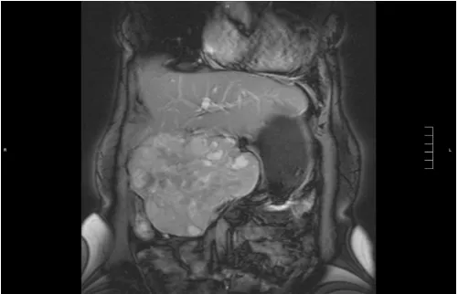
institution with persistent abdominal pain for three years after a palliative cholecystojejunostomy for an “inflammatory myofibroblastic pseudotumor of the pancreas” followed by 16 cycles of adjuvant chemotherapy with doxorubicin and ifosfamide. Her symptoms included epigastric pain, jaundice, and decreased appetite. Laboratory analysis was notable only for slight elevation of transaminases (ALT 42 IU/L, AST 30 IU/L). MRI showed a well circumscribed, oval heterogeneous mass 8.9x8.3 cm, in the head of the pancreas, displacing the common bile duct, portal vein and duodenum (Figure 1). Obliteration of the SMV with collateral formation and dilation of the pancreatic duct were noted. An exploratory laparotomy with pancreaticoduodenectomy was performed with resection of SMV and reimplantation into the IVC. Pathology showed a 9.5 cm pancreatic SPT without invasion. No lymph nodes were involved in tumor and immunohistochemical staining was positive for vimentin with patchy staining for synaptophysin and chromogranin. Her postoperative course was uncomplicated and she was discharged home on postoperative day 7. At three months follow up she was without complaints. She was unfortunately lost to follow up after as she returned home to Puerto Rico (Table 1).
Case 2: A 17yearold female presented with
episodic postprandial right upper quadrant pain. Cholecystitis was suspected and an ultrasound was performed showing cholelithiasis and an incidental
pancreatic tail mass eight cm in size. MRI was subsequently performed showing an 8.8x5.8 cm mass in the pancreatic tail with involvement of the splenic artery causing splenomegaly and gastric varices. Endoscopic ultrasound with fine needle aspiration was performed finding a solid and cystic mass in the pancreatic tail consistent with SPT. Laparoscopic subtotal pancreatectomy, splenectomy and cholecystectomy were performed demonstrating a 12.5 cm SPT with extrapancreatic extension. Immunostains were notable for diffusely positive vimentin, CD10 and progesterone, with focal positivity for cytokeratin and synaptophysin. No lymph nodes were involved with the tumor. Her postoperative course was uncomplicated and she was discharged home on postoperative day9. She has done well at nine months follow up with no signs of tumor recurrence (Table 1).
Case 3: A 52yearold female with a four month
history of postprandial discomfort and nausea with emesis was found to have an incidental 1.6 cm pancreatic body mass on CT scan performed for elevated liver function tests. Endoscopic ultrasound with biopsy was performed showing a 1.6 cm hypoechoic mass in the midbody of the pancreas with central calcification. Fine needle aspiration was suggestive of SPT. Laparoscopic distal pancreatectomy with enbloc splenectomy was performed demonstrating a two cm SPT without lymphovascular invasion. Immunostains were strongly positive for CD 10, vimentin and progesterone, with weak positivity for synaptophysin. No immediate complications were observed and she was discharged on postoperative day 2. No further therapy was required and she was without evidence of recurrence at two year follow up (Table 1).
Case 4: A 75yearold female presented with a one
year history of a palpable mass in her right upper quadrant associated with occasional stabbing abdominal pain. She had a history of an open cholecystectomy 30 years prior with the incidental finding of an unresectable “calcified tumor” between the pancreas and the liver. CT scan was performed showing a 12 cm cystic and calcified tumor near the pancreatic head (Figure 2). An ultrasound guided biopsy was performed suggestive of a malignant neuroendocrine tumor. MRI at our institution demonstrated a 16.3x13.9 cm pancreatic head mass with multiple cystic areas and foci of hemorrhage. Displacement of the portal vein and common bile duct was seen without evidence of invasion. An exploratory laparotomy and pancreaticoduodenectomy was performed demonstrating a 15 cm pancreatic head mass consistent with SPT. Immunostains were strongly positive for vimentin and neuron specific enolase with focal reactivity for chromogranin and synaptophysin, carcinoembryonic antigen and CAM5.2. No lymph nodes were involved with tumor. No immediate complications were observed and she was discharged on postoperative day8. At five year follow up she has required no additional therapy and is without evidence of recurrent disease (Table 1).
outside physician with abdominal pain after exposure to a family member with mononucleosis. Further questioning revealed diarrhea and a 20 pound weight loss over 1–2 months. An ultrasound was performed showing splenic calcifications and subsequently a CT scan noting a 1.1 cm mass in the pancreatic head. CT guided biopsy was performed with tumor staining positive for alpha1antitrypsin, betacatenin, CD10, vimentin, progesterone receptor and CD56. This was consistent with a diagnosis of SPT. She was referred to our institution for resection. MRI was performed for preoperative planning which confirmed a 1.2 cm lesion in the pancreatic head with high signal intensity on T2 and contrast enhancement on delayed images consistent with her outside diagnosis. A replaced right hepatic artery was incidentally identified without evidence of
additional disease. A laparoscopic
pancreaticoduodenectomy was performed, finding a 1.2 cm pancreatic head SPT with 0/7 involved lymph nodes
Figure 1: Abdominal MRI coronal view of patient 1 demonstrating a heterogeneous, well circumscribed 8.3x7.7x8.9 cm mass in the head of the pancreas with internal cystic structure and high T1 signal intensity suggesting hemorrhagic debris, consistent with SPT.
Figure 2: Patient 4 abdominal MRI coronal view demonstrating a 16.3x10.2x13.9 cm lobulated, heterogeneous, but wellcircumscribed mass at the level of the pancreatic head containing multiple cystic areas and discrete foci of hemorrhage consistent with SPT.
Figure 3: Patient 5 gross specimen demonstrating a well circumscribed 1.2 cm nodule. Microscopy showed a fibrous capsule, histologically with mixed solid and cystic components consistent with SPT.
(Figure 3). No immediate complications were observed and she was discharged on postoperative day 6. At one month follow up she is progressing well, with plans to undergo a six month follow up MRI (table 1).
DISCUSSION
Solid pseudopapillary tumor (SPT) of the pancreas is a rare neoplasm with a generally good prognosis after complete surgical excision. Earlier detection and increased awareness of these neoplasms has led to their increased prevalence in recent years [15]. SPT has a particularly high prevalence in females younger than 35 years of age. In the largest retrospective review conducted to date including 718 patients with SPT, more than 90% of patients were female with an average age of 22 years [6–4]. Female predominance has been attributed to the proximity of primordial pancreatic cells to the ovarian ridge during development [16]. The differential diagnosis of suspicious neoplasms should include microcystic adenoma, mucinous cystic neoplasm, nonfunctioning islet cell tumor, pancreatic adenocarcinoma, pancreaticoblastoma, cystic degeneration of solid neoplasm and calcified hemorrhagic pseudocyst [10, 17].
Radiographic features include a well encapsulated, hypodense mass with various solid and cystic components on CT scan [12]. Ultrasound demonstrates a homogenous or heterogenous encapsulated mass composed of both solid echogenic and hypoechogenic components [1819]. MRI features include a well defined lesion with high or low signal intensity on T1 and high signal intensity on T2 [12]. MRI is preferable to CT scan for demonstrating the presence of a capsule, hemorrhage, or cystic degeneration [12].
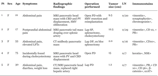
Table 1: Patient characteristics of all five patients.
Pt Sex Age Symptoms Radiographic
findings Operationperformed Tumorsize (cm)LN Immunostains
1 F 21 Abdominal pain MRI pancreatic head mass with CBD and PV displacement, SMV obliteration
Open PD with SMV resection and reimplantation
9.5 0/20 vimentin+, synaptophysin+, chromogranin+,
2 F 17 Postprandial abdominal
pain MRI pancreatic tail massdraping over splenic artery
Lap SP, splenectomy, cholecystectomy
12.5 0/29 vimentin+, CD 10, + PR+
3 F 53 Abdominal pain,
elevated LFTs CT midbody pancreaticmass Lap DP, en blocsplenectomy 2.0 0/16 vimentin+, CD10+,PR+
4 F 75 Incidentally found
during cholecystectomy MRI pancreatic headmass with PV and CBD displacement
Open PD 15 0/7 keratin+, NSE+
5 F 30 Abdominal pain,
diarrhea, weight loss CT/MRI pancreatic headmass, replaced right hepatic artery
Lap PD 1.2 0/7 vimentin+, PR,+ CD 10+, CD 56+, β catenin+, α1AT+
structures such as the mesenteric vessels, stomach, and duodenum may also be involved [10]. In the above study of 718 patients with SPT, pancreatic tail involvement was seen in 35.9% of patients, 34% of tumors were found in the pancreatic head, and 10.3% were found in the body and tail [6]. Extrapancreatic tumors were noted in approximately 1% of patients [6].
Grossly, the tumor is demarcated from adjacent pancreatic tissue by the presence of a fibrous capsule [10, 12]. Microscopic examination yields a mixed solid and cystic mass with hemorrhagic or necrotic cellular material in its center with lobules of solid tissue at its periphery [10]. Characteristic findings include the presence of solid areas alternating with pseudopapillary formations, foamy histiocytes, nuclear grooves and cytoplasmic globules [15].
Tumor markers observed in SPT including APT/β catenin, cyclin D1 and D3, vimentin, antitrypsin, NSE, and progesterone [12, 16, 20–24]. In a study by Kosmahl et al. the most consistent tumor markers present in 90% of their patients with SPT were vimentin, alpha1antitrypsin, NSE and progesterone [16]. Other authors have noted that immunoreactivity to βcatenin is found in the cytoplasm and nuclei of almost all SPT’s [23–24].
Malignant transformation occurs in 15% of adults and 13% of children, with a greater risk of malignant transformation noted in males and the elderly [12, 25]. Angioinvasion, perineural invasion and deep invasion of pancreatic tissue characterize malignancy [12]. Tumor recurrence has been found in 5–7% of patients after surgical resection [6, 15]. Metastases are most common
to liver, lymph nodes, and peritoneum and are found in 10–15% of patients [12, 15, 17, 26]. Poor prognostic factors include the presence of vascular invasion, tumor size greater than five cm and low nuclear grade [15, 26, 27]. Five year survival after complete resection is 94–100% [6, 15, 17].
Surgery is the only definitive treatment for SPT with a cure rate of greater than 95% with complete resection [28–34]. In contrast to other pancreatic tumors, aggressive surgical resection is warranted even in the presence of local invasion, recurrence, or limited metastases [27, 32–33, 35–36]. En bloc resection of tumor with formal lymphadenectomy can be undertaken in addition to resection of synchronous or metachronous metastases [14]. Surgical debulking can be effective for metastatic disease involving the liver [28–29, 37–39]. In contrast to other pancreatic tumors, invasion of the portal vein or superior mesenteric artery does not indicate tumor unresectability [40]. Pancreatic fistula is the most common complication after complete resection seen in approximately 7% of patients [11]. The role of adjuvant therapy in treatment of SPTs is unclear, with few studies demonstrating a role for gemcitabine and radiotherapy to downsize large tumor(s) or treat the rare case of unresectable disease [41–44]. The tumors’ high rate of resectability limits the need for adjuvant therapy [41].
CONCLUSION
Solid pseudopapillary tumors of the pancreas are a
rare but treatable pancreatic tumor. While clinical signs and symptoms are relatively nonspecific, characteristic findings on imaging and histology separate these tumors from the more malignant pancreatic tumors. Complete surgical excision is the treatment of choice and can be achieved through an open or minimal access technique.
********* Author Contributions
Cameron D Adkisson – Conception and design, Acquisition of data, Analysis and interpretation of data, Drafting the article, Critical revision of the article, Final approval of the version to be published
Adam S Harris – Conception and design, Acquisition of data, Analysis and interpretation of data, Drafting the article, Critical revision of the article, Final approval of the version to be published
Mellena D Bridges – Conception and design, Acquisition of data, Analysis and interpretation of data, Drafting the article, Critical revision of the article, Final approval of the version to be published Justin H Nguyen – Conception and design, Acquisition of data, Analysis and interpretation of data, Drafting the article, Critical revision of the article, Final approval of the version to be published
Horacio J Asbun – Conception and design, Acquisition of data, Analysis and interpretation of data, Drafting the article, Critical revision of the article, Final approval of the version to be published
John A Stauffer – Conception and design, Acquisition of data, Analysis and interpretation of data, Drafting the article, Critical revision of the article, Final approval of the version to be published
Guarantor
The corresponding author is the guarantor of Submission.
Conflict of Interest
The authors declare no conflict of interest. Copyright
© Cameron D Adkisson et al. 2012; This article is distributed under the terms of Creative Commons attribution 3.0 License which permits unrestricted use, distribution and reproduction in any means provided the original authors and original publisher are properly credited. (Please see www.ijhpd.com/copyright policy.php for more information.)
REFERENCES
1. Frantz VK. Tumors of the pancreas. In: Atlas of Tumor Pathology. Washington DC: Armed forces Institute of Pathology 1959:32–3.
2. Chen Z, Zhou GW, Zhou HJ, Peng, et al. Diagnosis and treatment of solidpseudopapillary tumors of the pancreas. Hepatobiliary and Pancreatic Dis Int
2005;4:456–9.
3. Pettinato G, Manivel JC, Ravetto C, et al. Papillary cystic tumor of the pancreas: a clinicopathologic study of 20 cases with cytologic, immunohistochemical, ultrastructural, and flow cytometric observations, and a review of literature. Am J Clin Pathol 1992;5:478–88. 4. Sun CD, Lee WJ, Choi JS, et al. Solid
pseudopapillary tumors of the pancreas: 14 years experience. ANZ J Surg 2005;75:684–9.
5. Kosmahl M, Peters K, Anlauf M, et al. Solid pseudopapillary neoplasms. Enigmatic entity with female preponderance. Pathologe 2005;26:41–5. 6. Papavramidis T, Papavramidis S. Solid
pseudopapillary tumors of the pancreas: review of 718 patients reported in English literature. J Am Coll Surg 2005;200:965–72.
7. Lack E, Levey R, Cassady J, et al. Tumors of the exocrine pancreas in children and adolescents: a clinical and pathologic study of eight cases. Am J Surg Pathol 1983;7:319–27.
8. Jaksic T, Yaman M, Thorner P, et al. A 20 year review of pediatric pancreatic tumors. J Pediatric Surg 1992;27:1315–7.
9. Grosfeld J, Vane D, Rescorla F, et al. Pancreatic tumors in childhood: analysis of 13 cases. J Pediatric Surg 1990;25:1057–62.
10. Dong D, Zhang, S. Solidpseudopapillary tumor of the pancreas: CT and MRI features of 3 cases. Hepatobiliary and Pancreatic Diseases International 2006;5(2):300–4.
11. Chen S, Zou S, Dai Q, Li H. Clinical analysis of solid pseudopapillary tumor of the pancreas: report of 15 cases. Hepatobiliary Panc Dis Int 2008;7(2):196–200.
12. Zhang H, Liang T, Wang W, et al. Diagnosis and treatment of solidpseudopapillary tumor of the pancreas. Pancreas 2006;5(3):454–8.
13. de Castro S, Singhal D, Aronson D, Busch O, Gulik T, et al. Management of solidpseudopapillary neoplasms of the pancreas: a comparison with standard pancreatic neoplasms. World J Surgery 2007;31:1129–34.
14. Tipton S, Smyrk T, Sarr M, et al. Malignant potential of solid pseudopapillary neoplasm of the pancreas. British Journal of Surgery 2006;93:733–7.
15. Martin R., Klimstra D, Brennan M, Conlon K. Solid pseudopapillary tumor of the pancreas: a surgical enigma? Annals of Surgical Oncology 2002;9(1):35–40.
16. Kosmahl M, Seada L, Janig U, et al. Solid pseudopapillary tumor of the pancreas: its origin revisited. Virchows Arch 2000;436:473–80.
17. Reddy S, Cameron J, Scudiere J, Hruban R, et al. Surgical management of solidpseudopapillary neoplasms of the pancreas (Franz or Hamoudi Tumors): a large single institutional series. Journal Am Coll Surg 2009;208(5):950–7.
18. D’Onofrio M, Malago R, Vecchiato F, et al. Contrast enhanced ultrasonography of small solid pseudopapillary tumors of the pancreas: enhancement pattern and pathologic correlation of 2 cases. J Ultrasound Med 2005;24:849–54.
19. Lee D, Ti B, Lim J, et al. Sonographic findings of solid and papillary epithelial neoplasm of the pancreas. J Ultrasound Med 2001;20:1229–32. 20. Miao J, Kusafuka T, Kuroda S, Yoneda A, at al.
accumulation in solid and cystic tumor of the pancreas associated with metastasis. Int J Mol Med 2003;11:461–4.
21. Abraham S, Klimstra D, Wilentz R, Yeo C, et al. Solid pseudopapillary tumors of the pancreas are genetically distinct from pancreatic ductal adenocarcinomas and almost aways harbor beta catenin mutations. Am J Pathol 2002;160:1361–9. 22. Santini D, Poli F, Lega S. Solidpapillary tumors of
the pancreas: histopathology. JOP 2006;7:131136. [3i]Andronikou S, Moon A, Ussher R. Peritoneal metastatic disease in a child after excision of solid pseudopapillary tumor of the pancreas: a unique case. Pediatr Radiol 2003;33:269–71.
23. Tanaka Y, Kato K, Notohara K, et al. Frequent B catenin mutation and cytoplasmic/nuclear accumulation in pancreatic solid pseudopapillary neoplasm. Cancer Res 2001;61:8401–4.
24. Abraham S, Klimstra D, Wilentz R, et al. Solid pseudopapillary tumors of the pancreas are genetically distinct from pancreatic ductal adenocarcinomas and almost always harbor b catenin mutations. Am J Pathol 2002;160:1361–9. 25. Machado C. M, Machado A. M, Bacchella T,
Jukemura J, et al. Solid pseudopapillary neoplasm of the pancreas: distinct patterns of onset, diagnosis, and prognosis for male versus female patients. Surgery 2008;29–34.
26. Kang C, Kim K, Choi J, Kim H, et al. Solid pseudopapillary tumor of the pancreas suggesting malignant potential. Pancreas 2006;32(3):276–80.
27. Nishihara K, Nagoshi M, Tsuneyoshi M, et al. Papillary cystic tumors of the pancreas: assessment of their malignant potential. Cancer 1993;71:8292. 28. Hassan I, Celik I, Nies C, et al. Successful treatment
of solidpseudopapillary tumor of the pancreas with multiple liver metastases. Pancreatology 2005;5:289–94.
29. Alexandrescu D, O’Boyle K, Feliz A, et al. Metastatic solidpseudopapillary tumor of the pancreas : clinicobiological correlates and management. Clin Oncol 2005;17:358–63.
30. Lam K, Lo C, Fan S. Pancreatic solidcysticpapillary tumor: clinicopathologic features in eight patients from Hong Kong and review of literature. World J Surg 1999;23:1045–50.
31. Salvia R, Bassi C, Festa L, et al. Clinical and biologic behavior of pancreatic solid pseudopapillary tumors: report on 31 consecutive patients. J Surg Oncol 2007;95:304–10.
32. Kaufman S, Reddick R, Stiegel M, et al. Papillary cystic neoplasm of the pancreas: a curable pancreatic tumor. World J Surg 1986;10:851–9. 33. Mao C, Guvendi M, Domenico D, et al. Papillary
cystic and solid tumors of the pancreas: a pancreatic embryonic tumor? Studies of three cases and cumulative review of the worlds’ literature. Surgery 1995;11:821–8.
34. Lam KY, Lo C, Fan S. Pancreatic solidcystic papillary tumor: clinicopathologic features in eight patients from Hong Kong and review of literature. World J Surg 1999;23:1045–50.
35. Fried P, Cooper J, Balthazar E, et al. A role for radiotherapy in the treatment of solid and papillary neoplasms of the pancreas. Cancer 1985;56:2783–5. 36. Zinner M, Shurbaji, Cameron J. Solid and papillary
epithelial neoplasm of the pancreas. Surgery
1990;108:475–80.
37. deCastro S, Singhal D, Aronson D, et al. Management of solidpseudopapillary neoplasms of the pancreas: a comparison with standard pancreatic neoplasms. World J Surg 2007;31:1130–5.
38. Nagri S, Abdu A, Anand S, et al. Liver metastasis four years after Whipple resection for solid pseudopapillary tumor of the pancreas. JOP 2007;8:223–7.
39. Rebhandl W, Felderbauer F, Puig S, et al. Solid pseudopapillary tumor of the pancreas (Frantz tumor) in children: report of four cases and review of literature. J Surg Oncol 2001;20:221–4.
40. Jeng L, Chen M, Tang R. Solid and papillary neoplasm of the pancreas: emphasis on surgical treatment. Arch Surg 1993;128:433–6.
41. Maffuz A, Bustamante F, Silva J, TorresVargas S. Preoperative gemcitabine for unresectable,solid pseudopapillary tumor of the pancreas. Lancet Oncol 2005;6:185–6.
42. Matsunou H, Konishi F. Papillarycystic neoplasm of the pancreas: a clinicopathologic study concerning the tumor aging and malignancy of nine cases. Cancer 1990;65:283–91.
43. Maffuz A, Bustamante F, Silva J, TorresVargas S. Preoperative gemcitabine for unresectable, solid pseudopapillary tumor of the pancreas. Lancet 2005;6:185–6.