Khosropanah H., et al. J Dent Shiraz Univ Med Sci., 2018 March; 19(1): 19-27.
Original Article
The Impact of Calcium Hydroxide on the Osteoinductive Capacity of
Demineralized Freeze-Dried Bone Allograft: an
In-Vitro
Study
Hengameh Khosropanah 1, Nazila Lashkarizadeh 2, Maryam Ayatollahi 3, Maryam Kaviani 4, Zohreh Mostafavipour 5
1
Dept. of Periodontics, School of Dentistry, Shiraz University of Medical Sciences, Shiraz, Iran.
2 Dept. of Periodontics, School of Dentistry, Kerman University of Medical Sciences, Kerman, Iran. 3
Molecular Genetics and Stem Cell Biology, Transplant Research Center, Bone and Joint Diseases Research Center, Shiraz University of Medical Sciences, Shiraz, Iran.
4
Transplant Research Center, Shiraz University of Medical Sciences, Shiraz, Iran.
5 Dept. of Biochemistry, Shiraz University of Medical Sciences, Shiraz, Iran.
KEY WORDS
Calcium Hydroxide;
Stem Cells;
Allografts;
Bone Regeneration;
Received April 2016;
Received in Revised form February 2017; Accepted May 2017;
ABSTRACT
Statement of the Problem: A great challenge in periodontal therapy is the regen-eration enhancement of osseous defects through applying osteoinductive materials.
Demineralized freeze-dried bone allograft (DFDBA) has already been introduced
as an allograft with osteoconductive and variable osteoinductive properties.
Calci-um hydroxide [Ca(OH)2] is an available well-known material in dentistry, which
induces hard tissue formation.
Purpose: This study evaluated the efficiency of combination of DFDBA and Ca(OH)2 in improving the quality of osteoinduction of DFDBA.
Materials and Method: Human bone marrow-derived mesenchymal stem cells were taken from volunteers’ iliac crest. Cell proliferation was determined by MTT test at 18, 24 and 48 hours post-culture in 10 groups. The employed material were
0.5, 1.0 mg/ml Ca(OH)2 in two forms of suspension and pH-adjusted solution,
10mg/ml DFDBA per se and in combination with 0.5 and 1.0 mg/ml Ca(OH)2.
Mineralization was assessed by Alizarin red staining in 10 mg/ml DFDBA,
DFDBA+ 0.5 and 1 mg/ml Ca(OH)2 in solution and suspension forms. The data
were statistically analyzed by using one-way ANOVA and Tukey’s post-hoc test
(p< 0.05).
Results: The pH-adjusted solutions exhibited better cell proliferation compared with the suspension groups. The combination of 0.5mg/ml Ca(OH)2 solution and
DFDBA increased the cell proliferation and mineralization compared with
DFDBA per se (p= 0.033).
Conclusion: The combination of Ca(OH)2 with DFDBA improved the
osteoinduc-tivity of DFDBA.
Corresponding Author: Lashkarizadeh N., Dept. of Periodontics, School of Dentistry, Kerman
Univer-sity of Medical Sciences, Kerman, Iran. Email:nazilalashkarizadeh@yahoo.com Tel: +98-3432119021
Cite this article as: Khosropanah H., Lashkarizadeh N., Ayatollahi M., Kaviani M., Mostafavipour Z. The Impact of Calcium Hydroxide on the Osteoinductive Capacity of Demineralized
Freeze-Dried Bone Allograft: an In-Vitro Study. J Dent Shiraz Univ Med Sci., 2018 March; 19(1): 19-27.
Introduction
Periodontitis is an inflammatory disease initiated by the
presence of bacterial plaque, leading to the destruction
of periodontium (periodontal ligament, alveolar bone
and cementum) and eventually tooth loss. [1]
Regener-ating the periodontal defects as well as the osseous
re-construction of edentulous ridge to place the implants is
a major challenge in modern dentistry and can have great influence on the patient’s aesthetics, chewing abil-ity and qualabil-ity of life. Several approaches have been
introduced to improve the reconstruction of alveolar
The process of periodontal and bone regeneration
is generally complicated by different types of present
cells. Active regeneration is valuable through induction
of purposed cell population to differentiate or migrate to
the defect area, which is the migration of bone
progeni-tor cells to the injured site and differentiation of stem
cells to osteoblasts that leads to the regeneration of bone
structure. [4] Also, in the reconstruction of large bony
defects, there should be a graft material to provide both
scaffold and active signaling to the bone marrow
mes-enchymal stem cells (MSCs) inducing them to migrate
and differentiate. [4-5]
Among bone replacement grafts, autograft is
con-sidered the gold standard. [6] They are the only bone
grafts that provide all the three properties of an ideal
graft material including osteogenesis (to provide vital
osteoblasts), osteoinduction (induction of
osteoprogeni-tor cells to differentiate to osteoblasts) and
osteoconduc-tion (to provide scaffold). [6] However, owing to the
low level of patient acceptance of autografts, allografts
(grafts from the same species) have become the most
popular bone grafts in oral surgery. Allografts are being
processed as fresh-frozen or freeze-drying. [6]
Demineralized freeze-dried bone allograft
(DFDBA) is a type of allograft claimed to be superior
because of the osteoconductive and osteoinductive
properties. The osteoinductivity is obtained through
demineralization of the graft material and exposure of
the bone morphogenetic proteins (BMPs). These are a
group of growth factors inducing MSCs differentiation
into bone-producing osteoblast cells in bone matrix.
[7-8] Therefore, the demineralization is critical for
oste-oinductive properties of DFDBA, since it provides
ade-quate levels of BMPs and does not allow the mineral
content to drop to very low levels.
It is believed that a certain concentration of Ca2+ ions is needed for the osteoinductive properties of
DFDBA; therefore, addition of Ca2+ ions might provide the necessary Ca2+ ions for the optimum function of these allografts. [6, 9] Moreover, other factors such as
the donor’s age make the osteoinductivity of DFDBA
variable and unpredictable. [10] Therefore, the recent
studies aimed at improving the active bone and
perio-dontal regeneration investigated the addition of different
growth factors such as emdogain or rh-PDGF to
DFDBA. It was noted that although such additives
im-proved the regeneration, these growth factors are
expen-sive and not readily accessible. [11-12]
In contrast, calcium hydroxide, [Ca(OH)2] with
hard tissue formation and antimicrobial properties, is a
readily accessible material and quite well known in
den-tistry, particularly in endodontics, for pulp capping or
apexification. Furthermore, Ca(OH)2 is a strong base
with low solubility in water that forms suspension in
aqueous environment and releases hydroxyl and
calci-um ions over time. Most properties of the Ca(OH)2
come from this ionic dissociation. [13]
Based on in vitro studies, Ca(OH)2 increases the
recruitment, migration, and proliferation of periodontal
ligament (PDL) stem cells and promotes the
mineraliza-tion and cementogenesis. However, scholars do not
have consensus over its efficiency mainly because it is
dose-dependent. [14-15] Paula-Silva et al. [15] in their
in vitro study found that low dosage of Ca(OH)2 did not
influence the induction of mineralization and high
dos-age was cytotoxic to cells. [15] Bone marrow-derived
MSCs are a group of multipotent stem cells that are
sensitive to their local environment and differentiate
into different types of cells including periodontal
liga-ment-like or alveolar bone cell types. [16]
The present study evaluated the effect of adding
different doses of Ca(OH)2 in both solution and
suspen-sion forms to DFDBA on viability, proliferation and
differentiation of human bone marrow-derived
mesen-chymal stem cells (hBM-MSCs) into osteoblasts. The
significance of this study is that Ca(OH)2 is inexpensive,
abundant and easy to obtain compared with other
mate-rials. To the best of the authors’ knowledge, this is the
first study carried out on this issue. Moreover, the
con-troversies about the osteoinductivity of DFDBA
en-couraged the evaluation of in vitro behavior of
hBM-MSCs in the presence of DFDBA (Cenobone,
Deminer-alized Cortical Cancellous Powder; Tissue Regeneration
Corporation Co., Kish, Iran) as a popular allograft
mate-rial routinely used in periodontal and osseous
recon-structive surgeries.
Materials and Method
Isolation and culture of human MSCs
Human MSCs were obtained from 5 ml bone marrow
aspirated from iliac crest of normal donors within the
Khosropanah H., et al. J Dent Shiraz Univ Med Sci., 2018 March; 19(1): 19-27.
marrow to relatives after obtaining approval of Ethic
Committee. Written informed consent was also taken to
permit the analysis of the clinical data. Bone
marrow-derived MSCs were isolated from mononuclear cell
(MNC) layer using our previously method [17] which is
briefly mentioned.
Each aspirated sample was diluted (1:1) with Dul-becco’s modified Eagle’s medium (DMEM) (Invitro-gen; Merelbeke, Belgium) containing 10% fetal bovine
serum (FBS), 1% penicillin, 1% streptomycin, and 2
mM glutamine as the basal DMEM media. The cells
were layered over about 5 ml of Ficoll (Lymphoprep;
Oslo, Norway), then centrifuged at 338 g for 15 min to
obtain MNC layer. The MNC layer was seeded on
cul-ture flasks, and maintained at 37°C in 5% CO2
atmos-phere. In order to obtain the MSCs cells, the adhered
monolayer cells was detached and expanded for
succes-sive passages, and characterized.
Characterization of human MSCs
The cells viability was analyzed by using trypan blue
staining. Flow cytometric analysis for detection of
MSC-morphologic markers was also achieved
follow-ing the method used by Ayatollahi et al. [18]
Proliferation assay
The two types of employed Ca(OH)2 were suspension
(non-pH-adjusted) and solution (pH-adjusted). For the
suspension groups, 50 and 100mg Ca(OH)2 (Merck;
Germany) were added to 10 ml normal saline, yielding
5- and 10-mg/ml concentration of Ca(OH)2. Then, they
were diluted in the culture medium in a 1:10 ratio to
obtain 0.5- and 1-mg/ml doses of Ca(OH)2. In the
solu-tion group, the powder was first dissolved in HCl, then,
NaOH was added and the pH was adjusted to 7.4 via a
pH meter.
Accordingly, 10 designed study groups were as
follows; group 1: control group (MSCs in culture
medi-um with no treatment), group 2: 0.5mg/ml Ca(OH)2
solution, group 3: 0.5mg/ml Ca(OH)2 suspension, group
4: 1mg/ml Ca(OH)2 solution, group 5: 1mg/ml Ca(OH)2
suspension, group 6: 10mg/ml DFDBA, group 7: 10mg/
ml DFDBA+ 0.5mg/ml Ca(OH)2 solution, group 8: 10
mg/ml DFDBA+ 0.5mg/ml Ca(OH)2 suspension, group
9: 10mg/ml DFDBA+ 1mg/ml Ca(OH)2 solution and
group 10: 10mg/ml DFDBA+ 1mg/ml Ca(OH)2
suspen-sion. Each experiment was repeated 4 times.
Cell proliferation in each of the above groups was
assessed by using Thiazolyl Blue Tetrazolium bromide
(MTT) (Sigma-Aldrich Co.; St.Louis, MO, USA) assay.
The hBM-MSCs (5.8×10³) were cultured with 200µL of
DMEM containing 10% FBS. After 3 days of culture,
treatment in the mentioned groups was done for 18, 24
and 48 hours. Finally, MTT assay was conducted via
0.5 mg/ml 3-((4,5-dimethyl thiazole-2-yl)-2,5-diphe-nyl
tetrazolium) bromide for 3 hours. Following the
for-mation of formazan solution in DMSO (dimethyl
sul-foxide), the absorbance of the solution was measured by
an absorbance microplate reader at 570 and 630nm as
thereferencewavelength(BioTekInstrumentsInc.;USA).
Osteogenic differentiation assay
The potential of human MSCs to differentiate into
oste-ogenic lineages was examined in the presence of
osteo-genic media in four groups by using Alizarin red
stain-ing (Sigma-Aldrich Co., USA) within 5, 12 and 14 days
after staining.The groups weregroup 1: MSCs in
oste-ogenic media containing DFDBA (10mg/ml), group 2:
MSCs in osteogenic media containing 0.5mg/ml
Ca(OH)2 (solution)+ DFDBA 10mg/ml, group 3: MSCs
in osteogenic media containing 0.5mg/ml Ca(OH)2
(suspension)+ DFDBA 10mg/ml, and group 4: MSCs in
osteogenic media (positive control).
The osteogenic media consisted of basal DMEM
media supplemented with 10 M/L dexamethasone, 10
mM/L glycerol phosphate, 3.7 g/L sodium bicarbonate
and 0.05 g/L ascorbic acid (all from Sigma-Aldrich; St.
Louis, USA). The osteogenesity was assessed by
Aliza-rin red staining. The morphologic differentiation of cells
and red mineralized nodule formation were observed by
using microscope (Nikon) under 10×40 magnification.
Statistical analysis
The triplicate samples obtained from different cases
were analyzed in duplicate. The statistical analyses were
performed by using SPSS software, version 16 (IBM;
USA). One-way ANOVA was used to analyze the data. Tukey’s post-hoc test was used for pair-wise compari-sons between the study groups. p< 0.05 was considered
to be statistically significant. Alizarin red staining
re-sults were presented qualitatively.
Results
Characterization of human MSCs
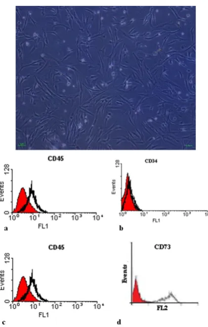
The rapidly grown fibroblast-like cells, which exhibited
expansion after inverted microscopic evaluation (Figure
1a). The viability assessed by trypan blue staining
anal-ysis showed 98-100% viability in human MSCs. The
flow cytometry determined the surface phenotype to be
positive for CD90 and CD73. Additionally, no cell
ex-pressed the hematopoietic markers CD45 and CD34
(Figure 1).
Figure 1: Characterization of human mesenchymal stem cells. Relatively homogenous fibroblast-like cells were obtained within 9-10 days after cell culture [A]. The surface phenotypic markers were positive for CD90, and CD73. Additionally, no cells expressed the hematopoietic markers CD45, CD34. The shaded area shows the profile of the negative control.
Cell proliferation analysis
There was a significant interaction effect between the
time and group, i.e., the culture duration and material
concentrations significantly affected the cellular
prolif-eration (p< 0.001). The cell proliferation was compared
among the groups at each time point of 18, 24 and 48
hours as demonstrated in Table 1.
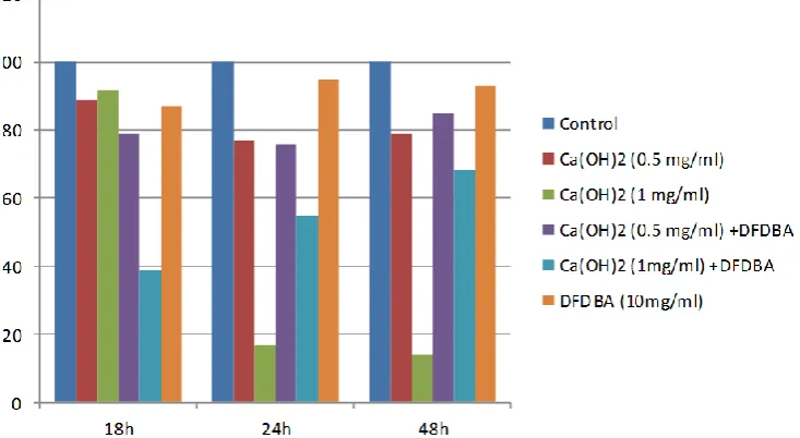
After 18 hours treatment
After 18 hours of treatment, the cell proliferation was
significantly lower in the group 10 compared with the
othergroups,whichwerenotsignificantlydifferent from
the control group.
Table 1: Bone morrow-derived mesenchymal stem cells viability in different study groups (significant differences have been marked)
Group Time (hr)
18h 24h 48h
1 0.98 ± 0.10a 0.96 ± 0.07a,b 0.96 ± 0.05a,b,c 2 0.8 ± 0.06a 0.91 ± 0.06a,b 0.93 ± 0.04a,b,c
3 0.88 ± 0.07a 0.74 ± 0.13b 0.76 ±0.05b,d,f
4 0.78 ± 0.02a 0.90 ± 0.05a,b 0.93 ± 0.01a,b,c
5 0.91 ± 0.05a 0.17 ± 0.02a 0.14 ± .03a,c
6 0.86 ± 0.08a 0.92 ± 0.02a,b 0.90 ± 0.04a,b,d 7 0.84 ± 0.17a 0.86 ± 0.13a,b 1.04 ± 0.05a,b,c,e 8 0.78 ± 0.04a 0.73 ± 0.22b 0.82 ± 0.06a,b,c,d,f 9 0.86 ± 0.09a 0.98 ± 0.04a,b 1.00 ± 0.10a,b,c,e 10 0.39 ± 0.08 0.53 ± 0.16b 0.66 ± 0.05b,c,d,e,f
a,b,c,d,e,f p< 0.05 as compared with group 10, 5, 3, 7, 8 and 9
respec-tively
After 24 hours treatment
The cell proliferation in groups 5 and 10 was
signifi-cantly lower than the control group. 1-mg/ml Ca(OH)2
solution yielded significantly higher cell proliferation
compared with the suspension form. Furthermore, cell
proliferation was significantly lower in 1 mg/ml
Ca(OH)2 suspension than in 0.5 mg/ml Ca(OH)2
sus-pension. Compared with 1 mg/ml Ca(OH)2 suspension
per se, the combination of 1 mg/ml Ca(OH)2 suspension
and DFDBA significantly increased the cell
prolifera-tion. However, the cell proliferation in DFDBA was
higher than its combination with 1 mg/ml Ca(OH)2
sus-pension.
After 48 hours treatment
The above-mentioned differences were still present
among the groups. Both 0.5 and 1 mg/ml Ca(OH)2
sus-pensions and DFDBA+ 1mg/ml Ca(OH)2 suspension
exhibited significantly lower proliferation in
compari-son with the control group. Both 0.5 and 1mg/ml
Ca(OH)2 solutions exhibited significantly higher cell
proliferation than the same doses in suspension form.
Furthermore, the combination of DFDBA with 0.5
mg/ml, Ca(OH)2 solution significantly increased the cell
proliferation in comparison with DFDBA per se. The
cell proliferation in groups 7 and 9 showed an ascending
trend from 18 to 48 hours to reach to a level higher than
the control group but the difference was not statistically
significant (Figures 2 and 3).
Osteogenic differentiation assay
Qualitatively, Alizarin red staining results showed
oste-ogenic differentiation in BM-MSCs in four designed
groups. The morphologic differentiation of cells and red
Khosropanah H., et al. J Dent Shiraz Univ Med Sci., 2018 March; 19(1): 19-27.
Figure 2: percentage change of proliferation and viability in different groups as compared with the control in different time points. DFDBA per se, Ca(OH)2 solution per se, and in combination with DFDBA
cope. It was noted that only MSCs in osteogenic media
containing Ca(OH)2 in the solution group qualitatively
exhibited mineralization in comparison with Ca(OH)2 in
suspension form and DFDBA groups (Figure 4).
Discussion
Results of the current study revealed that all the
Ca(OH)2 groups in solution form with an adjusted pH
(7.4) had a better proliferation viability in comparison
with the suspension groups (non-pH-adjusted),
indicat-ing an improved cellular function in the presence of a
proper pH.
Ca(OH)2 suspension in 1.0 mg/ml dose was
cyto-toxic to the cells and significantly decreased the cell
proliferation compared with the baseline. Combination
of 1 mg/ml Ca(OH)2 suspension with DFDBA increased
the viability of cell proliferation. This was probably due
to the buffering ability of DFDBA through a reduction
in Ca(OH)2 pH.
Narita et al. [19] demonstrated that dentine
pow-der and hydroxyapatite (both comprised of mineralized
compounds similar to DFDBA) had buffering effect on
Ca(OH)2; this is beneficial when using a mixture of
DFDBA and Ca(OH)2.
The results of the present study were consistent
with some previous studies. Narita et al. [19] used
dif-ferent doses of Ca(OH)2 at different pH levels. They
concluded that high dose of Ca(OH)2 (2.5 mg/ml) was
cytotoxic to the primary osteoblast cells (in both
pH-adjusted and non-pH-pH-adjusted groups); whereas, in low-
Figure 4: Osteogenic differentiation assay. Alizarin red staining results qualitatively showed osteogenic differentiation in marrow-derived mesenchymal stem cells in four designed groups. a: The red mineralized nodule formation were observed in osteogenic media containing DFDBA, b: Ca(OH)2 solution + DFDBA, c: Ca (OH)2 suspension + DFDBA. d: The MSCs in osteogenic media was cultured
as positive control.
er doses (0.25 and 0.025mg/ml), proliferation was
com-parable to the control group. In addition, 0.25mg/ml
Ca(OH)2 had better mineralization potency and this was
significantly higher for 7.4 pH in comparison to 8.5 pH.
[19]
Paula-Silva et al. [15] evaluated the effect of
dif-ferent concentrations of Ca(OH)2 on cementoblastic
differentiation of PDL mesenchymal cells, observed that
Ca(OH)2 with the concentration below 15mM (1.1mg/
ml) led to cell viability of more than 90%. A 10-mM
(0.74mg/ml) suspension of Ca(OH)2 decreased the cell
proliferation at post-culture 48-72 hours; meanwhile, it
led to significant cementoblastic differentiation of PDL
cells, increasing mineralization. In their study, the pH
was adjusted to 7.4 by the buffering agents of culture
environment and 5% CO2 incubation. [15]
Ji et al. [20] evaluated the effect of low doses of
Ca(OH)2 (1-10 µg/ml) on cell proliferation of
periodon-tal stem cells. They concluded that low doses of
Ca(OH)2 positively affected the proliferation of pulp
and PDL-derived stem cells. They also found that the
combination of 10µg/ml Ca(OH)2 and osteogenic media
increased the mineralization in vitro in comparison with
each of them per se. [20] This was not consistent with
the study conducted by Narita et al.[19] who found that
low dose of Ca(OH)2 (0.025 mg/ml) did not influence
the mineralization. This could be due to applying
Ca(OH)2 with osteogenic media, as well as different in
vitro conditions in different studies.
The present study, in line with Narita et al. [19]
and Paula-Silva et al., [15] showed that the pH-adjusted
solution of Ca(OH)2 yielded better outcomes compared
with the suspension form. They found that the Ca(OH)2-
induced mineralization was strictly dose-dependent and
responsible for osteoblast differentiation through
providing extracellular and intracellular Ca2+ ions which led to the activation of calcium/ calmodulin pathway.
[15, 19] However, previously the induction of hard
tis-sue formation by Ca(OH)2 was mostly attributed to its
alkaline pH (hydroxyl ions).
The elevated pH by Ca(OH)2 may not be a
prob-lem in pulp-capping or apexification, so that applying
Ca(OH)2 adjacent to the pulpal structure leads to the
necrosis (2mm), followed by an inflammation zone and
eventually hard-tissue formation. Yet, the elevated pH
should be considered in periodontal regeneration in
or-der not to disturb the cell viability, migration, and
dif-ferentiation. [13] The main and interesting result of the
study was that combination of 0.5 mg/ml pH-adjusted
solution of Ca(OH)2 and DFDBA significantly
in-creased the proliferation and mineralization compared
with the DFDBA per se. Further studies are suggested
to test it in vivo.
Moghadam et al. [21] evaluated the effect of
add-ing 0.15% by weight Ca(OH)2 to demineralized bone
material (DBM) gel on bone defects in rabbit calvaria.
They found no beneficial effect in adding Ca(OH)2 to
Khosropanah H., et al. J Dent Shiraz Univ Med Sci., 2018 March; 19(1): 19-27.
dose or form of Ca(OH)2, which is highly
dose-dependent regarding both cell proliferation and
induc-tion of hard tissue formainduc-tion.
In the present study, the results of cell
prolifera-tion in DFDBA group revealed that 10mg/ml
concentra-tion of DFDBA caused lower cell proliferaconcentra-tion than the
control group, and did not induce mineralization. This
was in agreement with Carnes et al.’s study [22] on 2T9
cells (preosteoblastic cell line derived from transgenic
mice calvariae). They reported that the exposure of
the-se cells to DFDBA did not result in mineralization.
However, exposure of these cells to soluble BMP-2 led
to increased mineralization and alkaline phosphatase
activity. Accordingly, they concluded that DFDBA was
not likely to liberate soluble BMPs towards
osteoinduc-tion, or in vivo studies might be more appropriate to
determine the osteoinductive properties. [22]
Vaziri et al. [23] in a study on the evaluation of
osteoinductive property in three different commercial
brands of DFDBA (one of them Cenobone- the same
brand as the present study) reported that 8 and 16 mg/ml
DFDBA decreased the proliferation of osteoblast-like
cells. However, in contrast to the present study, they
observed increased mineralization in 16mg/ml DFDBA.
[23] These differences could be due to different cell
lines and different doses administered.
In order to adopt the appropriate dose of DFDBA
in in vitro assessment, the present study followed Vaziri
et al. [23] and adopted 16mg/ml at the beginning.
How-ever, this dose resulted in very low cell viability and
proliferation in hBM-MSCs. Hence, 10mg/ml was
cho-sen according Dereka et al.’s study. [24] Other factors
likely to be involved in the variability of osteoinductive
property of DFDBA were the donor’s age, improper
processing of DFDBA, and not exposing adequate
amount of osteoinductive portions such as BMPs or
inadequate Ca2+ ions remained in its structure. [9, 25] This study observed that the low solubility of
Ca(OH)2 led to the formation of a suspension when
mixing Ca(OH)2 with culture medium. Therefore,
Ca(OH)2 was used in two forms of suspension and
solu-tion. The amount of ionic dissociation in the suspension
form was different from the solution form. The ions in
the suspension form were released into the environment
over time in a non-linear manner; while, in the solution
form, Ca(OH)2 was solved by applying acid, yielding a
consistent solution with a higher level of Ca2+ ions available.
The present study showed that combination of
Ca(OH)2 solution and DFDBA enhanced the
osteoin-ductive activity. However, regarding its use in clinical
conditions, especially in regeneration of osseous
de-fects, one should consider the following issues. First, an
appropriate combination is required to provide adequate
Ca2+ ions during the active phase of osseous regenera-tion in vivo in the presence of blood and inflammatory
exudate. Second, proper pH for regeneration is
neces-sary as alterations in pH may impair the regeneration
process. [2] There is a specific kind of oil-based
Ca(OH)2 called Osteora (previously Osteoinductal),
which is introduced for periodontal purposes considered
to have a milder pH increment. There are controversies
about the impact of this product on improving the
peri-odontal regeneration and whether it could release the
appropriate content of Ca2+ ions in favor of increasing mineralization or not. [14, 26] Therefore, the impact of
adding this product to DFDBA on osteoinduction could
be assessed in future studies.
Despite the promising findings of this study, it still
has some limitations including the undetermined
con-tent of Ca2+ and OH- ions in different time spans in Ca(OH)2 suspension groups as well as the Ca
2+
content
in DFDBA group to verify the hypothesis of the study.
Despite the previously conducted studies on the
properties of Ca(OH)2 in cell proliferation and
osteo-blast differentiation, the strength of the present study
lied in evaluation of the most effective dosages of
Ca(OH)2 with respect to the previous studies.
Further-more, the current study evaluated both solution and
sus-pension forms in order to investigate different aspects of
this issue and to examine the previously proposed
hy-potheses regarding the mechanism of mineralization
induction by Ca(OH)2. Besides, the recruited cell line
was hBM-MSCs, which are one of the most important
cells involved in the process of bone regeneration.
Conclusion
Within the limitations of this study, it seems that the
addition of 0.5mg/ml Ca(OH)2 solution to DFDBA is
beneficial for improving cell proliferation as well as
osteoblast differentiation compared with DFDBA per
Owing to the availability and low price of
Ca(OH)2, it is recommended that the manufacturers
consider Ca2+ content of DFDBA and apply DFDBA in intraosseous defects or bone reconstruction surgeries in
the combination form to provide appropriate content of
Ca2+ ions.
Acknowledgements
The authors thank the Vice-Chancellery of Shiraz
Uni-versity of Medical Science for supporting this research
(grant#93-01-03-8172). This article was based on the
thesis by Dr. Nazila Lashkarizadeh (thesis# 1734).The
authors would also like to thank Dr. Ehya Amalsaleh for
improving English in the manuscript and Dr. Vossough
from the Dental Research Development Center of the
School of Dentistry for the statistical analysis.
Conflict of Interest
The authors of this manuscript certify that they have no
conflict of interest.
References
[1] Araújo AC, Gusmão ES, Batista JE, Cimões R. Impact of
periodontal disease on quality of life. Quintessence Int.
2010; 41: e111-e118.
[2] Murphy KG, Gunsolley JC. Guided tissue regeneration
for the treatment of periodontalintrabony and furcation
defects. A systematic review. Ann Periodontol. 2003; 8:
266-302.
[3] Darby I, Chen ST, Buser D. Ridge preservation
tech-niques for implant therapy. Int J Oral Maxillofac
Im-plants. 2009; 24 Suppl: 260-271.
[4] Yamada Y, Ueda M, Naiki T, Takahashi M, Hata KI,
Nagasaka T. Autogenous injectable bone for regeneration
with mesenchymal stem cells and platelet-rich plasma:
Tissue-engineered bone regeneration. Tissue Engin.
2004; 10: 955-964.
[5] Yoshida T, Washio K, Iwata T, Okano T, Ishikawa I.
Current status and future development of cell
transplanta-tion therapy for periodontal tissue regeneratransplanta-tion. Int J
Dent. 2012; 2012: 307024.
[6] Kao ST, Scott DD. A review of bone substitutes. Oral
Maxillofac Surg Clin North Am. 2007; 19: 513-521.
[7] Miron RJ, Zhang YF. Osteoinduction: a review of old
concepts with new standards. J Dent Res. 2012; 91:
736-744.
[8] Li H, Pujic Z, Xiao Y, Bartold PM. Identification of bone
morphogenetic proteins 2 and 4 in commercial
deminer-alized freeze-dried bone allograft preparations: pilot
study. Clin Implant Dent Relat Res. 2000; 2: 110-117.
[9] Zhang M, Powers RM Jr, Wolfinbarger L Jr. Effect(s) of
the demineralization process on the osteoinductivity of
demineralized bone matrix. J Periodontol. 1997; 68:
1085-1092.
[10]Wei L, Miron RJ, Shi B, Zhang Y. Osteoinductive and
Osteopromotive Variability among
DifferentDemineral-ized Bone Allografts. Clin Implant Dent Relat Res. 2015;
17: 533-542.
[11]Fujioka-Kobayashi M, Schaller B, Zhang Y, Kandalam
U, Hernandez M, Miron RJ. Recombinant human bone
morphogenetic protein (rhBMP)9induces osteoblast
dif-ferentiation when combined with demineralized
freeze-dried bone allografts (DFDBAs) or biphasic calcium
phosphate (BCP). Clin Oral Investig. 2017; 21:
1883-1893.
[12]Miron RJ, Bosshardt DD, Laugisch O, Dard M, Gemperli
AC, Buser D, et al. In vitro evaluation of demineralized
freeze-dried bone allograft in combination with enamel
matrix derivative. J Periodontol. 2013; 84: 1646-1654.
[13]Mohammadi Z, Dummer PM. Properties and applications
of calcium hydroxide in endodonticsand dental
trauma-tology. Int Endod J. 2011; 44: 697-730.
[14]Eick S, Strugar T, Miron RJ, Sculean A. In vitro-activity
of oily calcium hydroxide suspension on microorganisms
as well as on human alveolar osteoblasts and periodontal
ligament fibroblasts. BMC Oral Health. 2014; 14: 9.
[15]Paula-Silva FW, Ghosh A, Arzate H, Kapila S, da Silva
LA, Kapila YL. Calcium hydroxide promotes
cemento-genesis and inducescementoblastic differentiation of
mesenchymal periodontalligament cells in a CEMP1- and
ERK-dependent manner. Calcif Tissue Int. 2010; 87:
144-157.
[16]Stockmann P, Park J, von Wilmowsky C, Nkenke E,
Felszeghy E, Dehner JF, et al. Guided bone regeneration
in pig calvarial bone defects using autologous
mesen-chymal stem/progenitor cells- a comparison of different
tissue sources. J Craniomaxillofac Surg. 2012; 40:
310-320.
[17]Ayatollahi M, Geramizadeh B, Zakerinia M, Ramzi M,
Yaghobi R, Hadadi P, et al. Human Bone
Marrow-deriv-ed Mesenchymal Stem Cell: A Source for Cell-BasMarrow-deriv-ed
Khosropanah H., et al. J Dent Shiraz Univ Med Sci., 2018 March; 19(1): 19-27.
[18]Ayatollahi M, Soleimani M, Geramizadeh B, Imanieh
MH. Insulin-like growth factor 1 (IGF-I) improves
hepat-icdifferentiation of human bone marrow-derived
mesen-chymal stem cells. Cell Biol Int. 2011; 35: 1169-1676.
[19]Narita H, Itoh S, Imazato S, Yoshitake F, Ebisu S. An
explanation of the mineralization mechanism in
osteo-blastsinduced by calcium hydroxide. Acta Biomater.
2010; 6: 586-590.
[20]Ji YM, Jeon SH, Park JY, Chung JH, Choung YH,
Choung PH. Dental stem cell therapy with calcium
hy-droxide in dental pulp capping. Tissue Eng Part A. 2010;
16: 1823-1833.
[21]Moghadam HG, Sándor GK, Holmes HH, Clokie CM.
Histomorphometric evaluation of bone regeneration
us-ing allogeneic and alloplastic bone substitutes. J Oral
Maxillofac Surg. 2004; 62: 202-213.
[22]Carnes DL J, Fontaine JDL, Cochran D, Mellonig J,
Keogh B, Harris S. Evaluation of 2 novel approaches for
assessing the ability of demineralized freeze-dried bone
allograft to induce new bone formation. J Periodontolo.
1999; 70: 353-363.
[23]Vaziri S, Vahabi S, Torshabi M, Hematzadeh S. In vitro
assay for osteoinductive activity of
differentdemineral-ized freeze-dried bone allograft. J Periodontal Implant
Sci. 2012; 42: 224-230.
[24]Dereka XE, Markopoulou CE, Mamalis A, Vrotsos IA.
Effect of rhBMP-7 combined with two bone grafts on
humanperiodontal ligament cell differentiation. Growth
Factors. 2009; 27: 274-279.
[25]Schwartz Z, Mellonig J, Carnes D, Fontaine JDL,
Cochran DL, Dean DD. Ability of commercial
deminer-alized freeze-dried bone allograft to induce new bone
formation. J Periodontol. 1996; 67: 918–926.
[26]Stratul SI, Schwarz F, Becker J, Willershausen B,
Scule-an A. Healing of intrabony defects following treatment
with an oilycalcium hydroxide suspension
(Osteoinduc-tal). A controlled clinical study. Clin Oral Investig. 2006;