SH Naser mostofy,et al
Study of the Effect of GapSeal on
Mi-crogap and Microleakage in Internal Hex
Connection After Cyclic Loading
SH Naser mostofy
1, E Jalalian
2, N Valaie
3, Z Mohtashamrad
4,
A Haeri
5, T Bitaraf *
61-Assistant professor, Prosthodontics Dept, Dental Faculty, Tehran Medical Sciences, Islamic Azad University, Tehran, Iran 2-Assistant professor, Prosthodontics Dept, Member of implant research center, Dental Faculty, Tehran Medical Sciences, Islamic Azad University, Tehran, Iran
3- Faculty member of Thalasemia Research center, Mazandaran ,Iran 4- Dentist, Tehran, Iran.
5- Postgraduate student, Oral and maxillofacial surgery Dept, Dental Faculty, Tehran Medical Sciences, Islamic Azad Uni-versity, Tehran, Iran
6- Assistant professor, Dental Implant Research Center, Dental Faculty, Tehran Medical Sciences, Islamic Azad University, Tehran, Iran
ABSTRACT ARTICLE INFO
Article History
Received: May 2019 Accepted: Apr 2019 ePublished: Jun 2019
Corresponding author:
T Bitaraf, Assistant pro-fessor, Dental Implant Research Center, Dental Faculty, Tehran Medical Sciences, Islamic Azad University, Tehran, Iran
Background and Aim: Formation of microgaps between the fixture and abutment
surfaces is still one of the major problems that may lead to mechanical and biological
failure and inflammation around the implant. In this study, the effect of GapSeal on
the prevention of liquid leakage and microgap in internal hex connection was inves-tigated.
Materials and Methods: In this experimental study, sixteen internal hex implants (BioHorizons) were used in two groups. All implant-abutment assemblies were
mounted in acrylic molds. GapSeal was inserted into the implants in the case group.
All specimens were given a torque of 30 Ncm. Then, 1,200,000 cycles with a 100-N force and frequency of 1 Hz were applied to all samples. The samples were immersed in a methylene blue solution for microleakage evaluation. Microgap was randomly measured at six areas using scanning electron microscopy (SEM). Data were analyzed by SPSS 22 software using t-test.
Result: The size of microgap was 3.04±0.54 µm in the control group and 0.99±0.39 µm in the case group, which was three times larger in the control group; the t-test
showed that this difference was significant (P<0.000). In the control group, all sam -ples (100%) showed leakage in the internal hex connection while in the case group, none of the samples (0%) showed leakage; Fischer’s exact test showed that the
differ-ence was statistically significant (P<0.0001).
Conclusion:According to the results of this study, it can be concluded that GapSeal
reduces microgap and microleakage in the case group compared to the control group.
Keywords:Dental Implant-Abutment Design, Dental Leakage/Microbiology, Si-loxanes, Dental Implants, Dental Leakage/Prevention and Control
J Res Dentomaxillofac Sci 2019;4(3):36-42. DOI: 10.29252/jrdms.4.3.36
Original Article
Introduction:
Formation of microgaps between the implant
fixture and abutment surfaces is still one of the
major problems that may lead to mechanical and
biological failure and inflammation around the
implant.(1,2) Implant failure rates in different
stud-ies have been reported to be less than 10%.(3,4)
For the first time, Donley and Gillette (1991)
investigated the possibility of microorganism
infiltration at the implant-abutment connec -tion.(5)
implant and abutment, and the amount of force used to tighten the abutment.(6)
The consequences of such gaps are divided into two groups: 1) biological problems, includ-ing peri-implant mucositis, peri-implantitis, cr-estal bone resorption, and halitosis, and 2) me-chanical problems, including abutment screw loosening and fracture, abutment fracture, and even fracture of the implant body.(7)
Microleakage occurs in both directions: from the internal parts of the implant to the outside and vice versa. Some methods have been reported to prevent or reduce leakage and bacterial contami-nation at the implant-abutment interface, such as the use of sealants, decontamination of the im-plant’s internal cavity, the use of shape memory alloys and various connection geometries. Sub-stances used for sealing the gap include silicone
washer, chlorhexidine thymol varnish, and Gap -Seal.(8-10)
Various studies have shown fluid flow and
bacterial accumulation around the implant-abut-ment junction regardless of the type of connec-tion (external or internal); gaps of up to 49 µm have been reported.(11-17) Studies have shown a
lower rate of microgap and leakage with inter-nal implant-abutment connection compared to the external ones.(9) The effect of sealants on
mi-crogap and microleakage reduction between fix -tures and abutments in internal connections under dynamic loading has been limitedly studied. In 2014, Nayak et al reported the lowest bacterial
growth rate in the GapSeal group compared to
O-ring.(8) They showed that the use of GapSeal
at the inner surface of the fixture before torque -ing reduces microleakage. In the cited study, the samples were evaluated statically.(8)
In 2018, Ozdiler et al showed that sealants, contrary to different taper angles of conical im-plants, reduce bacterial leakage at the internal conical implant-abutment interface under a dy-namic load of 50 N and 500,000 cycles.(11) In the
present study, the specimens underwent cyclic loading with more force compared to previous studies in the physiological range (200,000 to 1,200,000 cycles, 15 to 160 N), evaluating the
ef-fect of GapSeal equivalent to 48 months of chew
-ing force inside the mouth.(11)
In this study, the effect of GapSeal and its con -trol group on preventing liquid leakage and mi-crogaps in internal hexagon connection after dy-namic loading with a 100-N force and 1,200,000 cycles was investigated.
Materials and Methods
In this in-vitro, experimental study, sixteen implant-abutment assemblies were divided into two groups of 8 samples each. According to the results of a study by Rismanchian et al, (18) us-ing two-sample t-test power analysis option of PASS 11 software (NCSS, LLC. Kaysville, UT,
USA), and considering α=0.05, β=0.02, a mean
gap difference between the two groups equal to
19 μm, and standard deviations (SD) equal to 25 and 8 μm, the minimum number of samples in
each group was calculated to be 8 samples. The implant system used was the BioHorizons sys-tem (Birmingham, AL 35244, Vereinigte Staaten) with internal hexagonal connection. Internal hex-agon implants with a length of 10.5 mm and a diameter of 4 mm were used. Straight abutments with a length of 6 mm and a collar height of 1 mm
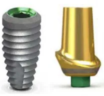
were fixed on the fixtures (Figure 1).
Figure 1. BioHorizons internal hexagon fixture and abutment
Implants were mounted in transparent auto-polymerizing acrylic resin blocks (Moravia, To-kyo, Japan) with a circular cross-section and a di-ameter of 34 mm and a height of 19 mm using a
parallelometer (Hahnenkratt, Berlin, Germany).
(16) For the preparation of acrylic resin, an
appro-priate powder/liquid ratio was used according to the manufacturer’s instructions for all samples.
A surveyor (J.M. Ney Co., Bloomfield, CT, USA) was used to mount the fixtures inside the
acrylic mold in a completely perpendicular posi-tion (a 90-degree angle relative to the horizon). After the setting of the acrylic resin, all speci-mens were prepared for testing.(16)
In the case group, the internal parts of the implants were thoroughly cleaned with alcohol according to the manufacturer's instructions.
GapSeal (Hager&Werken, Duisburg, Germany)
was poured to the maximum capacity of the in-ternal space of the abutments in the case group as instructed by the manufacturer to prevent air entrapment. Then, straight abutments were
fixed on all samples; the abutment screws were
torqued with a force of 30 Ncm using a digital torque meter (Lutron Electronic Enterprise Co. Ltd., Taipei, Taiwan) according to the implant manufacturer's instructions. In order to
compen-sate for the settling effect, five minutes later, the
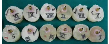
abutment screw was re-tightened using the digi-tal torque meter with a force of 30 Ncm (Figure 2).(16,17)
Figure 2. Case and control samples mounted in resin blocks
Microleakage measurement:
Methylene blue solution (Sigma-Aldrich, St. Louis, USA) was used to evaluate the mi-croleakage. For this purpose, the upper contact surface of the case and control abutments was
first sealed with a layer of rose wax and a layer
of nail varnish to prevent methylene blue from penetrating the abutments from above. The methylene blue solution was prepared according to the manufacturer’s instructions. All samples were then immersed in the solution and incu-bated for 24 hours at 37°C.(7)
To measure the microleakage, implant-abut-ment assemblies were cut (Mecatome T-201A, Presi, Paris, France) from the middle along the axial axis by a high-precision diamond wheel
(Strauen Minitorm, Barcelona, Spain; Figure 3), and dye penetration at the implant-abutment interface was reported at two levels using scan-ning electron microscopy (SEM; Neon 40 with
Gemini® column, Zeiss, Oberkochen, Germany) at x200 magnification.
Microgap measurement:
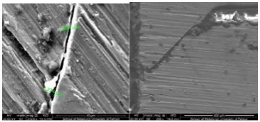
To measure the microgap, images were taken from the specimens sectioned at six areas, which were randomly selected (Figure 4; three points on the right and three points on the left) under the
SEM at a voltage of 20 kV at ×2000 magnifica -tion, and measurements were made.(16)
The data collected from this study were en-tered into SPSS software (Version 22; SPSS Inc., Chicago, IL, USA) for analysis using t-test and Fischer’s exact test.
Resutl:
This study was performed on 16 samples
includ-ing 8 samples with GapSeal (case group) and 8 samples without GapSeal (control group). The
implants used were 10 mm in length and 4 mm in diameter. In this study, abutments with a length of 6 mm and a collar height of 1 mm were used. The size of microgap was 3.04±0.54 µm in the control group and 0.99±0.39 µm in the case group, which was three times higher in the con-trol group, and the t-test showed that this
differ-ence was statistically significant (P<0.000; Table
1; Figure 5).
Table 1. Comparison of microgaps in the control
and case groups according to the use of GapSeal
Microgap
Group
SD ± Mean
) m µ (
Minimum(µm)
Maximum ) m µ (
GapSeal Without
(Control) 3.04±0.54 4.2392.166 (Case)
GapSeal
With 0.99±0.39 1.4250 Difference Amount Three-fold
Percentage 67%
Result P<0.000
Figure 4. Microgap in the group with GapSeal (left) and in the group without GapSeal (right) un-der the scanning electron microscope (SEM; x2000)
Figure 5. Seal of the implant-abutment in-terface by GapSeal (left) and penetration of methylene blue solution into the implant-abutment interface of one of the control samples (right)
The amount of microleakage between the fix -ture and the abutment in internal hex connection
according to the use of GapSeal is presented in
Table 2 and shows that in the control group, all samples (100%) had leakage, whereas in the case group, none of the samples (0%) showed leakage. Fischer’s exact test showed that this difference
was statistically significant (P<0.001; Table 2).
Table 2. Distribution of samples according to
microle-akage divided by GapSeal
Microleakage
Group No Yes Total
GapSeal Without
(Control) 80 8
(Case) GapSeal
With 08 8
value
-P P<0.001
Discussion:
This study was performed to investigate the
effect of GapSeal on microleakage and microgap
between the implant and abutment with internal hex connection in 8 case and 8 control samples.
The results showed that the use of GapSeal de -creased the microgap and microleakage in inter-nal hex implant-abutment connection in the case group compared to the control group.
According to previous studies, leakage of liquids and bacteria at the implant-abutment in-terface depends on various factors, such as the implant-abutment union, the micromovement
between the components, and the final abutment
torque. The shape of the abutment-implant junc-tion and dynamic loading can increase micro-movement and create a pumping effect, affecting leakage of bacteria and liquids.(7,11,19)
in some previous studies, the amount of leak-age has been studied statically. In 2011, Lorenzo-ni et al showed that microleakage and gap at the implant-abutment interface were present in two types of hexagonal implants; however, the sam-ples were not cyclically loaded.(16) In 2014, Smith
static conditions.(6)
In 2012, Rismanchian et al evaluated micro-gap and microbial leakage in static conditions at the interface of four abutments of the Strau-mann system.(18) Their results showed that the
use of different types of abutment affected the average size of microgaps and the mean number of leaked colonies (colony-forming unit [CFU]/
ml) throughout the fixture and abutment junction within the first 5 hours, but it has no significant
effect on the microleakage at 24 hours, 48 hours, and 14 days.(18)
Failure to use cyclic loading makes the
gener-alizability of the results difficult.
Numerous articles have pointed to the importance of leakage under loading conditions. According to various studies, cyclic loadings increase the microgap size at the implant-abutment interface, especially in implants with external hexagonal connections.(9,20-22)
In the present study, the samples underwent cyclic loading to simulate clinical conditions, and
the findings of the present study, indicating the
presence of microleakage in the space present at the abutment-implant interface, are in line with previous studies. The results of studies that used bacteria to measure microleakage are more con-sistent with reality compared to the use of meth-ylene blue.(6,11)
In the present study, the size of microgap
was 0.99±0.39 µm in the GapSeal group and
3.04±0.54 µm in the control group. Bacteria ranged in size from 0.2 to 5 µm. Jansen et al sug-gest that the microgap between the implant and abutment can be as large as 49 µm, indicating that even the smallest microgaps are 0.4 to 10 times larger than bacteria.(16,23)
In a study by Smith and Turkyilmaz, no rela-tionship was found between the size of the micro-gap and bacterial leakage.(6)
In 2015, Martin-Gili et al investigated fluid
leakage in external and internal connections of screw abutments.(9)
The size of microgap in the cited study was 2.34 µm in the internal hex connection after cyclic loading and 4.01 µm in the external hex connec-tions. The size of microgap in the internal
con-nection was significantly smaller compared to the
external one. As the number of mechanical cycles
increased, the size of microgaps increased due to titanium deformation. Methylene blue leakage was higher in the external connection.(9)
GapSeal is a silicone and bacteriostatic gel for
implant cavity seal, which reduced microgap and microleakage in internal hex implant-abutment connections in the present study. The results of the previous studies that follow are in line with the present study. In 2018, Ozdiler et al showed that sealants (silicone sealant and chlorhexidine), unlike the taper angle, reduce bacterial leakage at the conical internal implant-abutment interface under dynamic loading with a 50-N force and 500,000 cycles.(11) In the present study, the
speci-mens were loaded with more cycles and higher forces in the physiological range (200,000 to 1,200,000 cycles, 15 to 160 N); the results were consistent with the results of the study by Ozdiler et al.(11)
Nayak et al (2014) evaluated the sealing
abil-ity of O-ring polysilicon and GapSeal to prevent microleakage between the fixture and abutment
in static conditions and showed that the use of
GapSeal at the inner surface of the fixture before
torqueing reduced the microleakage rate. (8)
The c-clamp was used to fasten the abutment
screw without mounting the fixture, whereas in the present study, the fixtures were mounted in
molds containing acrylic resin, which were more
similar to the fixture inserted in the patient’s jaw.
In an in-vivo study in 2014, Pimentel et al used silicone membranes to seal the microgap between the implant and abutment with external hexago-nal connection.(21)
Application of silicone membrane reduced
bacterial strains that infiltrated the
implant-abutment assembly after 30 and 90 days using DNA chain polymerization technique but did
not completely prevent bacterial infiltration. Dis
-advantages of silicon membranes include film
thickness and early decomposition in the mouth.
GapSeal is also a silicone gel that disintegrates
over time. Further studies are needed to evaluate
the life span of GapSeal.
This double-blind study was performed with
sufficient number of samples in each group. The
ef-fects on biological and biomechanical factors that affect the success of implant treatment. One of the limitations of this study was the impossibil-ity of placing the samples in the methylene blue solution in a cyclic loading device. Future studies can investigate the microleakage in different types of abutment-implant systems, even under different cyclic loading forces and lateral cyclic loading with different abutment lengths.
Conclusion:
According to the results of this study, it can be
concluded that GapSeal decreases microgap and
microleakage in internal hex implant-abutment connection in the case group compared to the control group.
Acknowledgements:
We would like to acknowledge the support of the
Research Deputy of the Faculty of Dentistry of Azad University of Medical Sciences, Tehran, Iran. It should be noted that this study is the result of a thesis with design code 154/T.
References:
1. Sahin C, Ayyildiz S. Correlation between microleakage and screw loosening at implant-abutment connection. J Adv Prosthodont. 2014 Feb;6(1):35-8.
2. Yeo IS, Lee JH, Kang TJ, Kim SK, Heo SJ, Koak JY, et al. The effect of abutment screw length on screw loosening in dental implants with external abutment connections after ther-mocycling. Int J Oral Maxillofac Implants. 2014 Jan-Feb;29(1):59-62.
3. Sakaguchi RL, Borgersen SE. Nonlinear con-tact analysis of preload in dental implant screws. Int J Oral Maxillofac Implants. 1995 May-Jun;10(3):295-302.
4. Norton MR. Assessment of cold welding properties of the internal conical interface of two commercially available implant systems. J Pros-thet Dent. 1999 Feb;81(2):159-66.
5. Donley TG, Gillette WB. Titanium endosse -ous implant-soft tissue interface: a literature re-view. J Periodontol. 1991 Feb;62(2):153-60. 6. Smith NA, Turkyilmaz I. Evaluation of the sealing capability of implants to titanium and zir-conia abutments against Porphyromonas
gingi-valis, Prevotella intermedia, and Fusobacterium nucleatum under different screw torque values. J Prosthet Dent. 2014 Sep;112(3):561-7.
7. Scarano A, Assenza B, Piattelli M, Iezzi G,
Leghissa GC, Quaranta A, et al. A 16-year study of the
microgap between 272 human titanium implants and their abutments. J Oral Implantol. 2005;31(6):269-75.
8. Nayak AG, Fernandes A, Kulkarni R, Ajantha GS, Lekha K, Nadiger R. Efficacy of antibacterial sealing
gel and O-ring to prevent microleakage at the implant abutment interface: an in vitro study. J Oral Implantol. 2014 Feb;40(1):11-4.
9. Martin-Gili D, Molmeneu M, Fernandez M, Pun
-set M, Giner L, Armengou J, et al. Determination of fluid leakages in the different screw-retained implant-abutment connections in a mechanical artificial mouth.
J Mater Sci Mater Med. 2015 Jul;26(7):211.
10. Berberi A, Maroun D, Kanj W, Amine EZ, Philippe
A. Micromovement Evaluation of Original and Com-patible Abutments at the Implant-abutment Interface. J Contemp Dent Pract. 2016 Nov 1;17(11):907-913.
11. Ozdiler A, Bakir-Topcuoglu N, Kulekci G, Isik-Ozkol G. Effects of Taper Angle and Sealant Agents
on Bacterial Leakage Along the Implant-Abutment Interface: An In Vitro Study Under Loaded Condi-tions. Int J Oral Maxillofac Implants. 2018 Sep/ Oct;33(5):1071-77.
12. Tripodi D, D’Ercole S, Iaculli F, Piattelli A,
Perrot-ti V, Iezzi G. Degree of bacterial microleakage at the
implant-abutment junction in Cone Morse tapered im-plants under loaded and unloaded conditions. J Appl Biomater Funct Mater. 2015 Dec 18;13(4):e367-71.
13. do Nascimento C, Miani PK, Pedrazzi V, Gon -çalves RB, Ribeiro RF, Faria AC, et al. Leakage of saliva through the implant-abutment interface: in vitro evaluation of three different implant connections un-der unloaded and loaded conditions. Int J Oral Maxil-lofac Implants. 2012 May-Jun;27(3):551-60.
14. Dias EC, Bisognin ED, Harari ND, Machado SJ,
da Silva CP, Soares GD, et al. Evaluation of implant-abutment microgap and bacterial leakage in five exter -nal-hex implant systems: an in vitro study. Int J Oral Maxillofac Implants. 2012 Mar-Apr;27(2):346-51.
15. Coelho PG, Sudack P, Suzuki M, Kurtz KS, Roma
-nos GE, Silva NR. In vitro evaluation of the implant
abutment connection sealing capability of different im-plant systems. J Oral Rehabil. 2008 Dec;35(12):917-24.
16. Lorenzoni FC, Coelho PG, Bonfante G, Carvalho
RM, Silva NR, Suzuki M, et al. Sealing Capability and SEM Observation of the Implant-Abutment Interface. Int J Dent. 2011;2011:864183.
17. Piattelli A, Scarano A, Paolantonio M, Assenza
microbial penetration in the internal part of cement-retained versus screw-cement-retained implant-abutment con-nections. J Periodontol. 2001 Sep;72(9):1146-50. 18. Rismanchian M, Hatami M, Badrian H,
Kha-lighinejad N, Goroohi H. Evaluation of microgap
size and microbial leakage in the connection area of 4 abutments with Straumann (ITI) implant. J Oral Im-plantol. 2012 Dec;38(6):677-85.
19. Scarano A, Valbonetti L, Degidi M, Pecci R, Piat-telli A, de Oliveira PS, et al. Implant-Abutment Con-tact Surfaces and Microgap Measurements of Differ-ent Implant Connections Under 3-Dimensional X-Ray Microtomography. Implant Dent. 2016 Oct;25(5):656-62.
20. da Silva-Neto JP, Nóbilo MA, Penatti MP,
Sima-moto PC Jr, das Neves FD. Influence of methodo -logic aspects on the results of implant-abutment in-terface microleakage tests: a critical review of in vitro studies. Int J Oral Maxillofac Implants. 2012 Jul-Aug;27(4):793-800.
21. Pimentel AC, Manzi MR, Sartori SG, da Graça Naclério-Homem M, Sendyk WR. In vivo effective -ness of silicone gel sheets as barriers at the inner mi-crogap between a prosthetic abutment and an exter-nal-hexagon implant platform. Int J Oral Maxillofac Implants. 2014 Jan-Feb;29(1):121-6.
22. Pereira J, Morsch CS, Henriques B, Nascimento RM, Benfatti CA, Silva FS, et al. Removal Torque
and Biofilm Accumulation at Two Dental
Implant-Abutment Joints After Fatigue. Int J Oral Maxillofac Implants. 2016 Jul-Aug;31(4):813-9.
23. Jansen VK, Conrads G, Richter EJ. Microbial leakage and marginal fit of the implant-abutment
interface. Int J Oral Maxillofac Implants. 1997 Jul-Aug;12(4):527-40. Erratum in: Int J Oral Maxillofac Implants. 1997 Sep-Oct;12(5):709.
Please cite this paper as: