ORIGINAL ARTICLE
Primary outcomes of the VIDI study: phase 2,
double-masked, randomized, active-controlled
study of ASP8232 for diabetic macular edema
Quan Dong Nguyen
1*, Yasir J. Sepah
1,2, Brian Berger
3, David Brown
4, Diana V. Do
1, Alberto Garcia‑Hernandez
5,
Sunil Patel
6, Firas M. Rahhal
7, Yevgeniy Shildkrot
8and Ronny W. Renfurm
5on behalf of the VIDI Research Group
Abstract
Background: ASP8232 is a potent and specific small molecule vascular adhesion protein‑1 (VAP‑1) inhibitor. This study evaluated the effect of ASP8232 on excess retinal thickness when given alone or in combination with ranibi‑ zumab in patients with center‑involved diabetic macular edema (CI‑DME).
Methods: This was a phase 2a, placebo and sham‑injection controlled, double‑masked, randomized, parallel‑group clinical trial. Participants were patients with CI‑DME and central subfield thickness (CST) ≥ 375 µm in the study eye as assessed by spectral domain optical coherence tomography. Eligible patients were randomized to (1) daily oral ASP8232 40 mg monotherapy; (2) combination therapy of daily oral ASP8232 40 mg and monthly intravitreal ranibi‑ zumab 0.3 mg; or (3) monthly intravitreal ranibizumab 0.3 mg monotherapy. The treatment period was 12 weeks. CST and best corrected visual acuity (BCVA) were assessed at baseline and at Weeks 2, 4, 8, 12, 16 and 24. The primary out‑ come was the mean percent change from baseline in excess CST at Week 12. Secondary outcomes were BCVA, safety and tolerability, and pharmacokinetic and pharmacodynamic characteristics of ASP8232.
Results: After 12 weeks, the mean (95% confidence interval) percent change in excess CST was 11.4% (− 15.0%, 37.8%) in the ASP8232 group, − 61.7% (− 86.1%, − 37.2%) in the ASP8232/ranibizumab group, and − 75.3% (− 94.8%,
− 55.8%) in the ranibizumab group. The change from baseline in the two ranibizumab arms was statistically signifi‑ cant (P < 0.001) as was the difference between the ranibizumab groups and the ASP8232 group (P < 0.001). Mean (SD) increase in BCVA score from baseline was 3.1 (7.3) in the ASP8232 group, 5.2 (7.1) in the ASP8232/ranibizumab group, and 8.2 (9.5) in the ranibizumab group. The increase from baseline in BCVA score was statistically and clinically signifi‑ cant in the ranibizumab group compared with the ASP8232 group (P= 0.015). ASP8232 resulted in near complete inhibition of plasma VAP‑1 activity whilst ranibizumab had no effect.
Conclusions: Near complete inhibition of plasma VAP‑1 activity with ASP8232 had no effect on CST in patients with CI‑DME. Furthermore, combination therapy did not provide additional benefit to treatment with ranibizumab alone, which significantly reduced CST and improved BCVA.
Trial registration clinicaltrials.gov; NCT02302079. Registered on November 26, 2014
Keywords: ASP8232, VAP‑1 inhibitor, Center‑involved diabetic macular edema, Central subfield thickness, Ranibizumab, Clinical trial
© The Author(s) 2019. This article is distributed under the terms of the Creative Commons Attribution 4.0 International License (http://creat iveco mmons .org/licen ses/by/4.0/), which permits unrestricted use, distribution, and reproduction in any medium, provided you give appropriate credit to the original author(s) and the source, provide a link to the Creative Commons license, and indicate if changes were made. The Creative Commons Public Domain Dedication waiver (http://creat iveco mmons .org/ publi cdoma in/zero/1.0/) applies to the data made available in this article, unless otherwise stated.
Open Access
*Correspondence: ndquan@stanford.edu
1 Byers Eye Institute, Stanford University School of Medicine, 2370 Watson Court, Suite 200, Palo Alto, CA 94303, USA
Background
Diabetic retinopathy (DR) is a common complication of diabetes mellitus that leads to loss of vision and blindness among working age adults [1–3]. During progression of DR, patients can develop diabetic macular edema (DME), which is characterized by the thickening of the macula caused by the breakdown of the blood-retinal barrier and consequent retinal vascular hyperpermeability [3]. In 2010, the global prevalence of DR among adults with diabetes mellitus aged 20–79 years was estimated to be 34.6% for any DR and 6.81% for DME [4]. DME is the leading cause of vision loss among patients with DR. It is associated with the type of diabetes, and increases with the duration and severity of disease [5, 6]. Other signifi-cant risk factors common to DR and DME include hyper-glycemia and hypertension [4]. DME negatively impacts patients’ health-related quality of life and represents an economic burden due to the increased use of healthcare resources by affected patients [7, 8].
While there is no curative treatment available for DME, laser photocoagulation represents an effective treatment to preserve vision. However, this treatment modality is limited by its inability to restore vision once it has been lost [9]. The current standard of care for DME includes intravitreal anti-vascular endothelial growth factor (VEGF) antibodies and corticosteroids [9]. Clinical stud-ies have confirmed that monthly intravitreal treatment with the anti-VEGF antibody ranibizumab can improve vision, with up to 45% of patients gaining ≥ 15 letters
in best-corrected visual acuity (BCVA) after 24 months [10, 11]. Similar improvements were found after treat-ment with the anti-VEGF antibodies aflibercept [12–14] and with bevacizumab, an approved treatment for colon cancer that is commonly used off label for DME [9, 15]. Despite the proven efficacy of VEGF inhibitors, the requirement of frequent injections causes a high rate of treatment discontinuation among patients with DME and represents a major limitation [16]. Moreover, the pres-ence of potential side effects and the significant propor-tion of patients who do not respond to treatment [10, 12] suggest that there remains a need for the development of improved therapies for DME.
Vascular adhesion protein-1 (VAP-1) belongs to the family of copper-containing amine oxidase/semicar-bazide-sensitive amine oxidases that catalyze the oxida-tive deamination of primary amines with subsequent production of aldehyde, ammonium, and hydrogen peroxide, which are involved in oxidative stress and are cytotoxic. VAP-1 is expressed in vascular endothelium (including renal and retinal capillaries), smooth muscle cells, hepatic stromal cells and adipocytes [17, 18]. The enzymatic action stimulates leukocyte trafficking to the interstitium and therefore has a pro-inflammatory action
[17, 18]. After inflammatory stimulus, a soluble form of VAP-1, which retains its enzymatic activity, is released into circulation from the endothelial cells. Elevated levels of soluble VAP-1 have been found in the serum of dia-betic patients [19] and in the vitreous fluid of patients with proliferative DR (PDR) compared with non-PDR patients [20, 21]. Recently, it was shown that VEGF induces soluble VAP-1 release in retinal capillaries and thereby induces increased production of reactive oxygen species [22]. In experimental models of uveitis and DR in rats, VAP-1 was suggested to play a significant role in leucocyte recruitment into retinal endothelium [23–25]. Such evidence suggests that inhibition of VAP-1 may rep-resent a novel therapeutic strategy for DR and DME.
ASP8232 is a potent and specific small molecule VAP-1 inhibitor. In a streptozocin-induced rat model of DME, ASP8232 inhibited plasma VAP-1 activity and improved retinal hyperpermeability and plasma total antioxidant status. In combination with intravitreal anti-rat VEGF antibody, ASP8232 was more effective in reducing ocu-lar hyperpermeability compared with either the anti-rat-VEGF antibody or ASP8232 alone (data on file).
In multiple pharmacology and safety/toxicity experi-mental studies, ASP8232 had low acute toxicity and showed no effect on central nervous system, cardiovas-cular, and respiratory functions or fertility and early embryonic development, and had no genotoxic or terato-genic potential. ASP8232 was tested in two phase 1 stud-ies in healthy subjects, patients with renal impairment, and patients with type 2 diabetes mellitus. ASP8232 was administered in doses up to 6000 mg as single dose and up to 800 mg as multiple doses in healthy subjects. In subjects with renal impairment and diabetes, ASP8232 was administered in a dose of 150 mg for 4 weeks (unpub-lished data from first-in-man study, NCT02218099). ASP8232 was safe and well-tolerated, and pharmacoki-netic and pharmacodynamic modelling and simulations suggested that a daily dose of 40 mg would be safe and would deliver complete inhibition of VAP-1.
The VIDI study (VAP-1 Inhibition in DME) was a phase 2 study designed to evaluate ASP8232 safety and effect on excess retinal thickness when given alone or in combination with the anti-VEGF agent, ranibizumab, in patients with CI-DME.
Methods
Study design
Institutional Review Board (IRB)/Ethics Commit-tee approval was obtained and the study was conducted in accordance with the Declaration of Helsinki, Good Clinical Practice guidelines, and the Health Insurance Portability and Accountability Act; all patients provided informed consent. An independent academic reading center, the Ocular Imaging Research and Reading Center (OIRRC, Sunnyvale, California), served as the Reading Center for the VIDI study; the OIRRC received, stored, processed, and graded all images from the study.
The study was divided into 3 periods: a screening period of 1–4 weeks; a treatment period of 12 weeks, and a follow-up period of 12 weeks. Patients were ran-domized (1:1:1) to one of three treatment groups: (1) ASP8232 monotherapy: oral ASP8232 40 mg once daily + sham intravitreal injections once per month; (2) combination therapy of ASP8232 and ranibizumab: oral ASP8232 40 mg once daily + ranibizumab (0.3 mg) intra-vitreal injections once per month; and (3) ranibizumab monotherapy: oral ASP8232-matched placebo once daily + ranibizumab (0.3 mg) intravitreal injections once per month.
During the treatment period, study participants received ASP8232 (40 mg) or placebo once daily from Day 1–84 and an intravitreal injection of ranibizumab or sham on Days 1, 29, and 57. The dose of 40 mg ASP8232 was selected based on previous studies that showed that this dose was safe and was predicted to achieve maximal VAP-1 inhibition over a 24-h period with once daily dos-ing. All study participants and site staff were masked to all treatments, except for the injecting ophthalmologist, who was not involved in any other study assessments at the study site.
Participants
Patients were eligible if they were aged 18–85 years with type 1 or type 2 diabetes mellitus with the following specific characteristics: glycated hemoglobin (HbA1c)
≤ 12%; central subfield thickness (CST) ≥ 375 µm in the study eye; early treatment diabetic retinopathy study (ETDRS) BCVA letter score ≤ 73 (Snellen 20/40) and
≥ 24 (Snellen 20/320) in the study eye. Patients with any of the following characteristics were excluded from study participation: macular edema or decrease in BCVA due to a cause other than DME; presence of any other ocular disease in the study eye that may have caused substan-tial reduction in BCVA; significant macular ischemia or other retinal inflammatory or active periocular or ocular infection; history of noninfectious uveitis, high myopia (− 8 diopter or more correction), pars plana vitrectomy, any ocular surgery, YAG (yttrium–aluminum-garnet) capsulotomy, panretinal scatter photocoagulation, or focal laser within 3 months of study enrollment; history
of intravitreal, subtenon, or periocular, non-sustained release, steroid therapy within 3 months before the study; history of intravitreal sustained release dexamethasone therapy within 6 months of study enrollment; history of intravitreal sustained release fluocinolone within 3 years of study enrollment or history of prior treatment with intravitreal VEGF treatment within 8 weeks of study enrollment.
Study drug
ASP8232 was provided as 40 mg capsules and ASP8232-matched placebo was provided as matching capsules of microcrystalline cellulose. The capsules were taken in the morning with or without food. Ranibizumab was admin-istered as intravitreal injections of 0.05 mL of a 6 mg/mL ranibizumab aqueous solution containing 10 mM histi-dine HCl, 10% α,α-trehalose dihydrate, 0.01% polysorbate 20 with pH 5.5. Sham intravitreal for ranibizumab was supplied as an empty vial. An anesthetic was adminis-tered in the study eye before each intravitreal injection (including the sham intravitreal injection). The sham injection consisted of the (unmasked) injecting ophthal-mologist pressing an empty syringe against the surface of the eye to mimic an intravitreal injection. Ranibizumab was injected intravitreally according to the instructions of the manufacturer. Rescue therapy could be consid-ered, under the discretion of the evaluating investigator, if a patient experienced a decrease in ETDRS-BCVA of
≥ 15 letters from baseline or a decrease in ETDRS-BCVA of ≥ 10 letters and an increase in CST of ≥ 75 µm. Res-cue therapy in the study eye could only be considered after completion of the assessments at Week 4 and was managed by the injecting ophthalmologist. Patients who received rescue therapy with laser photocoagulation were permitted to continue the study; patients who required any other rescue therapy were discontinued from the study treatment. The randomization list and study medi-cation mask were generated and maintained by an inter-active website response system.
Assessments
independent academic centralized reading center, the OIRRC, for grading and analyses conducted by masked graders. Samples for the assessment of ASP8232 concen-trations in plasma were collected during visits on Days 15, 29, 57 prior to oral dosing (approximately 24 h after the previous day dose), and on Days 85, 113, and 169 (approximately 24 h, 4 weeks, and 12 weeks post-dose). Samples for the assessment of ASP8232 concentrations in aqueous humor from the anterior chamber of the study eye were collected on Days 1, 28, and 85. Concentrations of ASP8232 were measured using a validated liquid chro-matography-tandem mass spectrometry method. Sam-ples for VAP-1 concentrations and VAP-1 activity were collected during each visit.
Outcome parameters
The primary efficacy endpoint was the percent change from baseline (%CFB) to Week 12 [end of treatment (EoT)] in excess CST in the study eye, as assessed by SD-OCT. Excess CST was defined as the difference between measured CST and the predefined “normal” value of 320 μm [26, 27]. Secondary endpoints included the abso-lute change from baseline in CST in the study eye at each visit, and the change from baseline in ETDRS-BCVA score in the study eye at each visit. Pharmacokinetic and pharmacodynamic end points included ASP8232 concen-trations in plasma and anterior chamber aqueous humor and VAP-1 concentration and activity in plasma and in aqueous humor. Safety and tolerability of ASP8232 was assessed by monitoring the nature, frequency, and sever-ity of systemic and ocular treatment-emergent adverse events (TEAEs), as well as vital signs, clinical laboratory assessments, and electrocardiograms (ECGs).
Statistical methods
The VIDI study was designed to reject that the %CFB in excess CST is ≤ 10%, assuming a true %CFB in excess CST of 40% with a standard deviation of 65%. Assuming a drop-out rate of 15%, the total number of patients to be randomized to have an 80% power was planned to be 84 (28 per group). The efficacy analyses were conducted using the full analysis set (FAS), which included all ran-domised participants who received at least one dose of the study drug and had an efficacy measurement at base-line and at least one at post basebase-line. The safety analysis set included all participants who received at least one dose of study drug and was used for the demographics and baseline characteristics and also the safety analyses. The pharmacokinetic analysis set included participants from the safety analysis set population for whom suf-ficient plasma concentration data were collected (i.e. at
least one plasma concentration with a recorded dosing time prior to the sample collection). All data analyses were conducted using Statistical Analysis Software® ver-sion 9.3 on UNIX.
Efficacy analysis was performed on %CFB in excess CST and change from baseline in ETDRS-BCVA score at 12 weeks. The hypothesis test on the %CFB in excess CST was based on the upper bound of a 2-sided 80% confidence interval (CI) constructed using the t-dis-tribution. If this upper bound was lower than − 10%, then the null hypothesis of no significant effect was rejected, and the correspondent alternative hypothesis was accepted. In addition, a secondary analysis was performed using a mixed model for repeated measures including treatment group, visit and treatment group by visit interaction, as fixed class factors, and baseline excess CST as continuous covariate. Observations after treatment discontinuation were excluded from the pri-mary analysis but reported separately as part of the post-treatment follow-up period.
Results
Patient disposition and baseline characteristics
All study patients were consecutively screened and ran-domized across the participating sites. Of 240 patients who signed informed consent, 96 were randomized to receive ASP8232 monotherapy (N = 32), ASP8232/
ranibizumab combination therapy (N = 33), or
ranibi-zumab monotherapy (N = 31). A total of 29 patients
(90.6%) in the ASP8232 group, 31 patients (93.9%) in the ASP8232/ranibizumab group and 27 patients (87.1%) in the ranibizumab group completed the study. All 96 randomized patients were included in the safety analysis set; 95 patients were included in the FAS (ASP8232 group, n = 32; ASP8232/ranibizumab group,
n = 32; ranibizumab group, n = 31). Table 1 and
Addi-tional file 1: Table S1 summarize the patient demo-graphics and baseline characteristics.
Overall, 15.6% and 18.8% of patients in the safety analysis set received previous and concomitant eye medications to the study eye, respectively. Treatment groups were well-balanced with respect to clinically important medical history, and the median treatment duration was similar across treatment groups and ranged between 84.0–84.5 days. A minority of patients also had a fellow eye enrolled (ASP8232 group, n = 7;
ASP8232/ranibizumab group, n = 3; ranibizumab
group, n = 4). The mean absolute CST values at baseline
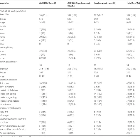
Table 1 Demographics and baseline characteristics (safety analysis set)
Parameter ASP8232 (n = 32) ASP8232/ranibizumab
(n = 33) Ranibizumab (n = 31) Total (n = 96)
Sex
Male 17 (53.1) 15 (45.5) 16 (51.6) 48 (50.0)
Female 15 (46.9) 18 (54.5) 15 (48.4) 48 (50.0)
Ethnicity
Not Hispanic or Latino 21 (65.6) 18 (54.5) 24 (77.4) 63 (65.6)
Hispanic or Latino 11 (34.4) 15 (45.5) 7 (22.6) 33 (34.4)
Race
White/Caucasian 26 (81.3) 27 (81.8) 22 (71.0) 75 (78.1)
Black/African American 4 (12.5) 5 (15.2) 5 (16.1) 14 (14.6)
Asian 1 (3.1) 0 1 (3.2) 2 (2.1)
American Indian/Alaskan native 0 0 2 (6.5) 2 (2.1)
Other 1 (3.1) 1 (3.0) 1 (3.2) 3 (3.1)
Age, y
Mean (SD) 61.5 (8.1) 59.8 (9.2) 63.4 (8.6) 61.5 (8.7)
Median 61.5 60.0 65.0 62.0
Range 47–82 30–81 45–78 30–82
Age group (years)
≤ 64 22 (68.8) 22 (66.7) 14 (45.2) 58 (60.4)
≥ 65 10 (31.3) 11 (33.3) 17 (54.8) 38 (39.6)
EudraCT age category (years)
≥ 18 to ≤ 64 22 (68.8) 22 (66.7) 14 (45.2) 58 (60.4)
≥ 65 to ≤ 84 10 (31.3) 11 (33.3) 17 (54.8) 38 (39.6)
≥ 85 0 0 0 0
Weight, kg
Mean (SD) 87.66 (19.79) 92.68 (25.33) 92.42 (20.37) 90.92 (21.91)
Median 86.85 83.60 88.20 86.40
Range 53.2–139.5 61.4–157.7 55.9–140.9 53.2–157.7
Height, cm
Mean (SD) 165.74 (9.13) 167.53 (11.31) 167.58 (8.78) 166.95 (9.76)
Median 166.82 167.64 165.10 165.55
Range 146.05–182.88 149.86–190.50 152.40–187.96 146.05–190.50
BMI, kg/m2
Mean (SD) 31.92 (6.99) 32.70 (6.80) 33.06 (7.76) 32.56 (7.13)
Median 29.40 30.50 31.10 30.60
Range 21.5–51.1 24.5–54.5 21.8–53.3 21.5–54.5
Study eye
Left 12 (37.5) 20 (60.6) 14 (45.2) 46 (47.9)
Right 20 (62.5) 13 (39.4) 17 (54.8) 50 (52.1)
Number of qualified eyes
1 25 (78.1) 30 (90.9) 27 (87.1) 82 (85.4)
2 7 (21.9) 3 (9.1) 4 (12.9) 14 (14.6)
CST, study eye (µm)
Mean (SD) 535.8 (117.9) 508.2 (104.3) 501.6 (105.5) 515.3 (109.3)
Median 522.0 489.0 488 490.0
Range 367–809 356–827 348–824 348–827
CST stratification (µm)
≤ 500 16 (50.0) 18 (54.5) 16 (51.6) 50 (52.1)
Primary endpoint: percent change from baseline in excess CST
In the ASP8232 group, the mean %CFB in excess CST to Week 12/EoT in the study eye was 11.4% (95% CI,
− 15.0%, 37.8%; P = 0.108), and therefore the study did not meet the primary endpoint. The %CFB in the ranibi-zumab group was − 75.3% (95% CI, − 94.8%, − 55.8%;
P < 0.001) after 12 weeks. The mean %CFB in excess CST in the study eye in the ASP8232/ranibizumab group showed a somewhat lesser treatment response compared
with the ranibizumab group [− 61.7% (95% CI, − 86.1%,
− 37.2%); P < 0.001] (Fig. 1). ANCOVA analysis of the %CFB in excess CST at Week 12/EoT showed no signifi-cant difference between the ASP8232/ranibizumab and the ranibizumab group; however, both groups showed a significant difference compared with the ASP8232 group (P < 0.001). In the fellow eye, the %CFB was not statisti-cally significant in any of the 3 treatment groups (data not shown).
Data are presented as n (%) unless otherwise noted
BCVA best-corrected visual acuity, BMI body mass index, DPP-4 dipeptidyl peptidase 4 inhibitors, ETDRS early treatment diabetic retinopathy study Table 1 (continued)
Parameter ASP8232 (n = 32) ASP8232/ranibizumab
(n = 33) Ranibizumab (n = 31) Total (n = 96)
ETDRS-BCVA, study eye (letters)
Mean (SD) 59 (10.1) 59.9 (10.8) 57.7 (14.7) 58.9 (11.9)
Median 61.5 63.0 64.0 63.0
Range 34–78 32–73 9–75 9–78
Iris color
Blue 7 (21.9) 5 (15.2) 6 (19.4) 18 (18.8)
Green 1 (3.1) 1 (3.0) 1 (3.2) 3 (3.1)
Brown 20 (62.5) 25 (75.8) 17 (54.8) 62 (64.6)
Hazel 4 (12.5) 2 (6.1) 6 (19.4) 12 (12.5)
Other 0 0 1 (3.2) 1 (1.0)
Smoking history
Never 22 (68.8) 20 (60.6) 20 (64.5) 62 (64.6)
Current 2 (6.3) 1 (3.0) 2 (6.5) 5 (5.2)
Former 8 (25.0) 12 (36.4) 9 (29.0) 29 (30.2)
Smoking duration, y
n 10 13 11 34
Mean (SD) 21.8 (15.8) 20.5 (11.1) 18.5 (11.8) 20.2 (12.5)
Median 29.5 20.0 20.0 20.0
Range 0–42 2–35 1–40 0–42
Diabetic medication
Metformin 16 (50.0) 15 (45.5) 16 (51.6) 47 (49.0)
DPP‑4 inhibitors 5 (15.6) 6 (18.2) 2 (6.5) 13 (13.5)
Insulin for inhalation 1 (3.1) 3 (9.1) 6 (19.4) 10 (10.4)
Insulin, fast‑acting 11 (34.4) 10 (30.3) 14 (45.2) 35 (36.5)
Insulin, long‑acting 15 (46.9) 16 (48.5) 17 (54.8) 48 (50.0)
Insulins/combinations 14 (43.8) 8 (24.2) 15 (48.4) 37 (38.5)
Sulfonylureas 11 (34.4) 10 (30.3) 11 (35.5) 32 (33.3)
Previous eye medications
Study eye 5 (15.6) 5 (15.2) 5 (16.1) 15 (15.6)
Fellow eye 5 (15.6) 6 (18.2) 8 (25.8) 19 (19.8)
Ocular interventions, study eye
Focal laser 7 (21.9) 6 (18.2) 4 (12.9) 17 (17.7)
Panretinal photocoagulation 5 (15.6) 4 (12.1) 5 (16.1) 14 (14.6)
Cataract/Phacoemulsification 4 (12.5) 3 (9.1) 8 (25.8) 15 (15.6)
Change in absolute CST
The mean (SD) absolute CST change from baseline was − 123.1(112.3) μm in the ranibizumab group after 12 weeks of treatment, whereas no change was observed in the ASP8232 group. The change from baseline to Week 12 in the ASP8232/ranibizumab group was comparable with the ranibizumab group (Table 2).
CST decreased quickly after the first injection of ranibizumab in the study eye in both ranibizumab and ASP8232/ranibizumab groups, whereas no changes were observed in the ASP8232 treated eyes at any visit. CST values remained stable for up to 12 weeks after the last injection in the ranibizumab group, whereas values tended to return to baseline levels in the ASP8232/ ranibizumab group (Fig. 2, Table 3).
The mean change from baseline in absolute CST values in the qualified fellow eye at Week 12/EoT was − 82 μm,
− 15.3 μm and − 55 μm in the ASP8232, ASP8232/
ranibizumab, and ranibizumab groups, respectively (Fig. 3).
Effect on ETDRS‑BCVA
The mean (SD) change from baseline in ETDRS-BCVA score to Week 12/EoT was 3.1 (7.3) in the ASP8232 group, 5.2 (7.1) in the ASP8232/ranibizumab group, and 8.2 (9.5) in the ranibizumab group. The change in ETDRS-BCVA score was significantly higher in the ranibizumab group than in the ASP8232 group (P= 0.015) (Fig. 4). In the ranibizumab treated eyes,
ETDRS-BCVA showed an increase by Week 2 and then continued to improve until week 12/EoT. In the ASP8232 group a small increase in ETDRS-BCVA was observed at week 2, and then ETDRS-BCVA remained at the same level in the ASP8232 group. During the follow-up period, ETDRS-BCVA remained stable until 12 weeks after the last injection in the ranibizumab
Fig. 1 Percent change (95% CI) from baseline in excess CST at EoT visit (FAS). CI, confidence interval; CST, central subfield thickness; EoT, end of treatment; FAS, full analysis set
Table 2 Change in absolute CST values from baseline to Week 12/EoT CIRC-values (FAS)
Data are presented as mean (SD)
CIRC central imaging reading center, CST central subfield thickness, EoT end of treatment, FAS full analysis set
ASP8232 (n = 32) ASP8232/ranibizumab (n = 32) Ranibizumab (n = 31)
Baseline, μm 535.8 (117.9) 511.8 (103.9) 501.6 (105.5)
Week 12 (EoT), μm 530.3 (127.4) 374.0 (105.6) 378.6 (107.5)
group and returned to baseline levels in the two remaining treatment groups. At week 2, the number of patients with 5-, 10-, and 15-letter gain in the ETDRS-BCVA was highest in the ranibizumab groups and at week 12, the percentage of patients with ≥ 5-letter
gain in ETDRS-BCVA was 48.1%, 64.5%, 69.0% in the ASP8232, ASP8232/ranibizumab, and ranibizumab group, respectively.
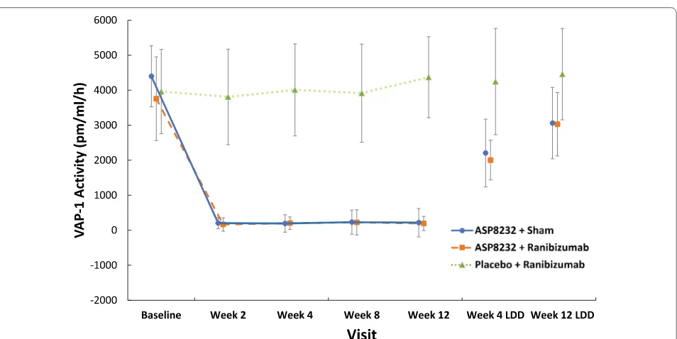
Pharmacokinetic and pharmacodynamic end points
Median trough plasma levels of ASP8232 ranged from 24.1 to 43.10 ng/mL during the treatment period. ASP8232 was still measurable in plasma 4 and 12 weeks after the last dose (Fig. 5). The median ASP8232 levels in the aqueous humor at 12 weeks was 2.98 ng/ml ranging from 0 to 31.1 ng/ml. The median plasma VAP-1 protein levels did not change throughout the study across treat-ment groups and ranged from 443 to 569 ng/ml. The median VAP-1 protein levels in aqueous humor did not change significantly from baseline to week 12 in any of the 3 treatment groups and ranged from 0.72 to 1.21 ng/ ml across treatment groups and time points. In the ASP8232 groups, plasma VAP-1 activity was nearly com-pletely inhibited throughout the treatment period, with median inhibition from baseline to week 12 ranging from 96.8% in the ASP8232/ranibizumab group to 97.3% in the ASP8232 group, indicating that ASP8232 fully inhib-its VAP-1 with or without ranibizumab. VAP-1 activity was ~ 50% of baseline within 4 weeks after the last dose of ASP8232 and remained between 70% and 85% of the baseline value within 12 weeks after the last dose. Plasma VAP-1 activity did not change in the ranibizumab group
Fig. 2 Mean (95% CI) change from baseline in absolute CST values in the study eye (FAS). CI, confidence interval; CST, central subfield thickness; FAS, full analysis set; LDD, last dose date; LOCF, last observation carried forward
Table 3 Absolute and percent change from baseline in excess CST to Weeks 2, 4, and 8 (FAS)
CST central subfield thickness, FAS full analysis set, SD standard deviation
ASP8232 ASP8232/ranibizumab Ranibizumab
Baseline
n 32 32 31
Mean (SD) 215.8 (117.9) 191.8 (103.9) 181.6 (105.5) Week 2
n 31 32 30
Mean (SD) 230.9 (134.3) 62.3 (109.1) 71.5 (85.3) %CFB 16.3 (39.4) − 69.3 (55.4) − 57.6 (51.9) Week 4
n 32 32 31
Mean (SD) 223.1 (130.6) 76.7 (115.0) 88.8 (109.0) %CFB 11.6 (44.1) − 56 (57.0) − 50.2 (64.4) Week 8
n 32 32 29
Fig. 3 Mean (95% CI) change from baseline in absolute CST values in the fellow eye (FAS). CI, confidence interval; CST, central subfield thickness; FAS, full analysis set; LDD, last dose date; LOCF, last observation carried forward
(Fig. 6). In the aqueous humor, the median inhibition of VAP-1 activity was 53.2% in the ASP8232 group com-pared with 35.4% and 27.9% in the ASP8232/ranibizumab and ranibizumab groups, respectively.
Safety results
The incidence of overall and ocular TEAE was similar across treatment groups, and most were
mild-to-moderate in severity (Table 4; Additional file 2: Table S2). The most common TEAEs were conjunctival hemorrhage [ASP8232/ranibizumab, n = 3 (9.1%)],
wors-ening of type 2 diabetes mellitus [ASP8232, n = 2 (6.3%);
ASP8232/ranibizumab, n = 3 (9.1%)] and diabetic retinal
edema [ASP8232, n = 3 (9.4%); ASP8232/ranibizumab,
n = 1 (3.0%); ranibizumab, n = 2 (6.5%)] (Table 4). A
small number of patients reported drug-related TEAE
Fig. 5 Mean plasma concentration of ASP8232 (pharmacokinetic analysis set). LDD, last dose date
[ASP8232, n = 2 (6.3%); ASP8232/ranibizumab, n = 3 (9.1%); and ranibizumab, n = 3 (9.7%)]. Drug-related ocular TEAEs included retinal disorder [ASP8232, n = 1 (3.1%); ASP8232/ranibizumab, n = 1 (3.0)], metamor-phopsia [ranibizumab, n = 1 (3.2%)] and photophobia [ranibizumab, n = 1 (3.2%)]. The number of patients reporting serious TEAEs was generally low [ASP8232 n = 3 (9.4%); ASP8232/ranibizumab, n = 1 (3.0%); and ranibizumab, n = 3 (9.7%)] and none of these events were considered to be related to the study drug.
Three patients experienced TEAEs leading to discon-tinuation of the study drug. In the ASP8232 group, one patient experienced progression of DME that required rescue treatment and was considered possibly drug-related, and another discontinued due to a diagnosis of prostate cancer diagnosed 1 month after randomization,
which was considered not related to the study drug. In the ASP8232/ranibizumab group, one patient discontin-ued due to progression of metastatic malignant mela-noma that had been diagnosed 3 years prior to the start of the trial. The patient had been in remission at enroll-ment and the condition progressed during the study; this TEAE was not considered to be related to the study drug. No clinically significant differences were observed in the change from baseline for clinical laboratory assessments, vital signs, and ECGs between treatment groups, and no deaths occurred during the study. Intraocular pressure was assessed as a safety measure across the treatment arms. Median levels were comparable across treatment groups and time points and ranged from 14 to 17 mmHg with no significant changes (> 10 mmHg after baseline) reported.
Table 4 Overview of TEAEs (safety analysis set)
Data are presented as n (%)
DM diabetes mellitus, E number of events, TEAEs treatment-emergent adverse events a Systemic TEAEs include all non-ocular TEAEs
ASP8232 (N = 32) ASP8232/ranibizumab
(N = 33) Ranibizumab (N = 31)
n (%) E n (%) E n (%) E
Overall TEAEs 21 (65.6) 43 17 (51.5) 50 19 (61.3) 51
Ocular TEAEs 10 (31.3) 14 13 (39.4) 21 12 (38.7) 23
Systemic TEAEsa 17 (53.1) 29 13 (39.4) 29 15 (48.4) 28
Drug‑related TEAEs 2 (6.3) 6 3 (9.1) 3 3 (9.7) 3
Drug‑related ocular TEAEs 1 (3.1) 1 1 (3.0) 1 2 (6.5) 2
Drug‑related systemic TEAEs 2 (6.3) 5 2 (6.1) 2 1 (3.2) 1
Serious TEAEs 3 (9.4) 3 1 (3.0) 1 3 (9.7) 3
TEAEs leading to permanent discontinuation of study drug 2 (6.3) 2 1 (3.0) 1 0 0
Drug‑related TEAEs leading to permanent discontinuation of study
drug 1 (3.1) 1 0 0 0 0
TEAEs reported in ≥ 5% of patients in any treatment group System organ class
Preferred term (MedDRA v15.1)
Endocrine disorders 0 2 (6.1) 0
Hypothyroidism 0 2 (6.1) 0
Eye disorders 8 (25.0) 13 (39.4) 12 (38.7)
Conjunctival hemorrhage 0 3 (9.1) 0
Diabetic retinal edema 3 (9.4) 1 (3.0) 2 (6.5)
Retinal aneurysm 0 0 2 (6.5)
Retinal exudates 2 (6.3) 2 (6.1) 1 (3.2)
Visual acuity reduced 0 2 (6.1) 1 (3.2)
Vitreous floaters 1 (3.1) 2 (6.1) 1 (3.2)
Vitreous hemorrhage 0 2 (6.1) 2 (6.5)
Gastrointestinal disorders 1 (3.1) 2 (6.1) 1 (3.2)
Vomiting 0 2 (6.1) 0
Metabolism and nutrition disorders 3 (9.4) 3 (9.1) 3 (9.7)
Discussion
VAP-1 is an endothelial adhesion molecule with semicar-bazide-sensitive amine oxidase activity that is expressed in the endothelial cells of the retinal vessels [19]. During inflammation, VAP-1 acts with other leukocyte adhe-sion molecules to recruit inflammatory cells [19]. Since the VAP-1 enzymatic activity results in the production of toxic metabolites including hydrogen peroxide and alde-hydes, which are involved in cellular oxidative stress, it has been postulated that increased levels of VAP-1 may contribute to the development of DR.
The VIDI study assessed the clinical efficacy of the spe-cific VAP-1 inhibitor ASP8232 on excess retinal thickness when administered alone or in combination with the anti-VEGF drug ranibizumab in patients with CI-DME. After 12 weeks of treatment, a statistically significant decrease from baseline in mean excess CST of 75.3% was observed in the ranibizumab group whilst no change was observed in the ASP8232 group. The effect in the combination treatment group was numerically less (but not statisti-cally significant) compared with ranibizumab alone and ASP8232 did not provide any additional benefits on CI-DME. Similarly, changes from baseline in absolute CST of 123.1 μm and 137.8 μm were observed in the ranibizumab and ASP8232/ranibizumab group, respectively, whereas no change was observed in the ASP8232 group.
After 12 weeks of treatment, the mean ETDRS-BCVA score in the study eye increased from baseline by 3.1, 5.2, and 8.2 letters in the ASP8232, ASP8232/ranibizumab, and ranibizumab groups, respectively. These data con-firm results from previous studies of ranibizumab and demonstrated that treatment with ranibizumab is effec-tive in improving ETDRS-BCVA in DME. ASP8232 alone provided limited improvement in ETDRS-BCVA of only three letters, which is not considered to be of clinical relevance. Interestingly, the outcomes in the combina-tion group were less favorable compared with the ranibi-zumab group, which confirms that ASP8232 does not provide relevant improvement in ETDRS-BCVA. The minor effects in the qualified fellow eye suggest some effect on CST but because of the very low number of eyes, these data should be interpreted with caution.
In the ASP8232 groups, VAP-1 activity in plasma was greatly inhibited throughout the treatment period and remained below the baseline value within 12 weeks after the last dose, whereas ranibizumab had no effect on VAP-1 activity, as expected. This confirms that the dose of 40 mg ASP8232 once daily was sufficient to completely inhibit VAP-1 in plasma. VAP-1 protein levels and activ-ity in the aqueous humor were much lower than plasma VAP-1 protein levels and activity; the median concentra-tion of ASP8232 in the aqueous humor was only about 11.5% of plasma trough levels.
It is not known whether VAP-1 plays a role in DR at the level of the retinal vasculature or at the vitreo-reti-nal interface. We measured VAP-1 levels and activity in the anterior chamber as a possible representation of the activity in the vitreous. However, in this study, VAP-1 activity in the aqueous humor was below the detection limits at all points. Therefore, we cannot draw any con-clusions regarding its activity.
Although the VIDI Study did not meet its primary end-point, we have demonstrated that VAP-1 activity can be inhibited with ASP8232. Recently, VAP-1 inhibition dem-onstrated a significant benefit in diabetic nephropathy, an end-organ diabetic complication of diabetes (manuscript accepted). Therefore, it would not be appropriate to dis-regard the VAP-1 pathway in diabetic macular edema and/or diabetic retinopathy based solely on the VIDI study results. Further studies evaluating different modes of delivery and/or concentrations of VAP-1 inhibition for eye diseases such as DME and DR may be warranted to further elucidate this potentially important target of pathophysiology.
Other studies evaluating combination treatment have yielded similar results, suggesting that VEGF is the major driver in the pathophysiology of DME [28]. Another VAP-1 inhibitor is currently being investigated in patients with DR in the absence of center-involved macular edema (www.clini caltr ials.gov; NCT03238963). It is conceivable that the presence of significant CI-DME requires inhibition of VEGF, whilst DR could be reduced by VAP-1 inhibition alone.
Our study had several strengths. Patients were ran-domly assigned to treatments, and all staff at the inves-tigative sites and the sponsor were masked to treatment allocation. A trained and experienced injecting ophthal-mologist performed all intravitreal and intravitreal sham injections and had no other involvement in the trial. Standardized and state-of-the-art measures of efficacy and safety were assessed, and study staff were trained before activation of the site. All images were reviewed and assessed by an independent and experienced reading center and formalized reading protocols were utilized. One potential limitation of our study was that the study duration may have been too short to show additional benefit of the combination treatment. Moreover, VAP-1 protein levels and activity were only measured in the aqueous humor and not in the vitreous.
Conclusions
unexpected adverse events were reported. VAP-1 inhi-bition may not impact DR efficacy; future studies inves-tigating this will provide more insight. The proper route of delivery of ASP8232 for ocular diseases should also be reassessed, as local delivery may be more appropriate than systemic administration.
Additional files
Additional file 1: TableS1. Baseline characteristics (safety analysis set).
Additional file 2: TableS2. Ocular TEAEs (safety analysis set).
Abbreviations
%CFB: percent change from baseline; BCVA: best corrected visual acuity; BMI: body mass index; CI: confidence interval; CI‑DME: center‑involved diabetic macular edema; CST: central subfield thickness; DME: diabetic macular edema; DPP‑4: dipeptidyl peptidase 4 inhibitors; DR: diabetic retinopathy; ECG: elec‑ trocardiogram; ETDRS: early treatment diabetic retinopathy study; EOT: end of treatment; FA: fluorescein angiography; FAS: full analysis set; OIRRC : Ocular Imaging Research and Reading Center; PDR: proliferative diabetic retinopathy; SD‑OCT: spectral domain‑optical coherence tomography; TEAE: treatment‑emergent adverse event; VAP‑1: vascular adhesion protein‑1; VEGF: vascular endothelial growth factor; VIDI: VAP‑1 Inhibition in DME; YAG : yttrium–aluminum‑garnet.
Acknowledgements
Editorial and logistical support was provided by OPEN Health Medical Com‑ munications (Chicago, IL).
Authors’ contributions
Concept and design: QDN, YJS, BB, DB, YS, RWR. Data acquisition: QDN, YJS, BB, DB, DVD, AGH, SP, FMR, YS, RWR. Data analysis/interpretation: QDN, YJS, BB, DB, AGH, YS, RWR. Drafting of the work or revising for important intellectual con‑ tent: QDN, YJS, BB, DB, DVD, AGH, SP, FMR, YS, RWR. Final approval of the work to be published: QDN, YJS, BB, DB, DVD, AGH, SP, FMR, YS, RWR. Agreement to be accountable for all aspects of the work: QDN, YJS, BB, DB, DVD, AGH, SP, FMR, YS, RWR. All authors read and approved the final manuscript.
Funding
This study was funded by Astellas Pharma, Inc., Northbrook, IL. The funder of the study had a role in the study design, and was responsible for the collec‑ tion, analysis, and interpretation of data, writing the report, and the decision to submit the paper for publication. All authors had full access to the study data in preparing the manuscript and the corresponding author had final responsibility for the decision to submit for publication.
Availability of data and materials
Access to anonymized individual participant level data collected during the trial, in addition to supporting clinical documentation, is planned for trials conducted with approved product indications and formulations, as well as compounds terminated during development. Conditions and exceptions are described under the Sponsor Specific Details for Astellas on www.clini cal st udyda tareq uest.com. Study‑related supporting documentation is redacted and provided if available, such as the protocol and amendments, statisti‑ cal analysis plan and clinical study report. Access to participant level data is offered to researchers after publication of the primary manuscript (if appli‑ cable) and is available as long as Astellas has legal authority to provide the data. Researchers must submit a proposal to conduct a scientifically relevant analysis of the study data. The research proposal is reviewed by an Independ‑ ent Research Panel. If the proposal is approved, access to the study data is provided in a secure data sharing environment after receipt of a signed Data Sharing Agreement.
Ethics approval and consent to participate
This study was approved by the Ethics Committee and all patients provided informed consent.
Consent for publication
Not applicable.
Competing interests
QDN: chaired the Steering Committee for the RISE and RIDE studies of ranibi‑ zumab for diabetic macular edema; Grants from Astellas, Genentech, and Regeneron; scientific advisory boards for Astellas, Genentech, and Regeneron. YJS: Research Support from Astellas, Genentech, Optovue; Personal Fees from Genentech/Roche and Optos. BB: Research Support from Genentech, Regen‑ eron, Allegro. DB: Grants from Astellas and Genentech/Roche, Regeneron, Bayer, Adverum, Allergan, Regenxbio, Clearside Biomedical, OHR, Heidelberg, Novartis, Thrombogenics, Ophthotech, NEI, Kanghong Biotech, Samsung, Ophthea, Apellis; Personal Fees from Genentech/Roche, Adverum, Allergan, Regenxbio, Clearside Biomedical, OHR, Bayer, Heidelberg, Novartis, Optos, Zeiss, Thrombogenics, Santen, Senju, Kanghong Biotech, Samsung, Apellis. DVD: Grants from Genentech, Regeneron, Santen. AGH: Personal Fees from Astellas; Employee of Astellas. SP: Grants from Genentech, Novartis, Ophtho‑ tech, Allergan, Opthea, Clearside, Samsung, Aerpio, Regeneron, Graybug and Taiwan Liposome Company. FMR: Nothing to disclose. YS: advisory board and research support from Castle Biosciences. RWR: Employee of Astellas.
Author details
1 Byers Eye Institute, Stanford University School of Medicine, 2370 Wat‑ son Court, Suite 200, Palo Alto, CA 94303, USA. 2 Ocular Imaging Research and Reading Center, Sunnyvale, CA, USA. 3 Retina Research Center, PLLC, 3705 Medical Parkway, Austin, TX 78705, USA. 4 Retina Consultants of Houston, Hou‑ ston, TX, USA. 5 Formerly With Astellas Pharma Europe BV, Sylviusweg 62, 2333 BE Leiden, The Netherlands. 6 Integrated Clinical Research, LLC, 5441 Health Center Drive, Abilene, TX 79606, USA. 7 Retina Vitreous Associates Medical Group, 9001 Wilshire Boulevard, Suite 301, Beverly Hills, CA 90211, USA. 8 Uni‑ versity of Virginia, 1300 Jefferson Park Avenue, Charlottesville, VA 22908, USA.
Received: 1 April 2019 Accepted: 4 June 2019
References
1. Ciulla TA, Amador AG, Zinman B. Diabetic retinopathy and diabetic macu‑ lar edema: pathophysiology, screening, and novel therapies. Diabetes Care. 2003;26(9):2653–64.
2. Ding J, Wong TY. Current epidemiology of diabetic retinopathy and diabetic macular edema. Curr Diabetes Rep. 2012;12(4):346–54. 3. Wang W, Lo ACY. Diabetic retinopathy: pathophysiology and treatments.
Int J Mol Sci. 2018;19(6):1816–30.
4. Yau JW, Rogers SL, Kawasaki R, et al. Global prevalence and major risk factors of diabetic retinopathy. Diabetes Care. 2012;35(3):556–64. 5. Boyer DS, Hopkins JJ, Sorof J, Ehrlich JS. Anti‑vascular endothelial growth
factor therapy for diabetic macular edema. Ther Adv Endocrinol Metab. 2013;4(6):151–69.
6. Klein R, Klein BE, Moss SE, et al. The Wisconsin epidemiologic study of diabetic retinopathy. IV. Diabetic macular edema. Ophthalmology. 1984;91(12):1464–74.
7. Chen E, Looman M, Laouri M, et al. Burden of illness of diabetic macular edema: literature review. Curr Med Res Opin. 2010;26(7):1587–97. 8. Shea AM, Curtis LH, Hammill BG, et al. Resource use and costs associ‑
ated with diabetic macular edema in elderly persons. Arch Ophthalmol. 2008;126(12):1748–54.
9. Ford JA, Lois N, Royle P, et al. Current treatments in diabetic macu‑ lar oedema: systematic review and meta‑analysis. BMJ Open. 2013;3(3):e002269.
10. Nguyen QD, Brown DM, Marcus DM, et al. Ranibizumab for diabetic macular edema: results from 2 phase III randomized trials: RISE and RIDE. Ophthalmology. 2012;119(4):789–801.
•fast, convenient online submission •
thorough peer review by experienced researchers in your field • rapid publication on acceptance
• support for research data, including large and complex data types •
gold Open Access which fosters wider collaboration and increased citations maximum visibility for your research: over 100M website views per year •
At BMC, research is always in progress.
Learn more biomedcentral.com/submissions
Ready to submit your research? Choose BMC and benefit from: 12. Brown DM, Schmidt‑Erfurth U, Do DV, et al. Intravitreal aflibercept for dia‑
betic macular edema: 100‑week results from the VISTA and VIVID studies. Ophthalmology. 2015;122(10):2044–52.
13. Wykoff CC, Le RT, Khurana RN, et al. Outcomes with as‑needed aflibercept and macular laser following the phase III VISTA DME trial: ENDURANCE 12‑month extension study. Am J Ophthalmol. 2017;173:56–63. 14. Midena E, Gillies M, Katz TA, et al. Impact of baseline central retinal thick‑
ness on outcomes in the VIVID‑DME and VISTA‑DME studies. J Ophthal‑ mol. 2018;2018:3640135.
15. Haritoglou C, Kook D, Neubauer A, et al. Intravitreal bevacizumab (Avastin) therapy for persistent diffuse diabetic macular edema. Retina. 2006;26(9):999–1005.
16. VanderBeek BL, Shah N, Parikh PC, Ma L. Trends in the care of dia‑ betic macular edema: analysis of a national cohort. PLoS ONE. 2016;11(2):e0149450.
17. Trivedi PJ, Tickle J, Vesterhus MN, et al. Vascular adhesion protein‑1 is elevated in primary sclerosing cholangitis, is predictive of clinical out‑ come and facilitates recruitment of gut‑tropic lymphocytes to liver in a substrate‑dependent manner. Gut. 2018;67(6):1135–45.
18. Weston CJ, Shepherd EL, Claridge LC, et al. Vascular adhesion protein‑1 promotes liver inflammation and drives hepatic fibrosis. J Clin Invest. 2015;125(2):501–20.
19. Luo W, Xie F, Zhang Z, Sun D. Vascular adhesion protein 1 in the eye. J Ophthalmol. 2013;2013:925267.
20. Murata M, Noda K, Fukuhara J, et al. Soluble vascular adhesion protein‑1 accumulates in proliferative diabetic retinopathy. Invest Ophthalmol Vis Sci. 2012;53(7):4055–62.
21. Abu El‑Asrar AM, Alam K, Garcia‑Ramirez M, et al. Association of HMGB1 with oxidative stress markers and regulators in PDR. Mol Vis. 2017;23:853–71.
22. Yoshida S, Murata M, Noda K, et al. Proteolytic cleavage of vascular adhe‑ sion protein‑1 induced by vascular endothelial growth factor in retinal capillary endothelial cells. Jpn J Ophthalmol. 2018;62(2):256–64. 23. Noda K, Miyahara S, Nakazawa T, et al. Inhibition of vascular adhe‑
sion protein‑1 suppresses endotoxin‑induced uveitis. FASEB J. 2008;22(4):1094–103.
24. Noda K, Nakao S, Zandi S, et al. Vascular adhesion protein‑1 regulates leukocyte transmigration rate in the retina during diabetes. Exp Eye Res. 2009;89(5):774–81.
25. Matsuda T, Noda K, Murata M, et al. Vascular adhesion protein‑1 blockade suppresses ocular inflammation after retinal laser photocoagulation in mice. Invest Ophthalmol Vis Sci. 2017;58(7):3254–61.
26. Grover S, Murthy RK, Brar VS, Chalam KV. Normative data for macular thickness by high‑definition spectral‑domain optical coherence tomog‑ raphy (spectralis). Am J Ophthalmol. 2009;148(2):266–71.
27. Ibrahim MA, Sepah YJ, Symons RC, et al. Spectral‑ and time‑domain optical coherence tomography measurements of macular thickness in normal eyes and in eyes with diabetic macular edema. Eye (Lond). 2012;26(3):454–62.
28. Campochiaro PA, Khanani A, Singer M, et al. Enhanced benefit in diabetic macular edema from AKB‑9778 Tie2 activation combined with vascular endothelial growth factor suppression. Ophthalmology. 2016;123(8):1722–30.
Publisher’s Note