R E S E A R C H
Open Access
Monitoring mitochondrial inner membrane
potential for detecting early changes in viability
of bacterium-infected human bone
marrow-derived mesenchymal stem cells
Mika Pietilä
1,2*, Kaarina Lähteenmäki
3, Siri Lehtonen
1,2, Hannu-Ville Leskelä
1,2, Marko Närhi
4, Maarit Lönnroth
3,
Jaana Mättö
3, Petri Lehenkari
1,2and Katrina Nordström
4Abstract
Introduction:One of the most challenging safety issues in the manufacture of cell based medicinal products is the control of microbial risk as cell-based products cannot undergo terminal sterilization. Accordingly, sensitive and reliable methods for detection of microbial contamination are called for. As mitochondrial function has been shown to correlate with the viability and functionality of human mesenchymal stem cells (hMSCs) we have studied the use of a mitochondrial inner membrane potential sensitive dye for detecting changes in the function of mitochondria following infection by bacteria.
Methods:The effect of bacterial contamination on the viability of bone marrow-derived mesenchymal stem cells (BMMSCs) was studied. BMMSC lines were infected with three different bacterial species, namely two strains of Pseudomonas aeruginosa, three strains ofStaphylococcus aureus, and three strains ofStaphylococcus epidermidis. The changes in viability of the BMMSCs after bacterial infection were studied by staining with Trypan blue, by
morphological analysis and by monitoring of the mitochondrial inner membrane potential.
Results:Microscopy and viability assessment by Trypan blue staining showed that even the lowest bacterial inocula caused total dissipation of BMMSCs within 24 hours of infection, similar to the effects seen with bacterial loads which were several magnitudes higher. The first significant signs of damage induced by the pathogens became evident after 6 hours of infection. Early changes in mitochondrial inner membrane potential of BMMSCs were evident after 4 hours of infection even though no visible changes in viability of the BMMSCs could be seen. Conclusions:Even low levels of bacterial contamination can cause a significant change in the viability of BMMSCs. Moreover, monitoring the depolarization of the mitochondrial inner membrane potential may provide a rapid tool for early detection of cellular damage induced by microbial infection. Accordingly, mitochondrial analyses offer sensitive tools for quality control and monitoring of safety and efficacy of cellular therapy products.
Introduction
Stem cell therapies have raised hopes for the develop-ment of viable alternatives for the treatdevelop-ment of many severe degenerative and inflammatory conditions that currently cannot be cured or alleviated by traditional medicine [1-5]. However, there are still many important
issues that need to be solved before the transfer of these advanced cellular therapies from the laboratory to clinics can be successfully achieved. One of the major challenges of such products and therapies is ensuring product safety while maintaining efficacy since viable stem cell-based products cannot undergo terminal sterility prior to implantation and use [6]. Accordingly, the development of aseptic manufacturing processes and process controls is pivotal to the successful translation of research into clinical practice [7]. In addition, the short shelf-life of * Correspondence: mika.pietila@aalto.fi
1
Institute of Biomedicine, Department of Anatomy and Cell Biology, Aapistie 7, P.O. Box 5000, FIN-90014, University of Oulu, Oulu, Finland
Full list of author information is available at the end of the article
products calls for the development of rapid methods to detect bacterial contamination in a matter of hours [8,9].
Even though the incidents of microbial contamination of cell products seem to be rare, the risk of contamina-tion is a critical issue since the consequences to patient health may be unpredictable and even devastating [10-16]. Currently, there is little or no evidence to pre-dict the long-term outcomes of possible infections after cell implantation. Therefore, uncertainties of possible further transmission of infectious agents from cell or tis-sue implants to patient exist. Moreover, microbial damage to stem cell products prior to implantation may well lead to unpredictable consequences in the efficacy of the implanted cells. Accordingly, strict requirements set by regulatory agencies for the manufacture of cell-based products, which must be performed according to the principles of good manufacturing practices or good tissue practices [17]. Even so, the most common site for contamination is thein vitro expansion and manipula-tion of cells [14,15], and the most frequent sources of contamination are human skin, the environment, cloth-ing, and gowning of hospital staff [12,15,18,19]. A vari-ety of different pathogens have been isolated from cellular products, and the most common is coagulase-negativeStaphylococcus spp. [10-16]. Moreover, a recent study has elucidated the ability of different pathogens, namelyS. aureus, S. epidermidis, andPseudomonas aer-uginosa, to adhere to a biomaterial surface in the pre-sence and abpre-sence of macrophages [20]. Despite the presence of macrophages, osteoblast-like cells lost the race of growth area on the biomaterial surface in the presence ofS. aureus orP. aeruginosa, but the cells sur-vived about 48 hours in the presence of S. epidermidis. These findings highlight the importance of microbial risk management in the prevention of biomaterial-asso-ciated infections.
Regulatory demands for the manufacture of stem cell-based medicinal products in the European Union dictate certain requirements for their quality, safety, and efficacy [21]. However, even though there are a number of estab-lished methods for the control of conventional biological medicinal products, most of them are poorly applicable to the control of the manufacturing processes of cell-based products. Moreover, methods that can be used are often laborious and time-consuming [22-25]. Therefore, one possible avenue to be explored in microbial risk manage-ment is the developmanage-ment of methods for monitoring of the mitochondrial status of the cells in the intended product. Mitochondria are the powerhouses of cell and are respon-sible for the main energy production and are also an important regulator of apoptosis [26-28]. In addition, it has recently been shown that, in stem cell biology, mito-chondria have a special role in the regulation of the ulti-mate fate of stem cells [29-33]. Accordingly, we have
focused on mitochondrial analysis as a tool for measuring the efficacy and potency of stem cell-based cellular pro-ducts intended for clinical applications [33]. Recently, we have shown that by analyzing mitochondrial inner mem-brane potential (ΔΨm) it is possible to detect early changes in viability and also to predict the osteogenic differentia-tion potency of human mesenchymal stem cells (hMSCs). We therefore postulate that monitoring ofΔΨmcould also be used as a tool to detect early changes in viability of infected hMSCs and thus monitor the microbiological safety of stem cell products.
In the present study, the viability of human bone mar-row-derived human mesenchymal stem cells (BMMSCs) was studied after deliberate and controlled cell culture infection in co-culture with eight different bacterial strains. The bacteria included two strains ofP. aeruginosa, three strains of Staphylococcus aureus, and three strains of Staphylococcus epidermidis.P. aeruginosa, which is a Gram-negative rod, and the Gram-positive cocciS. aureus andS. epidermidishave been shown to be common con-taminants of cell products and in the hospital environment [10-16,34]. Two of the bacterial strains had previously been isolated from discarded, contaminated batches of blood products, making them biologically relevant models for studying the contamination process of stem cell cul-tures. The effects of the bacterial strains on ΔΨm, cell morphology, and plasma membrane integrity of BMMSCs were studied.
Materials and methods
BMMSC isolation and culture
BMMSC lines 407, 412, and 470 were isolated from unaf-fected bone sites of patients who underwent surgery for osteoarthritis at Oulu University Hospital in Finland. These cell lines have been approved for research use by permission of the Northern Ostrobothnia Hospital District Ethical Committee. Samples from bone marrow were sus-pended in a proliferation medium containing alpha
mini-mum essential medium (aMEM) (Sigma-Aldrich, St.
Louis, MO, USA) buffered with 20 mM HEPES and con-taining 10% heat-inactivated fetal bovine serum (FBS), 100 U/mL penicillin, 0.1 mg/mL streptomycin, and 2 mM L-glutamine. The suspension was transferred into cell cul-ture flasks, and BMMSCs were allowed to attach for 48
hours at 37°C under 5% CO2and 20% O2. Nonattached
cells were removed by changing fresh medium, and attached cells were cultured at the bottom of the flask until they reached a 70% to 80% confluence. The medium was changed twice a week, and cells from passages 3 to 5 were used in experiments.
Flow cytometric analysis of surface antigens
Mesenchymal and Tissue Stem Cell Committee of the International Society for Cellular Therapy [35]. BMMSC lines 407, 412, and 470 were detached from culture flasks and suspended in phosphate-buffered saline (PBS) + 0.5% bovine serum albumin (BSA) (100,000 cells/mL). Conju-gated antibodies were incubated for 20 minutes at room temperature: CD90 (FITC; StemCell Technologies, Van-couver, BC, Canada), CD73 (PE; BD Biosciences, San Jose, CA, USA), HLA-ABC (APC; BD Biosciences), and CD105 (FITC; Abcam, Cambridge, UK). Negative surface antigens for hMSCs were incubated simultaneously as a group in the same sample: HLA-DR (PE; BD Biosciences), CD34 (PE; BD Biosciences), CD45 (PE; BD Biosciences), CD14 (PE; BD Biosciences), and CD19 (PE; BD Biosciences). After incubation, cells were washed with PBS + 0.5% BSA and analyzed by using a FACSCalibur instrument (Becton, Dickinson and Company, Franklin Lakes, NJ, USA), equipped with a laser emitting at 488, 633, and 407 nm. FITC, PE, and APC channels were used to detect the emission of conjugated surface antigens. Flow cytometric data were analyzed with FlowJo. Cell debris was gated out from all samples. On the basis of isotype controls and unlabeled cells, the gates for positive cells were defined.
Bacterial strains and cultivation condition
P. aeruginosaPA01 (VTT E-84219),S. aureusCowan I
(DSM 20372), and S. epidermidis ATCC 14990 were
obtained from commercial culture collections.S. aureus KK1089 andS. epidermidisKK1087 were isolated from discarded, contaminated batches of thrombocyte prepara-tions. The bacterial strains were stored in skim milk suspension at−80°C. Before co-culture experiments, bac-teria were plated onto trypticase soy agar (TSA) plates (Tammer Tutkan Maljat Ltd., Tampere, Finland) and cul-tivated at 37°C overnight. Following incubation, a single colony was inoculated into 5 mL of soy casein broth (Tammer Tutkan Maljat Ltd. Tampere). The bacterial sus-pensions were cultivated at 37°C overnight. The next morning, bacteria were pelleted by centrifuging at 2,465g for 10 minutes, washed twice with 10 mL of PBS, pH 7.2, and finally suspended in 1 mL of PBS. The bacterial sus-pensions were adjusted to an optical density of 1.0 at A600 nm by Shimadzu UV-1700 Pharma Spec spectrophot-ometer (Shimadzu Corporation, Kyoto, Japan). Based on previous experience, this corresponds to an order of mag-nitude of 108bacteria/mL. Viable counting of bacterial suspensions in different experiments showed that bacterial numbers ranged from 9 × 10⁷ to 3 × 10⁸ CFU/mL for S. aureusstrains, from 1 × 10⁸ to 5 × 10⁸ CFU/mL for S. epidermidisstrains, and from 4 × 10⁸to 7 × 10⁸CFU/ mL forP. aeruginosaPA01. Series of 10-fold dilutions of all bacterial suspensions were performed in PBS, and three dilutions of each bacterial strain were used to infect BMMSCs. Small bacterial inocula (approximately 101to
103CFU) were used in order to simulate the aseptic cell production process, in which contaminations are most likely to derive from infection with only a few bacterial cells, followed by bacterial multiplication in the nutrient-rich cell culture medium. Moreover, previous studies on the interaction of bacteria with MSCs or osteoblast-like cells have similarly used bacterial amounts ranging from 3 × 10²to 1 × 10³[20,36].
P. aeruginosaATCC9027,S. aureusATCC6538, andS. epidermidisATCC12228, which are available as certified reference material, were obtained from Microbiologics (Labema OY, Kerava, Finland as lyophilized preparations that contain a specified number of bacteria. The prepara-tions were dissolved in accordance with the instrucprepara-tions of the manufacturer. The number of recovered bacteria was verified by viable counting. The final amounts of bacteria used were 2 × 10²CFU/mL forP. aeruginosaATCC9027, 4 × 10²CFU/mL forS. aureusATCC6538, and 3 × 10² CFU/mL forS. epidermidisATCC12228.
Bacterial infection of BMMSCs
BMMSCs were first cultivated as described above with antibiotics and then were washed with PBS, trypsinized from culture flasks, and suspended in the expansion med-ium without antibiotics. BMMSCs were plated on clear 96-well plates for Trypan blue staining and microscope analysis and in parallel on black (clear bottom) 96-well plates forΔΨmanalysis. BMMSCs were plated as 10,000 cells per well and cultured overnight at 37°C under 5%
CO2 and 20% O2. Bacterial dilutions corresponding
approximately to 101, 102, or 103bacteria per well were inoculated, each individual strain separately, into the 96-well plates in which BMMSCs had been grown overnight. For theΔΨmanalysis, carbonyl cyanide m-chlorophenyl-hydrazone (CCCP) (Sigma-Aldrich; 10μM) was used as a positive control. All treatments were performed as four independent replicates. TheΔΨmanalysis was performed after 4 hours of infection and Trypan blue staining, and observation of cellular morphology was performed after 3, 4, 6, and 24 hours of infection.
Determination of bacterial growth
Analysis of mitochondrial inner membrane potential The infection of BMMSCs was terminated after 4 hours by removing the bacterium-containing medium and washing two times with PBS. After washing, the mitochondrial inner membrane potential sensitive dye enhanced 5,5’,6,6’ -tetrachloro-1,1’,3,3’-tetraethylbenzimidazolcarbocyanine iodide (JC-10) (Enzo Life Sciences, New York, NY, USA) was used. The JC-10 staining solution (5μM) was pre-pared by diluting the JC-10 stock solution (1.7 mM in DMSO) into the expansion medium. The staining solution was mixed well, added to the cells, and incubated for 60 minutes at room temperature. Following incubation, the staining solution was removed and PBS was added. Expan-sion medium in wells without cells was used as a back-ground, and JC-10 staining was performed as for the wells with BMMSCs. The JC-10 signal was detected by a fluor-escence plate reader (Victor2 1420 Multilabel counter; Wallac, now part of PerkinElmer Inc., Waltham, MA, USA). Fluorescence was excited at 488 nm, and the emis-sion was measured at 595 ± 42 nm and 535 ± 30 nm. The background fluorescences of 595 and 535 nm from wells without cells were subtracted from the analyzed samples. The 595 nm/535 nm emission ratio was determined from four different replicates, and the ratio of infected or CCCP-treated cells was compared with the control cells. CCCP was used to depolarizeΔΨm, which was seen as a decrease in the 595 nm/530 nm emission ratio.
Trypan blue staining
Trypan blue (Stem Cell Technologies, Vancouver, BC, Canada) was diluted 1:100 in PBS and added onto the cells. Trypan blue staining, together with photography, was performed after 3, 4, 6, and 24 hours of infection. Cells were studied by using an inverted microscope Leica DM IL LED (Leica, Wetzlar, Germany) and photographed with a Leica DFC 420 digital microscope camera.
Statistics
Statistical analysis and all diagrams were performed by using the OriginPro version 8 (OriginLab Corporation, Northampton, MA, USA). JC-10 data are presented as mean ± standard deviation (SD) of the results from four independent replicates. The significance level was deter-mined by two-samplettest. APvalue of less than 0.05 was considered statistically significant.
Results
Even low numbers of pathogens damage BMMSCs within 24 hours
Bacterial infection of BMMSCs was studied in three BMMSC lines, namely 407 (female donor, 57 years old), 412 (male donor, 51 years old), and 470 (female donor, 70 years old). All of the BMMSC lines expressed cell surface antigens characteristic for hMSCs as proposed by the
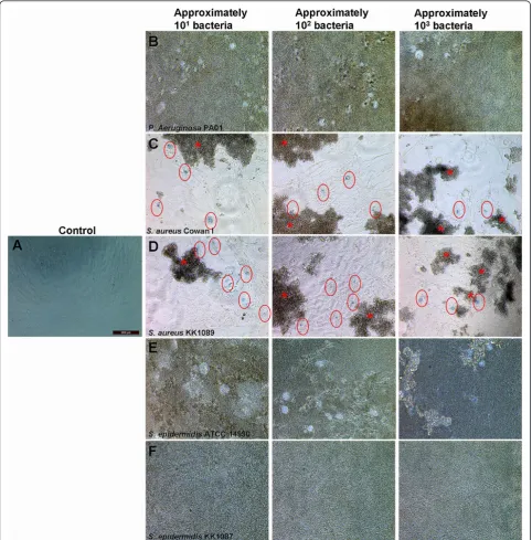
Mesenchymal and Tissue Stem Cell Committee of the International Society for Cellular Therapy [35] (Table 1). Bacteria were first incubated in three different amounts with BMMSCs (line 412). To determine the period of time that the bacterial contamination needs to induce total dis-sipation of BMMSCs even with the lowest bacterial load, we first decided to monitor the infection of BMMSCs for 24 hours. The effect of infection was compared with the uninfected control of BMMSCs (Figure 1a). The results show thatP. aeruginosaPA01 caused total destruction of BMMSCs within 24 hours of infection (Figure 1b).S.
aur-eusCowan I andS. aureusKK1089 produced enormous
aggregates, and cell death of BMMSCs was evident due to the appearance of Trypan blue-positive nuclei (Figure 1c, d).S. epidermidisATCC 1499 andS. epidermidisKK1087 also caused total dissipation of BMMSCs after 24 hours of infection (Figure 1e, f). All pathogens caused a similar destruction of BMMSCs regardless of the initial amount of inoculated bacteria. This experiment was repeated with the BMMSC lines 407 and 470 and the results were con-sistent (data not shown).
Decrease in viability of BMMSCs after 6 hours of infection The early effects of bacterial contamination on the viability of BMMSCs were studied by inoculating cells with approximately 103bacteria per well and monitoring the cellular changes during the first 6 hours. Inoculated patho-gens appeared to spend 3 hours in a lag phase most prob-ably adapting to new culture conditions as during that time there were no differences between control cells (Figure 2a) and bacterium-infected cells (Figure 2b-f, photographs on the left column). After 6 hours of infec-tion, however, changes in BMMSC viability became evident when compared with uninfected control cells (Fig-ure 2a, photograph on the right column).P. aeruginosa PA01 caused a clear decrease in viability as determined by Trypan blue staining, and morphological changes in the BMMSCs were evident (Figure 2b, photograph on the right column). The morphological changes were seen as a rounding of the cells and loss of the characteristic spindle-like cell morphology.S. aureusCowan I andS. aureus KK1089 promoted formation of small aggregates after 6 hours of infection, and some Trypan blue-positive nuclei were evident, whereas the morphology of BMMSCs appeared to stay normal (Figure 2c, d, photographs on right).S. aureusKK1089 showed the formation of aggre-gates in experiments in which lower numbers of bacteria (101
and 102) were initially seeded, but such aggregates
were not evident with S. aureus Cowan I (data not
nuclei, but no clear morphological changes were evident in BMMSCs when compared with the uninfected control BMMSCs (Figure 2e, f, photographs on the right column).
Pathogen-induced depolarization ofΔΨmprecedes major changes in cell morphology or decreases in viability of BMMSCs
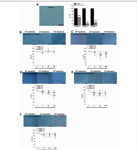
BMMSC lines 407, 470, and 412 were used to study the early effects of pathogen contamination on the state of mitochondrial energy of BMMSCs. Based on the above results with Trypan blue staining and analysis of cell mor-phology, which showed that changes begin to appear in the viability of BMMSCs after 6 hours of infection, the 4-hour time point was chosen forΔΨmanalysis. The aim was to investigate whether changes in mitochondrial energy state precede the changes that can be detected by traditional Trypan blue staining. In these experiments, CCCP treat-ment was used as a positive control to depolarize theΔΨm (Figure 3a). It was evident that theS. aureusstrains had a slightly different effect on the cells. Namely, withS. aureus Cowan I only, the BMMSC line 412 showed a significant decrease inΔΨmfrom 100% ± 5.8% to 84.9% ± 6.6% when the highest inoculum ofS. aureusCowan I was used and
theΔΨm of infected BMMSCs was compared with the
ΔΨmof control BMMSCs (P<0.05) (Figure 3b). Instead, S. aureusKK1089 induced a significant decrease inΔΨm under most experimental conditions in all three BMMSC lines even before any major changes in viability or mor-phology were evident (Figure 3c). The initial inoculum of 103ofS. aureusKK1089 caused significant depolarization ofΔΨmin all BMMSC lines from 100% ± 5.8% to 56.8% ± 7.4% (P <0.01) in BMMSC 407, from 100% ± 5.8% to 77.7% ± 2.6% (P<0.001) in BMMSC 412, and from 100% ± 5.8% to 85.3% ± 4.3% (P<0.01) in BMMSC 470.S. epider-midis ATCC 14990 and S. epidermidis KK1087 also induced significant depolarization ofΔΨm in all three BMMSC lines at all microbial loads when compared with uninfected control cells (Figure 3d, e). The initial bacterial load of 103 of S. epidermidisKK1087 caused dramatic decreases ofΔΨmin 407, 412, and 470 BMMSC lines from 100% ± 5.8% to 58.1% ± 20.1% (P<0.05), from 100% ± 5.8% to 73.6% ± 8.6% (P<0.01), and from 100% ± 5.8% to 81.1% ± 9.6% (P<0.05), respectively.P. aeruginosaPA01 caused depolarization of ΔΨm in all BMMSC lines at
higher bacterial loads. TheΔΨmdecreased from 100% ± 5.8% to 65.4% ± 11.9% in BMMSC 407 line (P<0.01), from 100% ± 5.8% to 89.1% ± 5.2% in BMMSC 412 line (P <0.05), and from 100% ± 5.8% to 90.0% ± 3.1% in BMMSC 470 line (P<0.01). However, the lowest bacterial loads did not induce a significant decrease ofΔΨmin the BMMSC line 407 when compared with control cells (Fig-ure 3f). The results clearly indicate that the depolarization ofΔΨmoccurred before major changes in morphology or in viability as determined by Trypan blue staining were evi-dent, except with the highest amount ofP. aeruginosa PA01 (Figure 3f). Trypan blue staining results are shown for the BMMSC line 412 only, but the results were identi-cal for the two other BMMSC lines. The results also show that the level of depolarization differed among the three BMMSC lines. Overall, cell line 407 showed the most dra-matic depolarization in response to all bacterial strains which caused significant depolarization ofΔΨm(all except S. aureusCowan I), and this line also showed the most dra-matic response to CCCP. The BMMSC line 412 showed a somewhat different response to CCCP when compared with the two other BMMSC lines.
All bacterial strains grew exponentially within 24 hours of infection, but there were some variations in bacterial growth during the first 4 hours of infection
Next, we determined bacterial growth in co-culture with BMMSCs after 4- and 24-hour infection. The initial inocu-lum of approximately 103bacteria was seeded onto the 407 BMMSCs. As BMMSCs have been reported to have anti-microbial effects [36,37], we inoculated bacteria for com-parison into cell culture medium not containing BMMSCs. Quantitative counting of bacteria showed that there was some variation in bacterial numbers in the initial inocula; however, the amounts of all strains were in the order of magnitude of 103. All bacterial strains showed exponential growth within 24 hours of infection when grown on top of the BMMSCs (Figure 4a). At 24 hours, efficient growth was also detected when bacteria were incubated in plain medium (data not shown). However, within the first 4 hours of infection, there were significant differences in growth between the different strains (Figure 4b). The amount ofP. aeruginosaPA01 increased 139 times during the first 4 hours of infection when grown on top of the
Table 1 Detailed characterization of human mesenchymal stem cell lines used in this work
hMSC line Positivity, percentage Age in years/Gender
CD90 CD105 CD73 HLA-ABC CD14, CD19, CD34, CD45, HLA-DR
407 98.2 99.8 100 99.5 1.7 57/Female
470 99.8 99.5 99.6 98.8 2.1 70/Female
412 100 99.6 99.9 99.4 1.1 51/Male
BMMSCs and 125 times in the medium only. On the other hand,S. aureusCowan I showed negligent growth during the first 4 hours of infection on top of the BMMSCs whereas in the plain medium the amount ofS. aureus
Figure 3 Depolarization ofΔΨmprecedes changes in morphology or plasma membrane integrity of bone marrow-derived
mesenchymal stem cells (BMMSCs). BMMSC lines 412, 407, and 470 were used to study the early changes inΔΨmand cell viability as
determined by Trypan blue after 4 hours of infection.(a)Trypan blue staining of control cells and response to CCCP (protonophore) that was used as a positive control.(b)Staphylococcus aureusCowan I caused slight depolarization ofΔΨm.(c)S. aureusKK1089 induced changes inΔΨm and Trypan blue after 4 hours.(d)Staphylococcus epidermidisATCC 14990 and(e)S. epidermidisKK1087 induced depolarization ofΔΨm.(f) Depolarization ofΔΨmand Trypan blue staining at 4 hours inPseudomonas aeruginosaPA01-infected BMMSCs. The fluorescent 595 nm/535 nm ratio of JC-10 was used to determineΔΨm, and the 595 nm/535 nm ratios of infected BMMSCs were compared with uninfected control cells. Results are indicated as mean ± standard deviation of four independent replicates. Magnification was 200 × in all panels. Red circles indicate Trypan blue-positive nuclei. Black squares indicate the aggregates produced byS. aureus. *P<0.05, **P<0.01, and ***P<0.001 in two-samplet test between control BMMSCs and infected BMMSCs.ΔΨm, mitochondrial inner membrane potential; CCCP, carbonyl cyanide
increase).S. epidermidisATCC14990 showed over three-fold increase in bacterial amounts when grown on top of the BMMSCs and over twofold increase in the medium only. Also,S. epidermidisKK1087 showed rapid prolifera-tion within the first 4 hours of infecprolifera-tion with CFU fold changes of nine in co-culture with BMMSCs and nine in medium only. The results show thatS. aureusCowan I in particular proliferates slowly during the first 4 hours of infection. This may explain the inability of the mitochon-drial assay to detectS. aureusCowan I-induced cellular stress within the first 4 hours of infection. We also deter-mined bacterial proliferation with the initial inocula of approximately 101and 102bacteria per well and noted that the bacterial growth curves were similar to those obtained with the highest inoculum (data not shown). The results also show that growth of theS. aureusstrains is signifi-cantly slower in the presence of BMMSCs than in plain medium, suggesting that antimicrobial effects from the cells may hinder their growth.
Mitochondrial assay showed sensitivity for reference strains used in microbial quality control of cellular products
Finally, we studied the potential ofΔΨmmeasurement to detect bacterium infection-induced changes in viability of BMMSCs by using certified reference microbial strains that are widely used in validation of quality control pro-cesses of cell therapy products. We measuredΔΨmand bacterial proliferation after infection of BMMSC 407
with the strains P. aeruginosaATCC9027, S. aureus
ATCC6538, andS. epidermidisATCC12228. Again, the
bacteria were seeded onto the 407 BMMSCs as well as into medium not containing BMMSCs, and bacterial growth was detected by counting CFU after 4 and 24 hours of infection. All bacterial strains grew exponen-tially within 24 hours of infection (Figure 5a). However, with reference strains, there were some variations in the proliferation within the first 4 hours of infection (Figure 5a, b). Interestingly, similar toS. aureus Cowan I and S. aureusKK1087,S. aureusATCC6538 grew to signifi-cantly higher numbers in the absence of BMMSCs (fold change of 43) than in the presence of cells (fold change of 7), again suggesting that BMMSCs may have antimi-crobial effects towardS. aureus(Figure 5b).P. aeruginosa ATCC9027 showed the highest growth with a fold change of close to 40 in both culture conditions, whereas the number ofS. epidermidisATCC12228 even declined within the first 4 hours of infection. On the other hand, even though there were differences in bacterial growth at the beginning, all bacterial strains, includingS.
epidermi-disATCC12228, grew efficiently during 24 hours of
infection (Figure 5c).S. aureusATCC6538 continued to show higher proliferation in medium only (fold change of 6 × 10⁵) when compared with the growth in co-culture
with BMMSCs (fold change of 2 × 10⁵) also during pro-longed incubation. Similarly,S. epidermidisATCC12228 proliferated more in medium only (fold change of 7 × 10⁵) than when cultivated on top of the BMMSCs (fold change of 2 × 10⁵) (Figure 5c).
In parallel with the determination of bacterial CFU, we performed the analysis ofΔΨmwithin 4 hours of infection. Results are represented as a mean ± SD of four indepen-dent replicates (Figure 5d). Again, CCCP was used as a positive control, and it induced total depolarization of
ΔΨmfrom 100% ± 7.2% to 10.8% ± 1.3% (P<0.001) when
compared with control BMMSCs. P. aeruginosa
ATCC9027, which showed rapid proliferation within the first 4 hours of infection, also induced significant depolari-zation ofΔΨmof BMMSCs from 100% ± 7.2% to 71.2% ± 3.7% (P<0.01). In addition,S. aureusATCC6538 induced the depolarization ofΔΨmof BMMSCs from 100% ± 7.2% to 74.9% ± 4.9% (P<0.05) when compared with control
BMMSCs. On the contrary,S. epidermidisATCC12228
caused only a slight depolarization ofΔΨmof BMMSCs with the change from 100% ± 7.2% to 91.9% ± 5.6%. Refer-ence strains behaved somewhat similarly as the strains used in earlier experiments: after 24 hours of infection, total dissipation of BMMSCs was evident withP.
aerugi-nosaATCC9027 andS. epidermidisATCC12228 whereas
S. aureusATCC6538 formed enormous aggregates (data not shown). Moreover, after 6 hours of infection,P. aeru-ginosaATCC9027-induced morphological changes in
BMMSCs andS. aureusATCC6538 formed aggregates as
did the two otherS. aureusstrains whereasS. epidermidis ATCC12228 did not induce any visible changes in BMMSCs (data not shown). These results indicate that the results from the mitochondrial assay were well in line with bacterial proliferation within the first 4 hours of infection. All together, the results with the reference strains support the results obtained with other bacterial strains.
Discussion
The risk of microbial contamination is well recognized in the propagation of cells for therapeutic use, but little has been published to demonstrate and characterize possible effects of contamination and infection on stem cell prop-erties and hence on the viability and ultimately on the safety monitoring of cell therapy products [10,12,14-16]. Accordingly, in the present study, we chose to focus on the quality control point of view of microbial risks during thein vitromanufacture of BMMSC-based cellular pro-ducts. The present study shows that infection of BMMSCs with even low numbers of bacteria inflicts
ser-ious damage on BMMSC cultures.P. aeruginosaPA01,
Figure 5Comparison ofΔΨmof bone marrow-derived mesenchymal stem cells (BMMSCs) and bacterial viability in co-culture of
BMMSCs with reference strainsPseudomonas aeruginosaATCC9027,Staphylococcus aureusATCC6528, andStaphylococcus epidermidis
ATCC12228. BMMSC line 407 was used to determine the bacterial growth kinetics within 4 and 24 hours of infection.P. aeruginosaATCC9027,S.
the viability of BMMSCs but behaved somewhat differ-ently by forming aggregates on the top of BMMSC monolayers. The exact content of such aggregates remains unknown at this time but is likely to be due to the coagulase protein ofS. aureuswhich reacts with pro-thrombin in blood and enables conversion of fibrinogen to fibrin, leading to blood clotting [38]. Remnants of coa-gulation cascade proteins in FBS may react withS. aureus coagulase and cause aggregation of bacteria. As opposed toS. aureus,S. epidermidisstrains are coagulase-negative, and this probably explains the absence of aggregates in samples infected withS. epidermidis.
Even thoughS. epidermidisgenerally may be regarded as less virulent thanS. aureusandP. aeruginosa[20], all three S. epidermidisstrains in our study showed definite deleter-ious effects on BMMSCs, as they also decreased theΔΨm and caused dissipation of BMMSCs. The results thus sug-gest that an opportunistic pathogen, such asS. epidermidis, is equally hazardous to stem cells as the more virulent pathogens. Overall, during prolonged incubation, all patho-gens in the present study promoted somewhat similar levels of cell destruction regardless of the amount of the initial inoculum.
Quantitative counting of bacteria showed thatP. aerugi-nosastrains proliferated actively from the beginning but that other strains showed lower growth. In particular, S. aureusCowan I showed almost negligent bacterial growth within the first 4 hours of infection and seemed to need more time to adapt to the BMMSC culture medium. Subsequently,S. aureusCowan I also began to cause a sig-nificant decrease in stem cell viability. The damage to BMMSCs was evident after 6 hours of infection as demon-strated by traditional live/dead assay, Trypan blue staining, and monitoring of cellular morphology. As expected, P. aeruginosaPA01 caused a clear decrease in viability of BMMSCs after 6 hours of infection and this is well in line with the rapid bacterial growth within the first 4 hours of infection. Also, other strains caused an increase in the number of Trypan blue-stained nuclei of BMMSCs within the first 6 hours of infection, even though the cellular damage was not as apparent as withP. aeruginosastrains. Accordingly, it seems that the signs of pathogen-induced cellular damage can be seen after 6 hours of infection with traditional viability methods. The result clearly demon-strates the risk associated with microbial contamination and infection. Therefore, methods for validation and qual-ity control of cell and tissue products call for systematic monitoring of microbial contamination but also for reli-able methods for monitoring the viability of the cell pro-duct and the possible deleterious effects of infection of stem cells during manufacturing.
Interestingly, the BMMSC line 407 appeared to show antimicrobial activity toward all threeS. aureusstrains studied. Proliferation ofS. aureusstrains was lower when
grown on top of BMMSCs than in the medium without the cells. Antimicrobial activity of BMMSCs has recently been reported in two studies [36,37]. Krasnodembskaya and colleagues [36] (2010) showed that hMSCs possess antimicrobial activity towardEscherichia coliK1,P. aeru-ginosa strain PA103, and S. aureus strain Newman. Moreover, they showed that intratracheal administration of hMSCs decreased bacterial growth in anin vivomouse
model of E. coli pneumonia. This was shown to be
mediated by the secretion of the antimicrobial peptide LL-37 [36]. Meisel and colleagues [37] (2011) postulated that hMSCs show antimicrobial activity towardS. aureus andP. aeruginosaand that the activity is enhanced when hMSCs are pretreated with interferon gamma (IFN-g). It was also shown that the antimicrobial function is mediated by indoleamine 2,3-dioxygenase (IDO) activity, which also plays a major role in immunosuppression activity of hMSCs, and is induced by IFN-g[39]. In our assay conditions, which did not include IFN-g, no signifi-cant effects towardP. aeruginosawere noted. However, our results withS. aureussupport earlier findings on the antimicrobial activity of MSCs.
Mitochondria are well-known targets of many bacterial toxins and compounds [40,41]. It has been demonstrated thatS. aureusdirectly targets host mitochondria and induces apoptosis througha-toxins and Panton-Valen-tine leukocidin [42,43]. In addition,P. aeruginosaandS. epidermidishave been shown to induce mitochondria-mediated apoptosis in infected host cells [44,45]. A rele-vant question, therefore, is whether changes inΔΨmare already present before plasma membrane breakout and morphological changes are detected. In the present study, we tested the suitability of mitochondrial analyses as a quality control tool for BMMSCs to detect microbial infection-induced cellular stress. To perform this experi-ment in accordance with current microbial quality con-trol methods, we also used reference strains which are used in routine quality control studies by cell and tissue
banks. All pathogens except S. aureus Cowan I and
more time for adapting to BMMSC culture conditions since they both did show exponential growth during a longer term of infection. Moreover, antimicrobial effects hindering the proliferation ofS. aureusCowan I in co-cul-ture with BMMSCs may partly explain the slow growth of S. aureusCowan I. In some cases, even the lowest num-bers of bacteria caused a significant decrease inΔΨm when compared with uninfected control stem cells. Thus, measurement ofΔΨmoffers a sensitive method for detec-tion of microbial damage to cell products. Interestingly, the three BMMSC lines showed different levels of depolar-ization ofΔΨmafter bacterial infection. The reason for such a difference remains speculative at this point and warrants further studies. However, we previously demon-strated that the level ofΔΨmcorresponds to the function and differentiation potency of BMMSCs [33]. Therefore, the development of rapid diagnostic tests based on mito-chondrial function is urgently called for to ensure the via-bility and potency of stem cell products.
Conclusions
To conclude, co-culture of BMMSCs in the presence of S. aureus,S. epidermidis, andP. aeruginosadamaged the viability of BMMSCs as measured byΔΨm. Such deleter-ious effects raise significant concerns for the safety and efficacy of stem cell products for use in clinical therapy. Additionally, a relatively short exposure to antibiotic-free medium can reveal a concealed and suppressed infection. Even low levels of bacterial contamination can be a major hazard for patients as such, and the deleterious effects of even short exposure to bacterial pathogens may seriously alter the efficacy of cellular therapy. On the other hand, by monitoring of the energy state of stem cell mitochondria, it is possible to detect changes in infected host cells even before any other changes are evident by Trypan blue stain-ing or microscopy for cell morphology. Accordstain-ingly, mito-chondrial analyses show strong potential for quality control and monitoring of safety and efficacy of cell-based products. In addition, in compliance with regulatory demands for microbial safety, techniques to detect low levels of bacterial contamination in a matter of hours are urgently called for and should be convenient to incorpo-rate into testing for mitochondrial function.
Abbreviations
αMEM: alpha minimum essential medium;ΔΨm: mitochondrial inner membrane potential; BMMSC: human bone marrow-derived mesenchymal stem cell; CCCP: carbonyl cyanide m-chlorophenylhydrazone; CFU: colony-forming unit; FBS: fetal bovine serum; hMSC: human mesenchymal stem cell; IDO: indoleamine 2,3-dioxygenase; IFN-γ: interferon gamma; JC-10: enhanced 5,5’,6,6’-tetrachloro-1,1’,3,3’-tetraethylbenzimidazolcarbocyanine iodide; PBS: phosphate-buffered saline; SD: standard deviation; TSA: trypticase soy agar.
Acknowledgements
The authors thank laboratory technologists Minna Savilampi and Henna Ek for their skillful contribution to this work. This study is funded by the
Academy of Finland, grant 122561, the Finnish Medical Association, and Maud Kuistila Memorial Foundation
Author details 1
Institute of Biomedicine, Department of Anatomy and Cell Biology, Aapistie 7, P.O. Box 5000, FIN-90014, University of Oulu, Oulu, Finland.2Institute of Clinical Medicine, Division of Surgery, University of Oulu and Clinical Research Centre, Department of Surgery and Intensive Care, Aapistie 5a, P.O. Box 5000, FIN-90014, Oulu University Hospital, Oulu, Finland.3Finnish Red Cross Blood Service, Kivihaantie 7, FI-00310, Helsinki, Finland.4Aalto School of Chemical Technology, Department of Biotechnology and Chemical Technology, Kemistintie 1A, P.O. Box 6100, 00076, Aalto, Finland.
Authors’contributions
MP carried out the cell and microbial culture, microbial infection of BMMSCs, and analysis of cellular viability; contributed to the study design; and wrote the manuscript. KL contributed to the study design and coordination, helped in experiments, participated in analysis of results, and helped to draft the manuscript. SL contributed to the study design, helped to draft the manuscript, established the BMMSC culture protocol, and helped in experiments. H-VL provided BMMSC material, contributed to the study design, and helped to draft the manuscript. MN contributed to the study design, helped to draft the manuscript according to microbial part, and contributed to the data analysis. ML isolated microbial material and contributed to the study design. JM contributed to the study design and coordination and helped to draft the manuscript. PL contributed to the study design and coordination, helped to draft the manuscript, and contributed to the data analysis. KN participated in study design and coordination and helped to draft the manuscript. All authors have read and approved the final manuscript.
Competing interests
The authors declare that they have no competing interests.
Received: 12 July 2012 Revised: 6 November 2012 Accepted: 29 November 2012 Published: 11 December 2012
References
1. Chang F, Ishii T, Yanai T, Mishima H, Akaogi H, Ogawa T, Ochiai N:Repair of large full-thickness articular cartilage defects by transplantation of autologous uncultured bone-marrow-derived mononuclear cells.J Orthop Res2008,26:18-26.
2. Dumas A, Moreau MF, Gherardi RK, Basle MF, Chappard D:Bone grafts cultured with bone marrow stromal cells for the repair of critical bone defects: an experimental study in mice.J Biomed Mater Res A2009,
90:1218-1229.
3. Le Blanc K, Frassoni F, Ball L, Locatelli F, Roelofs H, Lewis I, Lanino E, Sundberg B, Bernardo ME, Remberger M, Dini G, Egeler RM, Baciqalupo A, Fibbe W, Ringden O, Developmental Committee of the European Group for Blood and Marrow Transplant:Mesenchymal stem cells for treatment of steroid-resistant, severe, acute graft-versus-host disease: a phase II study.Lancet2008,371:1579-1586.
4. Loewenbruck K, Storch A:Stem cell-based therapies in Parkinson’s disease: future hope or current treatment option?J Neurol2011,258: S346-353.
5. Wollert KC, Meyer GP, Lotz J, Ringes-Lichtenberg S, Lippolt P, Breidenbach C, Fichtner S, Korte T, Hornig B, Messinger D, Arseniev L, Hertenstein B, Ganser A, Drexler H:Intracoronary autologous bone-marrow cell transfer after myocardial infarction: the BOOST randomised controlled clinical trial.Lancet2004,364:141-148.
6. Rayment EA, Williams DJ:Concise review: mind the gap: challenges in characterizing and quantifying cell- and tissue-based therapies for clinical translation.Stem Cells2010,28:996-1004.
7. Dietz AB, Padley DJ, Gastineau DA:Infrastructure development for human cell therapy translation.Clin Pharmacol Ther2007,82:320-324.
8. Ill CR, Dehghani H:Risk reduction in biotherapeutic products.Curr Opin Drug Discov Devel2009,12:296-304.
10. Klein MA, Kadidlo D, McCullough J, McKenna DH, Burns LJ:Microbial contamination of hematopoietic stem cell products: incidence and clinical sequelae.Biol Blood Marrow Transplant2006,12:1142-1149. 11. Hillyer CD, Josephson CD, Blajchman MA, Vostal JG, Epstein JS,
Goodman JL:Bacterial contamination of blood components: risks, strategies, and regulation: joint ASH and AABB.Hematology Am Soc Hematol Educ Program2003, 575-589.
12. Prince HM, Page SR, Keating A, Saragosa RF, Vukovic NM, Imrie KR, Crump M, Stewart AK:Microbial contamination of harvested bone marrow and peripheral blood.Bone Marrow Transplant1995,15:87-91. 13. Kamble R, Pant S, Selby GB, Kharfan-Dabaja MA, Sethi S, Kratochvil K,
Kohrt N, Ozer H:Microbial contamination of hematopoietic progenitor cell grafts-incidence, clinical outcome, and cost-effectiveness: an analysis of 735 grafts.Transfusion2005,45:874-878.
14. Vanneaux V, Fois E, Robin M, Rea D, de Latour RP, Biscay N, Chantre E, Robert I, Wargnier A, Traineau R, Benbunan M, Marolleau JP, Socie G, Larghero J:Microbial contamination of BM products before and after processing: a report of incidence and immediate adverse events in 257 grafts.Cytotherapy2007,9:508-513.
15. Smith D, Bradley SJ, Scott GM:Bacterial contamination of autologous bone marrow during processing.J Hosp Infect1996,33:71-76. 16. Rood IG, de Korte D, Ramirez-Arcos S, Savelkoul PH, Petterson A:
Distribution, origin and contamination risk of coagulase-negative staphylococci from platelet concentrates.J Med Microbiol2011,
60:592-599.
17. Gastens MH, Goltry K, Prohaska W, Tschope D, Stratmann B, Lammers D, Kirana S, Götting C, Kleesiek K:Good manufacturing practice-compliant expansion of marrow-derived stem and progenitor cells for cell therapy.
Cell Transplant2007,16:685-696.
18. Webb IJ, Coral FS, Andersen JW, Elias AD, Finberg RW, Nadler LM, Ritz J, Anderson KC:Sources and sequelae of bacterial contamination of hematopoietic stem cell components: implications for the safety of hematotherapy and graft engineering.Transfusion1996,36:782-788. 19. Wiener-Well Y, Galuty M, Rudensky B, Schlesinger Y, Attias D, Yinnon AM:
Nursing and physician attire as possible source of nosocomial infections.
Am J Infect Control2011,39:555-559.
20. Subbiahdoss G, Fernandez IC, Dominques JF, Kuijer R, van der Mei HC, Busscher HJ:In vitro interactions between bacteria, osteoblast-like cells and macrophages in pathogenesis of biomaterial-associated infections.
PLoSOne2011,6:e24827.
21. European Parliament and European Council:Directive 2001/83/EC of the European Parliament and of the Council.Brussel: European Parliament and Council; 2001:OJ 44(L311):67-128.
22. International Conference on Harmonisation:Note for guidance on specifications: test procedures and acceptance criteria for biotechnological/biological products (CPMP/ICH/365/96).London: European Medicines Agency; 1999.
23. International Conference on Harmonisation:Quality of biotechnological products: viral safety evaluation of biotechnology products derived from cell lines of human or animal origin (CPMP/ICH/295/95).London: European Medicines Agency; 2006.
24. Committee for Medicinal Product for Human Use:Guideline on human cell-based medicinal products (EMEA/CHMP/410869/2006).London: European Medicines Agency; 2008.
25. European Commission:Note for guidance on minimising the risk of transmitting animal spongiform encephalopathy agents via human and veterinary medicinal products (EMA/410/01 rev.3).Brussel: European Comission; 2011:OJ 54(C73):1-18.
26. Susin SA, Lorenzo HK, Zamzami N, Marzo I, Snow BE, Brothers GM, Mangion J, Jacotot E, Constantini P, Loeffler M, Larochette N, Goodlett DR, Aebersold R, Siderovski DP, Penninger JM, Kroemer G:Molecular characterization of mitochondrial apoptosis-inducing factor.Nature1999,
397:441-446.
27. Heiskanen KM, Bhat MB, Wang HW, Ma J, Nieminen AL:Mitochondrial depolarization accompanies cytochrome c release during apoptosis in PC6 cells.J Biol Chem1999,274:5654-5658.
28. Kroemer G, Reed JC:Mitochondrial control of cell death.Nat Med2000,
6:513-519.
29. Chen CT, Shih YR, Kuo TK, Lee OK, Wei YH:Coordinated changes of mitochondrial biogenesis and antioxidant enzymes during osteogenic
differentiation of human mesenchymal stem cells.Stem Cells2008,
26:960-968.
30. Pietilä M, Palomäki S, Lehtonen S, Ritamo I, Valmu L, Nystedt J, Laitinen S, Leskelä HV, Sormunen R, Pesälä J, Nordström K, Vepsäläinen A, Lehenkari P:
Mitochondrial function and energy metabolism in umbilical cord blood-and bone marrow-derived mesenchymal stem cells.Stem Cells Dev2012,
21:575-588.
31. Lonergan T, Brenner C, Bavister B:Differentiation-related changes in mitochondrial properties as indicators of stem cell competence.J Cell Physiol2006,208:149-153.
32. Mandal S, Lindgren AG, Srivastava AS, Clark AT, Banerjee U:Mitochondrial function controls proliferation and early differentiation potential of embryonic stem cells.Stem Cells2011,29:486-495.
33. Pietilä M, Lehtonen S, Närhi M, Hassinen IE, Leskelä HV, Aranko K, Nordström K, Vepsäläinen A, Lehenkari P:Mitochondrial function determines the viability and osteogenic potency of human mesenchymal stem cells.Tissue Eng Part C Methods2010,16:435-445. 34. Ensayef S, Al-Shalchi S, Sabbar M:Microbial contamination in the
operating theatre: a study in a hospital in Baghdad.East Mediterr Health J 2009,15:219-223.
35. Dominici M, Le Blanc K, Mueller I, Slaper-Cortenbach I, Marini F, Krause D, Deans R, Keating A, Prockop DJ, Horwitz E:Minimal criteria for defining multipotent mesenchymal stromal cells. The International Society for Cellular Therapy position statement.Cytothreapy2006,8:315-317. 36. Krasnodembskaya A, Song Y, Fang X, Gupta N, Serikov V, Lee JW,
Matthay MA:Antibacterial effect of human mesenchymal stem cells is mediated in part from secretion of the antimicrobial peptide LL-37.Stem Cells2010,28:2229-2238.
37. Meisel R, Brockers S, Heseler K, Degistirici Ö, Bulle H, Woite C, Stuhlsatz S, Schwippert W, Jäger M, Sorg R, Henschler R, Seissler J, Dilloo D, Daubener W:Human but not murine multipotential mesenchymal stromal cells exhibit broad-spectrum antimicrobial effector function mediated by indoleamine 2,3-isooxygenase.Leukemia2011,25:648-654. 38. Rivera J, Vannakambadi G, Höök M, Speziale P:Fibrinogen-binding
proteins of Gram-positive bacteria.Thromb Haemost2007,98:503-511. 39. Meisel R, Zibert A, Laryea M, Gobel U, Daubener W, Dilloo D:Human bone
marrow stromal cells inhibit allogenic T-cell responses by indoleamine 2,3-oxygenase-mediated tryptophan degradation.Blood2004,
103:4619-4621.
40. Arnoult D, Carneiro L, Tattoli I, Girardin SE:The role of mitochondria in cellular defense against microbial infection.Semin Immunol2009,
21:223-232.
41. Kozjak-Pavlovic V, Ross K, Rudel T:Import of bacterial pathogenicity factors into mitochondria1.Curr Opin Microbial2008,11:9-14. 42. Bantel H, Sinha B, Domschke W, Peters G, Schulze-Osthoff K, Jänicke RU:
alpha-Toxin is a mediator ofStaphylococcus aureus-induced cell death and activates caspases via the intrinsic death pathway independently of death receptor signaling.J Cell Biol2001,155:637-648.
43. Genestier AL, Michallet MC, Prevost G, Bellot G, Chalabreysse L, Peyrol S, Thivolet F, Etienne J, Lina G, Vallette FM, Vandenesch F, Genestier L:
Staphylococcus aureusPanton-Valentine leukocidin directly targets mitochondria and induces Bax-independent apoptosis of human neutrophils.J Clin Invest2005,115:3117-3127.
44. Jendrossek V, Grassme H, Mueller I, Lang F, Gulbins E:Pseudomonas aeruginosa-induced apoptosis involves mitochondria and stress-activated protein kinases.Infect Immun2001,69:2675-2683. 45. Pharmakakis NM, Petropoulos IK, Georgakopoulos CD, Vantzou CV,
Anastassiou ED, Mavropoulos A, Zolota V:Apoptotic mechanisms within the retina inStaphylococcus epidermidisexperimental endophthalmitis.
Graefes Arch Clin Exp Ophthalmol2009,247:667-674. doi:10.1186/scrt144