REVIEW
Probiotic engineering:
towards development of robust probiotic
strains with enhanced functional properties
and for targeted control of enteric pathogens
Moloko Gloria Mathipa and Mapitsi Silvester Thantsha
*Abstract
There is a growing concern about the increase in human morbidity and mortality caused by foodborne pathogens. Antibiotics were and still are used as the first line of defense against these pathogens, but an increase in the develop-ment of bacterial antibiotic resistance has led to a need for alternative effective interventions. Probiotics are used as dietary supplements to promote gut health and for prevention or alleviation of enteric infections. They are currently used as generics, thus making them non-specific for different pathogens. A good understanding of the infection cycle of the foodborne pathogens as well as the virulence factors involved in causing an infection can offer an alternative treatment with specificity. This specificity is attained through the bioengineering of probiotics, a process by which the specific gene of a pathogen is incorporated into the probiotic. Such a process will subsequently result in the inhibi-tion of the pathogen and hence its infecinhibi-tion. Recombinant probiotics offer an alternative novel therapeutic approach in the treatment of foodborne infections. This review article focuses on various strategies of bioengineered probiotics, their successes, failures and potential future prospects for their applications.
Keywords: Foodborne pathogens, Antibiotic resistance, Probiotics, Bioengineering
© The Author(s) 2017. This article is distributed under the terms of the Creative Commons Attribution 4.0 International License (http://creativecommons.org/licenses/by/4.0/), which permits unrestricted use, distribution, and reproduction in any medium, provided you give appropriate credit to the original author(s) and the source, provide a link to the Creative Commons license, and indicate if changes were made. The Creative Commons Public Domain Dedication waiver (http://creativecommons.org/ publicdomain/zero/1.0/) applies to the data made available in this article, unless otherwise stated.
Background
Poor hygiene and sanitation during food preparation can lead to the presence of different foodborne pathogens in food. Some of these pathogens or their toxins produced either before or after ingestion of such foods can either act locally within the gastrointestinal tract (GIT), lead-ing to development of illnesses, or disseminate to other parts of the body and damage cells/tissues and ultimately the immune system [1]. Incidences of foodborne illnesses are high in most developing countries as food control is a low priority issue due to limited funds. As a result of this, food pathogens are the leading cause of illnesses and death in these countries [2]. Most foodborne illnesses cause diarrhoea, which is the primary symptom. Most societies consider diarrhoea a normal, natural condition;
therefore, it goes unnoticed and/or untreated. Recently, the World Health Organization reported that of the 600 million global total cases of foodborne illness recorded in 2010, 550 million were due to infectious agents causing diarrhoea, of which 120 million and 96 million cases were caused by norovirus and Campylobacter spp., respec-tively. Diarrhoeal disease agents were responsible for approximately 55% (230,000 out of 420,000) deaths, with 59,000, 37,000, 35,000 and 26,000 deaths attributed to non-typhoidal Salmonella enterica, enteropathogenic E. coli (EPEC) and enterotoxigenic E. coli (ETEC), respec-tively [3]. These illnesses are not confined to developing countries. In the United States, foodborne pathogens cause an estimated 9.4 million illnesses, 55,961 hospitali-zations and 1351 deaths each year [4]. Enteric pathogens account for high morbidity and mortality and are con-sidered to be the fifth leading cause of death at all ages worldwide [5].
Open Access
*Correspondence: [email protected]
Probiotics have been used to restore the balance of the gut microbial ecosystem and control pathogenic infec-tions. They are defined as “live microorganisms that when administered in adequate amounts confer a health ben-efit on the host” [6]. Their administration assists in the prevention and control of foodborne illnesses, through a number of mechanisms including but not limited to, competitive exclusion of pathogens in the GIT, modu-lation of the host immune system and strengthening of the intestinal barrier [7–9]. Although probiotics have proven successful in the control of enteric pathogens, they do have limitations. They are generic in nature and often fail to inhibit the attachment of certain pathogens at specific sites of infection and induce low levels of an immune response [10]. A thorough understanding of the limitations of conventional probiotics, the behav-iour of the pathogens and the mechanisms by which they cause disease [11] provides possibilities to design new probiotic strains with desired characteristics and func-tionalities. Through genetic modification, novel bioen-gineered probiotic strains can be produced. Functioning of conventional probiotics in these novel strains can be strengthened to influence critical steps in pathogenesis. The strains can also be used to deliver drugs or vaccines, target a specific pathogen or toxin, mimic surface recep-tors and enhance an immune response within the host [12].
Probiotics
Probiotics include mainly bacteria from the genera Strep-tococcus, Enterococcus, Pediococci, Weissella and Lacto-coccus [13] but the most common ones used belong to
Lactobacillus and Bifidobacteria spp. These bacteria have met the criteria necessary to consider them as probiot-ics and they also have nutritional and therapeutic effects [14]. One of the criteria that bacteria must meet in order for them to be regarded as probiotics is that they have to be able to survive and thrive throughout the GIT condi-tions and confer their beneficial effects. It is therefore important to understand their mechanisms of action in order for them to be used both prophylactic and as treat-ment options for the different foodborne diseases. The presence of foodborne pathogens in the human GIT affects the balance of the “good to bad” microorganisms. Apart from the presence of the pathogens in the GIT, there are other factors that can affect the balance of the microorganisms in the host GIT. These factors include stress, illness or antibiotic treatment, which changes the balance in the GIT in favour of harmful bacteria [15, 16]. One of the characteristics of probiotics is that they are able to protect the host from microbial imbalance. Table 1 gives a summarized overview of the different
mechanisms by which probiotics exclude pathogens from the human GIT, which are discussed in more detail below.
Probiotics’ mechanisms of action against enteric pathogens
Competitive exclusion
Probiotics can exclude or reduce the growth of other microorganisms in the GIT either through competition for nutrients or adherence space [17–19]. Microorgan-isms in any environment require nutrients to multiply and either cause or alleviate infections. The GIT is well known for its abundance in nutrients, making it a suita-ble environment for microbial colonization. The potential of probiotics to outcompete pathogens for these nutri-ents favours their growth over that of the pathogens [20]. During competition for nutrients, probiotics produce metabolites such as volatile fatty acids reducing the pH of the GIT. The reduction in the pH of the GIT makes it an unfriendly environment for pathogens and will thus lead to their inhibition because most of them cannot grow at low pH [21, 22].
Competition for adherence space refers to the situation when the presence of probiotics blocks pathogenic bac-teria from colonizing favourite sites such as the intestinal villi, goblet cells and the colonic crypts [22]. Attachment to the surfaces of intestinal epithelial cells is a key patho-genic factor of enteric pathogens [23]. At the same time, colonization resistance, through which attachment and multiplication of the pathogens on the intestinal mucosal membrane is prohibited, is a critical function of the microbiota [24, 25]. Probiotics bind to intestinal cells via electrostatic interactions, steric forces or specific surface proteins. This affords them the ability to bind to these cells in high quantities [17, 26], thereby physically block-ing the sites, leavblock-ing no space for the pathogens to adhere and subsequently cause infection [27].
Probiotic LAB have a greater ability to adhere to the epithelial cells than pathogens [28]. Lactobacilli and bifi-dobacteria share carbohydrate-binding specificities with some enteropathogens [29]. Bifidobacterium bifidum and
Table 1 M echanisms of ac tion of pr obiotics M echanism of ac tion Pr obiotic bac teria Pa thogen Func tionalit y Ref er enc es Competitiv e ex clusion L. acidophilus ; L. johnsonii S. enteric a ser ovar Typhimur ium, Ent er opatho -genic E. c oli (EPEC ), Yersinia pseudotuber culosis , L. monoc ytogenes Inhibit ed adher
ence of pathogens t
o C aco -2 cells [ 33 ] L. c asei subsp . rhamnosus EPEC, Ent er ot oxigenic E. c oli (E TEC ), Klebsiella pneumoniae Inhibit ed adher
ence of pathogens t
o C aco -2 cells [ 33 ]
L. rhamnosus; L. acidophilus
E. c
oli
O157:H7
Inhibit
ed adher
ence of pathogens t
o
T-84 epi
-thelial cell; inhibit
ed colonization t
o C aco -2 cells [ 33 , 51 ] L. acidophilus H. p ylori Pr oduc
tion of lac
ticins A164 and BH5
[
37
]
Pr
oduc
tion of inhibit
or y substances L. lactis E. c oli , Salmonella Pr oduc
tion of alyt
eser
in-1a and A3APO
[ 31 ] B. longum Clostridium difficile , E . c oli Pr oduc
tion of bac
ter iocin [ 35 ] L. saliv arius L. monoc ytogenes Pr oduc
tion of bac
ter iocin Abp118 [ 35 ] L. sak e L. monoc ytogenes Pr oduc
tion of bac
ter iocin sak acin A [ 39 ] L. plantarum L. monoc ytogenes Pr oduc
tion of bac
ter iocins (plantar icins) [ 40 ] L. lactis C. difficile Pr oduc
tion of lac
ticin 3147
[
45
]
L. lactis
, L. c
asei, L. acidophilus
E. c
oli
O157:H7
Reduced the g
ro
wth of pathogen b
y lac
tic acid
pr
oduc
tion and pH r
educ tiv e eff ec t [ 33 ] Immune syst em modulation L. r euteri Salmonella Incr
eased the pr
oduc tion anti-Salmonella IgM [ 35 ] L. rhamnosus E. c oli O157:H7 Incr eased int estinal anti-E. c oli IgA r esponses
and blood leuk
oc yt e phagoc ytic ac tivit y [ 33 ] L. acidophilus , L. rhamnosus GG, L. johnsonii La1 S. T yphimur ium Incr
eased the pr
oduc
tion of
anti-Salmonella IgA [ 42 , 43 ] Impr ov ed bar rier func tion L. plantarum , L. rhamnosus GG E. c oli O157:H7 Sub ver
t the adher
ence of pathogen b
y incr
eas
-ing MUC2 and MUC3 in HT
-29 epithelial cell
line [ 45 , 47 ] L. acidophilus E. c oli Pr ot ec
ted against F-ac
tin r
ear
rangement, which
was induced in an epithelial cell line on expo
-sur
e t
o a pathogenic
E. c
oli
[
45
]
S. thermophilus; L. acidophilus
Ent er oin vasiv e E. c oli (EIEC ) Incr
eased transepithelial r
esistance
, maint
e-nance and enhancement of c
yt osk eletal and tight junc tional pr ot ein phosphor ylation in
HT29 and C
aco
-2 cell lines
[
60
and proliferation, and unavailability of adherent space in the GIT.
Production of inhibitory substances
In order to gain a competitive advantage when compet-ing for space and nutrients, microorganisms release anti-microbial compounds. Antianti-microbial compounds have a direct inhibition on several target pathogens [31]. The mechanisms used by probiotics to inhibit pathogenic bacteria are interconnected. As already mentioned, exclusion of pathogens occurs due to the ability of pro-biotics to secrete organic acids such as acetic and lactic acids [32]. The production of these organic acids leads to a decrease in the pH of the environment, making the microenvironment acidic thereby excluding pathogens that cannot survive acidic conditions [8]. The organic acids also have an effect on the pathogen metabolism and production of toxins, ultimately preventing disease.
The anti-pathogenic activity of probiotics is multifacto-rial [33]. In addition to the acids mentioned above, pro-biotics can produce other metabolites with antibacterial properties, such as H2O2 and bacteriocins, also referred to as non-lactic acid molecules [33–36, 41]. Bacteriocins are small antimicrobial peptides produced for bacterial competition in a natural ecosystem [31]. They may act as colonizing peptides by facilitating the introduction of probiotics into an already occupied niche on the intesti-nal epithelium. This competitive advantage allows for an increase in the density of probiotic bacteria on the sur-face of the host intestines [36]. They can also act as kill-ing peptides, by directly affectkill-ing pathogens. A study by Kim et al. [37] evaluated the antimicrobial activity of the bacteriocins: lacticin, pediocin and leucocin, produced by lactic acid bacteria against Helicobacter pylori. These bacteriocins were able to significantly inhibit the growth of H. pylori, with lacticin having the most inhibitory effect against this gut pathogen.
Lactobacillus acidophilus has been reported to produce metabolites such as acidophilin, lactocidin and acidolin [35], whereas bifidobacteria produces bacteriocin-like substances [38], all inhibiting bacteria such as Bacil-lus, Salmonella, Staphylococcus and E. coli, Clostridium perfringens, Listeria species, among others [35, 39, 40]. Fayol-Messaoudi et al. [41] reported that the antibacte-rial effects of the probiotic Lactobacillus that inhibited the growth and resulted in pathogen death were due to the synergistic action of lactic acid and the secreted non-lactic acid molecules. Certain probiotic strains can also stimulate the increase in the expression of host cell antimicrobial peptides. The intestinal cells of the host are able to produce defensins which can inhibit the func-tioning of pathogens thus aiding in the protection of the intestinal barrier [36].
Immune system modulation
Probiotics can displace pathogens through stimulation of host immunity [42]. There is considerable evidence to support the notion that probiotics displace pathogens in the GIT through stimulation of specific and non-specific immunity to inhibit bacteria causing intestinal diseases [43, 44]. They modulate the host’s immune system against the pathogens harmful antigens by the activation of lymphocytes and production of antibod-ies [45]. They can also stimulate the effects of different cells involved in innate and adaptive immunity, such as dendritic cells, macrophages, T cells and B cells, which enhances phagocytosis of gut pathogens [46]. Probiotic strains such as Lactobacillus rhamnosus and Lactoba-cillus plantarum adhere to gut-associated lymphoid tissue enhancing both systemic and mucosal immunity [9]. These probiotics strains enhance immunity by up-regulating production of intestinal mucins (MUC2 and MUC3), which disrupts the adherence of pathogens to the intestinal epithelium, consequently prevent-ing pathogen translocation. Furthermore, they induce expression of TGFβ and interleukins (IL-10 and IL-6) by epithelial cells, which enhances production and secre-tion of IgA [47].
Probiotics can be recognized by the immune system through pattern recognition molecules such as Toll-like receptors. This recognition can lead to various intracel-lular signal transduction cascades and enhancement or reduction of pro and anti-inflammatory cytokines. There are different studies supporting this evidence. In 2000, Fang et al. [43] divided 30 healthy volunteers into three different treatment groups, with each group consum-ing Lactobacillus GG, Lactococcus lactis and placebo (ethyl cellulose), respectively, for 7 days. All the treat-ment groups were given an attenuated Salmonella typhi
In addition to the above, probiotics can stimulate an anti-inflammatory response, which can be used as an approach to reduce inflammation caused by gastroenteri-tis, enterocolitis and irritable bowel syndrome [9]. An anti-inflammatory response is triggered when strains stimulate the activation of dendritic cells which secrete interleukin 10 (IL-10), a cytokine that plays a role in reducing inflam-mation. They also cause a decrease in the levels of proin-flammatory cytokines during inflammation [46].
Improved barrier function
The integrity of the intestinal barrier needs to be main-tained in order to prevent pathogens from reaching the intestinal cells, thereby leading to local and systemic infec-tions. Gut pathogens have the ability to disrupt the barrier when there is an imbalance in the microbial gut ecosystem [49]. It has previously been reported that consumption of probiotics can maintain the barrier function and mucosal integrity, prevent chronic inflammation, thereby protect-ing the host against infections [50]. Probiotics decrease paracellular permeability, providing innate defense against pathogens and enhancing the physical impediment of the mucous layer [51]. They are also able to repair this barrier after damage that may have been caused by gut pathogens. As an approach to repair the intestinal barrier, probiotics can stimulate mucous secretion, chloride and water secre-tion and the binding together of submucosa cells by tight junctional proteins [8].
Goblet cells express rod-shaped mucins (MUCs), which are either localized to the cell membrane or secreted into the lumen to form the mucous layer [52, 53]. There are 18 mucin-type glycoproteins that are expressed by humans [49]. In the human intestinal cell lines, Lactobacillus spe-cies increased mucin expression (MUC2 by Caco-2 cells; MUC2 and MUC3 by HT29), thus blocking cellular adhe-sion and invaadhe-sion by pathogenic E. coli [54, 55]. Madsen et al. [56] showed that the treatment of IL-10 gene-defi-cient mice with a combination probiotic VSL#3 (L. casei, L. plantarum, L. acidophilus, L. delbrueckii subsp. bulga-ricus, Bifidobacterium longum, B. breve, B. infantis, and Streptococcus salivarius subsp. thermophilus), resulted in normalization of colonic physiologic function and barrier integrity leading to significant improvement in histologic disease [57].
Tight junctions (TJ) form the continuous intercellu-lar barrier between epithelial cells, which is required to separate tissue spaces and regulate selective movement of solutes across the epithelium [58]. There are different proteins expressed on the TJ and the disruption of their expression leads to a dysfunctional epithelial barrier [57]. A study by Qin et al. [59] reported that L. acidophilus
increases the expression of occludin, a major component of TJ, in the gut mucosa of animals with cecal ligation
and perforation, leading to a reduced bacterial transloca-tion. A different study by Resta-Lenert and Barrett [60] reported that probiotic bacteria, specifically S. thermophi-lus and L. acidophilus, prevented reduction in the entero-invasive E. coli-induced phosphorylation of the proteins occludin and zonula occludens 1 (ZO-1), thereby pre-serving the TJ structure. Furthermore, Parassol et al. [61] showed that L. casei prevents the redistribution of the TJ protein ZO-1 away from the cell–cell contacts caused by infection with enteropathogenic E. coli.
The use of conventional probiotics for control of selected food pathogens
Due to the widespread use of antibiotics as therapeu-tic agents and the misuse of these antibiotherapeu-tics, there has been an increase in the antibiotic resistance of bacteria, an imbalance of normal microflora and the presence of drug residues in food products [41]. This brought about a requirement for new intervention in the treatment of bacterial pathogens, leading to an escalation in the research field of the beneficial microorganisms, i.e. pro-biotics. Prevention and treatment of infections caused by the different pathogens is one of the reasons why pro-biotics extensively studied [62]. When studying the pre-vention and treatment of pathogens, it is important to consider the complexity of the intestinal environment where a network of interactions among the microorgan-isms of the resident microbiota, epithelial and immune cells associated with the GIT, and nutrients exist [63, 64]. The epithelial and the immune cells play a role in the modulation of the immune functions and they pro-vide the first line of defense against the pathogenic bacte-ria. The resident microbiota have the ability to influence the composition and activity of the gut microbiota [62]. They also play a beneficial role in the treatment of dis-ease caused by foodborne pathogens [65, 66]. Different microorganisms infect different parts of the host GIT, for example, H. pylori, infects the gastric and duodenal mucosa, Salmonella spp. and Clostridium difficile cause inflammation in ileum and colon, while Shigella sp. clearly prefers the colonic mucosa [67].
Previous studies have shown the effects of probiotics, that when consumed as part of the daily diet, they can maintain the immune system in an active state and pre-vent different intestinal disorders [62]. Valdez et al. [68] reported that certain LAB probiotics inhibit apoptosis of macrophage infected with Salmonella preventing salmo-nellosis. Cano and Perdigón [69] studied the preventative measure of L. casei CRL 431 against S. serovar Typhimu-rium, reporting that administrating probiotics prevented
[62], where the preventative and continuous adminis-tration of probiotic L. casei CRL 431 against S. sero-var Typhimurium in a mouse model was studied. They reported that the study group fed the probiotic for 7 days before the introduction of the pathogen and post infec-tion experienced less severe infecinfec-tion compared to the control group which did not consume probiotics. They furthermore reported that 7-day administration of probi-otics post infection resulted in better protection against
Salmonella infection. They concluded that the continu-ous administration of the probiotic diminished counts of the pathogens in the intestine as well as their spread out-side this organ.
More studies have been conducted on different patho-gens to show the efficacy of probiotic strains. H. pylori
is a bacterium that plays a crucial role in the pathogen-esis of chronic active gastritis and peptic ulcer disease in both adults and children [70] with increasing amount of evidence supporting the hypothesis that it is an impor-tant co-factor in the development of gastric cancer [71].
H. pylori has been linked to cancer; however, there is no vaccine licensed to prevent infection with this organism [72]. There are different therapeutic approaches that are used to treat H. pylori, including but not limited to the commonly used triple therapy with proton pump inhibi-tor (PPI), clarithromycin and either amoxicillin or met-ronidazole or dual-therapy high-dosage amoxicillin and PPI; however, there have been reports that suggest that some patients still remain infected after administration of these treatment [73]. Administration of alternative compounds that may increase the efficacy of the treat-ment and/or reduce side-effects is of particular interest [72]. There is growing evidence from different studies emphasizing the efficacy of probiotics in the manage-ment of H. pylori infection targeting different aspects of this infectious disease [74, 75]. Cats et al. [74] inves-tigated whether readily available commercial prepara-tion containing L. casei inhibits the growth of H. pylori
in vitro. They reported that in vitro L. casei inhibits the growth of H. pylori; however, the probiotic cells have to be viable. In a different study, Bernet-Camard et al. [76] reported that probiotics such as L. johnsonii La1 (La1) or L. rhamnosus GG exert bacteriostatic or bactericidal activities against a wide range of pathogens, including H. pylori. Cruchet et al. [77] studied if the regular ingestion of a dietary product containing L. johnsonii La1 or L. par-acasei ST11 would interfere with H. pylori colonization in children. They concluded that regular ingestion of the dietary product containing L. johnsonii La1 may repre-sent an interesting alternative to modulate H. pylori colo-nization in children infected by this pathogen. Tursi et al. [78] demonstrated that a 10-day quadruple anti-helico-bacter therapy with ranitidine bismuth citrate (RBC) plus
proton pump inhibitors (PPI), amoxicillin and tinidazole obtains a high eradication rate, whereas supplementation with L. casei significantly increased the eradication rate of H. pylori infection. This study concluded that the sup-plementation of the therapy with the administration of probiotics showed a slight improvement in the eradica-tion of H. pylori. Probiotics can therefore be used as first course of anti-H. pylori treatment or can be used in con-jugation with the first-line therapeutic approaches.
Shigella is an antibiotic-resistant bacterium [79, 80] that has been reported to cause gastroenteritis-induced deaths in 3–5 million children aged less than 5 years in developing countries [81, 82]. The emergence of multiple drug resistance to cost-effective antimicrobials against
Shigella is a matter of concern in developing countries, and resistance pattern of this bacterium is the cause of numerous clinical problems worldwide [83]. Due to increased prevalence of its antibiotic resistance, the need for alternative treatment has therefore been deemed nec-essary. Zhang et al. [84] studied the antimicrobial activ-ity of the probiotics L. paracasei subsp. paracasei M5-L,
L. rhamnosus J10-L, L. casei Q8-L and L. rhamnosus GG (LGG) against Shigella sonnei. They reported that the tested lactobacilii strains showed strong antimicrobial activity against S. sonnei. In a study to screen for the anti-microbial activity of probiotics against S. sonnei, Zhang et al. [85] reported that L. johnsonii F0421 exhibited sig-nificant inhibitory activity and excluded, competed and displaced S. sonnei adhered to HT-29 cells. In a different study, Mirnejad et al. [83] evaluated the nature of anti-microbial substances and properties of L. casei against multi-drug-resistant clinical isolates of S. flexneri and
S. sonnei. Their results indicated that L. casei showed strong antimicrobial activity against S. flexneri and S. son-nei, and they attributed pathogen inhibition to produc-tion of organic acids by the test Lactobacillus. In another study, Zou et al. [86] studied the antimicrobial activity of nisin, a bacteriocin produced by L. lactis strains, against
L. monocytogenes, Staphylococcus aureus, Salmonella typhimurium and Shigella boydii. They reported that there was a decline in pathogen populations, which was ascribed to the changes in the fatty acid profiles, cell via-bility, membrane permeability and depolarization activity in response to nisin.
breast-fed infant faeces, with a potential antimicrobial activity against L. monocytogenes. These isolates actively inhibited L. monocytogenes by producing a heat-stable proteinaceous substance. Their study indicated that the use of bifidobacterial strains capable of competing with pathogenic organisms following the probiotic approach would advantageously improve intestinal bacterial ecol-ogy and provides a useful alternative strategy for inhibit-ing intestinal pathogens. In 2007, Corr et al. [91] studied the pretreatment of C2Bbe1 cells, a clone of the Caco-2 human adenocarcinoma cell line with strains of Bifido-bacterium and Lactobacillus to demonstrate that this can significantly interfere with subsequent invasion by
L. monocytogenes. They reported that the pretreatment of intestinal epithelial cells with probiotic bacteria prior to infection with L. monocytogenes EGDe resulted in a significant decrease in listerial invasion (60–90%). In yet another study testing for the antagonistic effect of Lac-tobacillus strains against E. coli and L. monocytogenes, it was reported that L. plantarum WS4174 exhibited a stronger inhibitory effect against the Gram-positive L. monocytogenes LMO26, possibly due to the accumulation of lactic acid and higher sensitivity of L. monocytogenes
to low pH [92].
Limitations of conventional probiotics
Although probiotics provide numerous benefits to the host, they do have certain limitations. Certain studies have provided evidence that probiotic strains may be inefficient or ineffective in response to specific gut patho-gens. Probiotics may release antimicrobial compounds that have a broad antimicrobial spectrum; however, reports have suggested that there are limitations in the success of probiotics targeting specific pathogens. There-fore, a cocktail of various probiotic strains would need to be produced in order to enhance the effects against dif-ferent pathogens within the gut [93].
Contrary to earlier reports that probiotics exhib-ited inhibitory effect against L. monocytogenes [90, 91], according to Koo et al. [94], probiotics have a limited suc-cess in preventing the attachment of L. monocytogenes
to intestinal monolayers. In their study, which used three experimental approaches of competitive exclusion, inhibition of adhesion or displacement, to determine whether selected lactobacilli would reduce adhesion of L.
monocytogenes to Caco-2 cells, they showed that the per-centages of L. monocytogenes adhesion in the presence and absence of probiotics were fairly similar. None of the lactobacilli and other LAB were able to significantly reduce adhesion or colonization on epithelial cells, even at higher numbers. Furthermore, an increase in the con-centration of the probiotic strain also failed to displace the attached L. monocytogenes. The data from the study
indicated the conventional LAB strains could not prevent adhesion of this pathogen.
Another report indicated that probiotics may also stimulate low levels of an immune response and low lev-els of an anti-inflammatory response [10]. L. salivarius
and B. infantis were orally administered to mice suffering from colitis. Results indicated that TGF-β levels in mice treated and untreated with probiotics remained the same. TGF-β is an anti-inflammatory cytokine, and the lev-els of this cytokine were not significantly increased but still maintained by L. salivarius; however, these were not maintained in the presence of B. infantis.
Most probiotics are administered as food or capsules; therefore, they have to be able to withstand both the technological and gastrointestinal stress factors. The broad mode of action of probiotics and the differences from one probiotic to another is also an obstacle in their efficacy. It has been reported that the beneficial attributes of one strain or a cocktail of strains may not be reproduc-ible and may vary from person to person [95]. In addition to that, the strain of the probiotic, the dosage, the route of administration, and the formulation of probiotic prep-aration can also affect their efficacy [94]. Taking these studies into consideration, it is evident that probiotics are still non-specific and non-discriminatory in their mode of action or ineffective in certain hosts [96].
The limitations discussed above introduce the need for more novel and innovative approaches in the use of probiotics for the prevention and treatment of foodborne pathogens. Previous literature has reported that the use of probiotics has been extended to deliver therapeutic and prophylactic molecules to the mucosal barrier of the host [94, 97, 98]. However, for that to be done success-fully, a thorough understanding of the behaviour of the pathogens and their disease mechanisms is needed [11]. Such knowledge can then be used to increase the efficacy of the probiotics and later use of a specific probiotic for a specific pathogen or toxin. Thus, novel probiotic strains with enhanced or even targeted probiotic functioning can be produced. Bioengineering techniques offers an opportunity for the design of such recombinant probiotic strains.
The concept of probiotic bioengineering or recombinant probiotics
can be used in the design and construction of new pro-biotic strains harbouring genes of interest derived from the pathogens. It allows for the production of proteins that were initially not present within the microorgan-ism. Virulence factors of the pathogens can be cloned and expressed into the probiotic strain and subsequent administration of the resultant recombinant probiotic strain will inhibit the development of infection and yield no clinical presentation of the symptoms. Furthermore, recombinant probiotics can be used to deliver drugs or vaccines, target specific pathogens or toxins, enhance an immune response and mimic cell surface receptors [100]. Most human receptors recognized by enteric pathogens or their toxins are well characterized. Also, by targeting a specific pathogen, this strategy deems the development of resistance to the vaccine or treatment unlikely. Bioen-gineering of probiotics is not entirely a new field, vari-ous researchers have reported on the successes attained with the use of such probiotics (Table 2). Culligan et al. [49] reported on the main advantages of using recombi-nant probiotics in the treatment of enteric infection. The next section of this review focuses on studies that were conducted on bioengineered probiotics aimed at improv-ing different functional properties of the conventional strains.
Applications of probiotic bioengineering
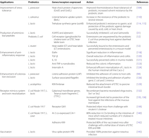
Improvement of stress tolerance
There has been an increase in the use of probiotics due to their known effects to confer beneficial health to the host. However, there are still problems frequently asso-ciated with the incorporation of probiotic strains into food products. These problems include but are not lim-ited to poor temperature, salt and oxygen tolerance of some species or strains. Different approaches including pre-adaptation to stress, the use of oxygen-impermeable containers, microencapsulation [101], incorporation of nutrients, and selection of stress-resistant strains have been used in an attempt to address these problems [102]. The use of bioengineering has been used in the field of stress adaptation, and there have been promising results.
The ability to confer additional stress tolerance in stress-sensitive cultures can lead to the development and delivery of novel probiotics with maximal therapeutic efficacy [103]. It has been reported that the two major heat shock proteins, GroES and GroEL, are essential for the survival of bacteria at all temperatures [104]. In a study by Desmond et al. [101], the effect of overexpres-sion of these heat shock protein chaperones (GroES and GroEL) in the probiotic L. paracasei NFBC338 was inves-tigated. Expression of these genes resulted in improved thermotolerance (heat tolerance) as well as increased solvent resistance by the probiotic strain. Furthermore,
they compared the survival of the non-adapted par-ent strain, stress adapted and the recombinant probiotic during exposure to heat stress. They reported that the recombinant probiotic survived 10- and 54-fold better than the stress-adapted and non-adapted parent strains, respectively.
The survival of pathogens is usually dependent on the different systems that can help them overcome the differ-ent stress conditions presdiffer-ent in the GIT. Three transport systems have to date been identified in L. monocytogenes
that have been linked to betaine and carnitine uptake [105, 106]. The first of these is a gene encoding the sec-ondary glycine betaine transporter, listerial betaine uptake system (BetL), which is linked to salt tolerance of
Listeria [107, 108]. It has been reported that disrupting BetL results in reduced growth at 37 °C in complex media of elevated osmolarity [107]. The reduction in the initial betaine uptake in the absence of BetL leads to dimin-ished intracellular solute pools [106], causing changes in the cell volume, intracellular solute concentration and the turgor pressure [109]. Sheehan et al. [110] studied the heterologous expression of the BetL into the probi-otic strain L. salivarius UCC118 using a nisin-controlled expression system. They reported that expression of BetL led to an increase in the resistance of the probiotic to several stresses (osmo, cryo, baro and chill), spray- and freeze-drying. Later in another study these researchers demonstrated that B. breve UCC2003 harbouring the betaine uptake (BetL) gene displayed an improved toler-ance to gastric juice and elevated osmolarity [111].
Trehalose is a non-reducing disaccharide ubiquitously distributed in nature and is well known for its role in pro-tecting cells against a variety of stresses [112]. In E. coli,
it is synthesized in response to high osmolarity [113]. Termont et al. [114] cloned the trehalose synthesis gene (ostAB) from E. coli into L. lactis and reported that there was an enhanced probiotic’s survival during freeze-dry-ing, in high bile concentrations and its resistance to gas-tric acid. In a different study, Carvalho et al. [115] studied the expression of the trehalose synthesis in the same pro-biotic L. lactis and reported that trehalose plays a definite role in the protection of this bacterium against damage caused by acid, cold or heat shock. These studies provide evidence that expression of genes from pathogenic spe-cies to improve stress tolerance of probiotics has been explored with promising results. However, further sci-entific assessment is still required to analyse the benefit of using these genes and interpretation by risk–benefit analysis [103].
Production of antimicrobial peptides
infections. Antimicrobial peptides (AMPs) have been explored as an alternative method for effective control of multi-drug resistant (MDR) pathogens [116]. As already mentioned, some probiotics produce several antimicro-bial compounds and peptides as a defense mechanism against pathogens [12] but they are not specific. Probiot-ics can therefore be used as candidates for the production and delivery of therapeutic antimicrobial peptides within the host GIT targeting a specific action or pathogen. The current methods for production of AMPs have been reported to have several limitations. Synthesis of pep-tides is not only expensive, but also time-consuming too; in some cases, the peptides eventually kill the producing
cells or are secreted as inclusion bodies. Oral administra-tion of the peptides subjects them to degradaadministra-tion before they can reach the target site. They are also difficult to administer systemically as they are rapidly identified and directed for restoration of the immune system before they can reach the site of infection. Therefore, an alterna-tive strategy will be to use probiotic strains to express the different AMPs resulting in a combination strategy where hosts will get the probiotic effects with the production of the different AMPs [116].
Volzing et al. [31] chose L. lactis as an ideal vehicle for production and delivery of AMPs to the site of GI infection due to its ability to survive within the human
Table 2 Applications of bioengineering
Applications Probiotics Genes/receptors expressed Action References
Improvement of stress
tolerance L. paracasei Heat shock protein chaperones (GroES and GroEL) Improved thermotolerance (heat tolerance) of probiotic; increased solvent resistance by the
probiotic strain
[104]
L. salivarius Listerial betaine uptake system
(BetL) Increase in the resistance of the probiotic to several stresses [110]
L. lactis Trehalose synthesis gene (ostAB) Enhanced probiotic’s resistance to gastric acid
protection of the probiotic against damage caused by acid, cold, or heat shock
[114, 115]
Production of
antimicro-bial peptides L. lactisProbiotic E. coli A3APO and alyteserinCell receptor (ganglioside) for Successfully inhibited E. coli and Salmonella [31]
cholera toxin or ETEC heat-labile toxin
Enterotoxins are sequestered by the probiotic
E. coli thus protecting host against diarrheal
infection
[12]
L. reuteri Heat-stable (ST) and heat-labile
(LT) enterotoxins Successfully bound to the enterotoxins and prevented enterotoxicity in a mouse model [12]
Enhancement of
anti-inflammatory response L. lactisL. lactis ElafinTGF-β Significant reduction in inflammationOverall reduction of inflammation and colitis [118][120]
L. lactis IL-10 Successfully prevented colitis in murine models [121]
L. lactis Anti-TNF-α nanobodies Reduced the colonic inflammation [123]
L. lactis Internalin A Enhanced efficient internalization of L. lactis in
the human intestinal cell line Caco-2 [124]
Enhancement of
coloniza-tion exclusion L. paracaseiL. lactis ListeriaSurface-associated flagellin adhesion protein (LAP) Inhibited the adhesion of Inhibited the binding and adhesion of patho-Listeria to host cells [94]
genic E. coli and S. enterica [126]
L. acidophilus K99 fimbriae Reduced the attachment of ETEC to porcine
intestinal brush border [129]
Receptor mimicry system and toxin neutralization
E. coli Nissle 1917; L.
lactis Galactosyl-transferase genes; Tetanus toxin fragment C
(TTFC)
Recombinant bacteria neutralized shiga toxins,
Stx1 or Stx2 [132]
Increased IgA levels led to protection of the host against the infections of the mucous membrane
[135, 136]
E. coli Nissle 1917 Receptor GM1 Protected infant mice from challenge with
virulent V. cholerae [139]
E. coli Nissle 1917; L.
casei AI-2 co-expressed CAI-1 80% reduction in Ctx binding to the intestines of mice which reduced numbers of V. cholerae in
treated mouse intestines
[140]
Adhesins K99 Protected 80% of the vaccinated mice after
challenge with a lethal dose of strains of ETEC K99 and K88
[142]
Vaccination L. lactis Virus spike protein VP8 Provided 100% protection against rotavirus
gastrointestinal tract and its amenability to heterologous gene overexpression. In their study, they engineered a L. lactis strain to inducibly express and secrete AMPs with high activity against Gram-negative pathogens, specifi-cally E. coli and Salmonella strains. The AMPs of inter-est, A3APO and alyteserin were selected and then cloned into L. lactis for the expression of the heterologous pep-tides. An expression cassette containing a codon-opti-mized sequence for alyteserin was fused with an Usp45 secretion signal sequence. This expression cassette was cloned under the control of a nisin inducible promoter and transformed into L. lactis. When the resultant L.
lactis recombinant strain was induced to express and secrete these peptides, and the effect of their expression on growth and viability of E. coli and Salmonella was tested, the results indicated successful inhibition of both these pathogens while viability of the host (i.e. the L. lac-tis expressing the peptides) was maintained. Inhibition of these pathogens by alyteserin was observed from concen-trations ranging from 0.125–1 mg/ml, while the L. lactis
strains remained viable when exposed to the alyteserin supernatant at 1 mg/ml. This system showed potential as a therapeutic alternative to antibiotics in order to target and inhibit Gram-negative bacteria.
Enhancement of anti‑inflammatory response
A group of chronic inflammatory disorders known as inflammatory bowel diseases (IBD) are responsible for the inflammation of the digestive tract. The two forms of the IBD are Crohn’s disease and ulcerative colitis, which are both characterized by an uncontrolled inflamma-tory response in the intestines [117]. The treatment of IBDs poses a challenge as the current treatment options are either costly or cause severe side-effects in patients. There have been a number of studies on the treatment of IBDs and recent research has reported that probiotic bacteria may counteract the chronic inflammatory pro-cess [118]. Elafin, is a protease inhibitor expressed in the intestinal epithelium, which contributes to the reduc-tion of inflammareduc-tion. During inflammareduc-tion, there is an increase in elastase and myeloperoxidase (MPO) activ-ity, elafin can inhibit the function of proteases, thereby reducing inflammation [119]. Bermúdez-Humarán et al. [118] bioengineered L. lactis to express elafin in mice suffering from colitis. The gene encoding for elafin was fused in frame with a gene encoding for a ribosome-bind-ing site and with an Usp45 secretion signal sequence and inserted into an expression vector. The recombinant plas-mid was thereafter transformed into L. lactis and expres-sion was induced under the control of a nisin-induced promoter. Colonic inflammation was then induced in mice with dextran sodium sulphate and then the mice were subsequently orally treated with either wild-type
or recombinant L. lactis. Analysis of mice colons for inflammation parameters such as colonic thickness, elastase activities and granulocyte infiltration after 7 days indicated that mice treated with recombinant L. lactis
secreting the elafin showed a significant reduction in all inflammation parameters. However, mice treated with wild-type probiotics did not show the same significant decrease in inflammation parameters, their response was similar to that of the control-untreated mice. Fur-thermore, comparison of efficiency of recombinant L. lactis secreting elafin to those expressing either the anti-inflammatory cytokine IL-10 or TGF-β1 (to be discussed next) showed that elafin-secreting strain was the most efficient. These results suggested that elafin was the most efficient anti-inflammatory molecule to be delivered by a probiotic strain at the mucosal surface in order to treat inflammation [118].
Chronic inflammation of IBD patients can also be reduced through the administration of anti-inflammatory cytokines such as interleukin 10 (IL-10). IL-10 plays a central role in down-regulation of inflammatory cascades and in the establishment of tolerance in the mucosa [9]. Interferons (IFN), including IFN-α and IFN-β, are widely expressed cytokines involved in innate responses and additionally, these cytokines have an immunomodulatory role in the anti-inflammatory host response. The use of probiotic bioengineering to treat IBD has been studied and it has been reported that this can indeed be used as an alternative. Several studies have been carried out with regard to probiotics expressing cytokines and other anti-inflammatory molecules such as IL-10 and TGF-β instead of elafin, using similar cloning procedures used for elafin. After transformation, recombinant probiotic strains were induced with nisin in order to either express IL-10 or TGF-β and orally administered to mice suffering from colitis. Recombinant L. lactis expressing TGF-β displayed beneficial effects by reducing MPO levels, overall reduc-ing inflammation and colitis in 40% of the mice. However, the protective effects against colitis were higher in mice treated with recombinant probiotics expressing elafin than those treated with probiotics expressing IL-10 [120]. Another study reported that intra-gastric administration of L. lactis expressing recombinant IL-10, a cytokine used in clinical trials for treatment of IBD, could successfully prevent colitis in murine models [121].
they constructed a recombinant L. lactis to produce anti-TNF-α nanobodies and reported that daily administra-tion of this strain reduced the colonic inflammaadministra-tion.
Enhancement of colonization exclusion
Enhancement of probiotic adhesion to the intestinal mucosal surface can be seen as a potential strategy in order to prevent adhesion and colonization of pathogenic bacteria. Strategies include using gene products of tar-get pathogens such as adhesins or secretory systems in probiotic bacteria to create a competitive environment for colonization [94]. A number of researchers investi-gated the efficiency of this approach in improvement of competitive exclusion by enhancing binding or adhesion efficacy of the probiotics to host cells. When internalin A from L. monocytogenes was cloned and expressed into the L. lactis strain, there was enhanced binding to human epithelial cells and bacterial internalization [124]. A more recent study, Koo et al. [94] developed a recombinant probiotic L. paracasei harbouring the Listeria adhesion protein (LAP) in order to control L. monocytogenes infec-tion. LAP interacts with a heat shock protein 60 recep-tor in host cells and promotes adhesion of Listeria to host cells. Conventional and recombinant probiotic L. para-casei were added to Caco-2 cell monolayers separately, thereafter these monolayers were Giemsa-stained. Pre-exposure of Caco-2 cell monolayers to recombinant L.
paracasei expressing LAP followed by the addition of L. monocytogenes led to a reduction of adhesion and trans-location of the pathogen. The wild-type probiotic strain had no significant reduction in the adhesion of the L. monocytogenes to the cell monolayer, while the recombi-nant strain resulted in a 60% reduction of adhesion.
It has been shown that flagellins from Bacillus cereus
are responsible for the adhesion of the bacteria to mucosal cells [125]. Gut pathogens may also use fimbriae or flagella which are extended appendages on the surface of the cell wall, to adhere to host cell receptors. There-fore, expression of these specific appendages in probiotic strains would allow them to bind to the intestinal epi-thelium, excluding pathogenic binding. Taking that into consideration, Sánchez et al. [126] cloned the surface-associated flagellin of B. cereus CH and expressed it in the probiotic L. lactis. The recombinant strain adhered strongly to the mucin-coated polystyrene plates in an in vitro experiment and competitively inhibited the bind-ing and adhesion of pathogenic E. coli and S. enterica.
Enterotoxigenic Escherichia coli (ETEC) K99 fim-briae have been reported to enhance the production of mucosal IgA and serum IgG1 fimbria-specific responses [127], thereby increasing the immune responses at mucosal surfaces such as the gastrointestinal (GI) tract, the respiratory tract, and the vaginal tract [128]. Chu
et al. [129] cloned and expressed the K99 fimbriae from ETEC into the probiotic L. acidophilus and reported that the recombinant L. acidophilus was able to reduce the attachment of ETEC to porcine intestinal brush border in a dose-dependent manner. The reduction of the adher-ence of the pathogen by the recombinant probiotic pre-vents the binding of the pathogen, therefore inhibiting the infection.
Receptor mimicry system and toxin neutralization
One mechanism that pathogens use to invade the host cells and cause infection is through the production of toxins. These pathogens secrete toxins, and sometimes express adhesins that bind to host cells via oligosaccha-ride receptors displayed on surface glycolipids or gly-coproteins. The interaction between the released toxin and the specific oligosaccharide receptors on the surface of the human intestinal cells is an essential step during pathogenesis [130]. Therefore, toxins or secretory sys-tems of pathogens may also serve as potential targets in development of therapeutics [131]. Taking this into con-sideration, it thus becomes apparent that interfering with the toxin receptor binding and adhesion can be used as a strategy to exclude the pathogen and subsequently minimize or control its infection [130]. A therapeutic strategy would be to express toxin receptors on the cell surface of probiotic strains in order to mimic the recep-tor [132]. This expression produces a lipopolysaccharide that mimics a host cell receptor, which, e.g. cholera toxin or ETEC heat-labile toxin could recognize and bind to. Therefore, upon infection, enterotoxins would bind to probiotics and become sequestered, protecting the host from a pathogenic infection [130]. That is, the toxin is sequestered when, instead of binding to the receptor on the surface of the host cell, it binds with high avidity to the receptor mimic expressed on the surface of the pro-biotic cell. This hinders the interaction between the toxin and the host cells, which is a crucial step in the disease process [130, 134]. Studies of probiotics expressing toxin receptor mimics were mostly biased towards impact on the disease progression without monitoring of the probi-otic-toxin complex. However, Paton et al. [130] reported that the receptor mimic probiotic was spontaneously eliminated from the GIT of mice a day or two after the end of its administration. Therefore, more studies track-ing the probiotic-toxin complex are required to establish their fate.
There are a number of pathogens that secrete these tox-ins and among them are, Vibrio cholerae, Shiga toxigenic
left untreated, these pathogens can cause severe bloody diarrhoea associated with haemorrhagic colitis [133]. In an earlier study by Paton et al. [134], the galactosyl-trans-ferase genes from Neisseria gonorrhoeae were cloned and expressed into a non-pathogenic E. coli. The results showed that the recombinant E. coli was 100% effective in treating mice infected with the normally fatal shiga toxigenic E. coli. Then later on in another study, these researchers cloned the glycosyltransferase gene, Neisseria meningitidis toxin-specific receptor, into the probiotic E. coli, creating a competitive environment for toxin binding to the host cells. Expression of these genes created a cell surface mimic of a shiga toxin receptor. This led to com-petitive exclusion of the pathogen by the probiotic and subsequently inhibiting its infection. This recombinant strain had a high binding capacity and efficacy in mouse models and was effective in neutralizing shiga toxin vari-ants (stx1 and stx2) [132]. Norton et al. [134] cloned and expressed a tetanus toxin fragment C (TTFC) in L. lactis.
They then reported that there were increased IgA levels in the host after oral administration of the recombinant probiotic, which led to protection of the host against the infections of the mucous membrane. These results were supported by other studies, where mice immunized with this recombinant probiotic showed more resistance to the lethal challenge with tetanus toxin than those that were not immunized [136, 137].
Pathogens are able to control the expression of their virulence genes by sensing signals from their own spe-cies, other bacteria or their environment, a phenomenon termed quorum sensing [12]. Interruption of quorum sensing of the pathogen can be used as an alternative strategy to control the pathogen. Cholera is a life-threat-ening gastrointestinal infection [138] that is caused by ingestion of water or food (usually undercooked shellfish) contaminated with V. cholerae [130]. Following inges-tion, V. cholerae passes through the stomach, colonizes the small intestine and then release cholera toxin (Ctx), which is responsible for its virulence. It has been hypoth-esized that neutralization of Ctx in the gut should pre-vent the disease from developing or at least speed up recovery from an established V. cholerae infection [130]. The cloning and expression of Ctx receptor into probiot-ics can therefore be used as an alternative strategy for the treatment of cholera. Focareta et al. [139] constructed a probiotic E. coli encoding receptor GM1 (to express the GM1 ganglioside) on its surface, which is capable of binding large amounts of Ctx and protecting infant mice from challenge with virulent V. cholerae. The resultant recombinant E. coli was capable of binding purified Ctx with high avidity and adsorbing >5% of its own weight of toxin in vitro. V. cholerae releases cholera autoinducer-1 (CAI-1) and autoinducer-2 (AI-2), which depending of
population density, can down- or up-regulate expression of virulence genes [12, 140]. Virulence genes involved are Ctx, which causes diarrhoea, and toxin-coregulated pilus (TCP), which facilitates attachment of vibrios to the intestinal wall. When cell densities are high, expression of genes encoding these virulence factors is reduced, while proteases expressed degrade the attachment matrix with consequent flushing out of the bacterial cells with diar-rhoeal fluids. In order to determine the possibility for use of bioengineered probiotic for control of cholera, Duan and March [140] constructed an AI-2 producing E. coli
Nissle that co-expressed CAI-1. They reported an 80% reduction in Ctx binding to the intestines of mice pre-treated with recombinant probiotic, which reduced the chances of infection. These results showed the poten-tial for use of bioengineered E. coli Nissle co-express-ing CAI-1 and AI-2 for the prevention or treatment of cholera.
Vaccination
Probiotics may induce low levels of the immune response. Therefore, probiotics can be bioengineered to deliver immunogenic molecules to the intestinal mucosal surface to enhance the immune response. Recombinant probi-otics can act as a vaccine arming the host immune sys-tem to deal with gut pathogens [141]. In order to exploit a safe and effective vaccine for the prevention against K99 infections of ETEC, Wen et al. [142] cloned and expressed ETEC adhesins K99 into the probiotic L. casei. They reported that there was an increase in the efficacy of the recombinant probiotic and that more than 80% of the vaccinated mice were protected after challenge with a lethal dose of standard strains.
consumers, a decreased risk in transmission of food-borne diseases and the stimulation of both innate and adaptive immunity [9].
Safety concerns regarding bioengineered probiotics
Bioengineered probiotics are increasingly being studied as vehicles that can express and target delivery of specific genes directed towards a specific foodborne pathogen. One of the main drawbacks of working with bioengi-neered probiotics is that they are classified as genetically modified organisms (GMO) [144]. The nature of such probiotics regarded as GMO presents a major limitation to their widely applications. It is well known that some consumers have ethical reasons for not consuming GMO for fear that such organisms may pose danger to one’s life [145]. Other concerns about GMO relate to their release into the environment and their survival and propagation in this environment, dissemination of antibiotic selec-tion markers or other genetic material to other organisms [146]. Introduction of the GMO into the environment can impact there directly by competing with natural spe-cies, or indirectly by changing the balance between native species [147]. However, these modified microorganisms have a great potential to address novel approaches for prevention and treatment of different human and ani-mal pathological conditions. It is therefore, important to establish criteria that can be used for the assessment of the environmental safety and tracing the fate of recom-binant DNA in vitro and in vivo, which are both of sig-nificant importance [148]. Hence, safety of these strains needs to be guaranteed in order for them not to possess antibiotic selection markers or to transfer genetically modified DNA to other bacteria [144]. Biological con-tainment systems can be used to prevent dissemination of genetic material to other bacteria and to prevent a sig-nificant uncontrolled increase of probiotic cells into the natural environment [145]. The organism is genetically programmed to only grow in the laboratory and to die in the natural environment [149]. The use of the thymidine-deficient strains is one of the promising strategies for bio-logical containment of bioengineered probiotics. In these strains, the gene of interest is cloned into the chromo-somal thymidylate synthase gene (thyA), which codes for production of thymine essential for growth of L. lactis. This disruption of the thyA gene makes the recombinant strain dependent on external supplementation of thymi-dine or thymine in the growth medium for growth and survival. Thymine is absent in the environment or its lev-els are limiting in vivo, and this ensures that the recombi-nant strain dies rapidly due to the absence of an essential growth component. In addition, chromosomal location
of the introduced gene provides stability and reduces the risk of horizontal gene transfer [146, 150].
When cloning and expressing the different virulent traits into probiotics, only traits that will not make the probiotics pathogenic should be used. It is also crucial that each bioengineered strain be carefully evaluated for virulence determinants and sensitivity to clinically rele-vant antibiotics before being deemed suitable as a probi-otic [151]. When cloning probiotics, therapeutic safety of recombinant probiotic carrier organisms is crucial, espe-cially when the strain has to be used in individuals who are already infected with a pathogen. The risk exposure determination, risk assessment and safety assessment are essential to ensure protection for the population against any unintended consequences of the use of probiotics [152].
Conclusions and future perspective
The rise in morbidity and mortality due to foodborne pathogens remains a serious concern worldwide and the need for an alternative strategy for the control and treatment of infections caused by pathogens is equally crucial. The application of probiotics in food for control of enteric pathogens has been explored and the otic market is growing worldwide. The ability of probi-otics to inhibit human enteric pathogen has been well researched and documented and this has led to their use as a therapeutic approach for treatment of enteric infec-tions. These studies showed both their successes and lim-itations, mainly highlighting the generic nature of their mode of action and their failure in controlling some spe-cific pathogens. These limitations can be overcome and functions of conventional probiotics enhanced to create a greater beneficial effect through the use of bioengineer-ing. The modification of conventional probiotics by use of bioengineering technology has a significant potential for design and development of novel therapeutic approaches for effective treatment of pathogens.
therapeutic approach for the prevention and treatment of foodborne infections. More studies targeting different virulence genes and pathogens, including the less studied and emerging ones, are necessary in order to establish the future of this field of research and determine how it will impact on the food and health industries.
In addition to the above, most bioengineered probiot-ics are designed to be orally administered; therefore, they must still be able to survive through both technological and gastrointestinal stresses. It is also crucial that these strains have scientifically validated health properties, demonstrated safety and good technological proper-ties to be produced on a large scale [147]. They should remain viable in large numbers so as to confer the benefi-cial effects to the host and should not develop unpleasant flavours or textures upon their incorporation into foods [148]. Furthermore, studies on bioengineered probiot-ics, specifically for targeted control of pathogens, have focused on the impact of the recombinant probiotic strain on the pathogen(s) of interest. The influence of administration of these probiotics on commensal bacte-ria or the whole microbiota has not been the subject of studies. These aspects should also be addressed in future studies on bioengineered probiotics.
Abbreviations
GIT: gastrointestinal tract; LAB: lactic acid bacteria; IgA: immunoglobulin A; IL-10: interleukin 10; RBC: ranitidine bismuth citrate; BetL: listerial betaine uptake system; AMPs: antimicrobial peptides; MDR: multi-drug-resistant; IBD:
inflam-matory bowel diseases; MPO: myeloperoxidase; LAP: Listeria adhesion protein;
ETEC: enterotoxigenic Escherichia coli; STEC: Shiga toxigenic Escherichia coli;
TTFC: tetanus toxin fragment C; GMO: genetically modified organisms; TCP:
toxin coregulated pilus; thyA: thymidylate synthase gene.
Authors’ contributions
MST conceived the original concept and outline for the review. MST and MGM collected information, drafted and edited the manuscript. Both authors read and approved the final manuscript.
Acknowledgements
Not applicable.
Competing interests
The authors declare that they have no competing interests.
Availability of data and materials
Data sharing not applicable to this article as no datasets were generated or analysed during the current study.
Funding
We are grateful for funding received from the National Research Foundation of South Africa and the University of Pretoria. Both funders were not involved in the design of the study and collection, analysis and interpretation of data and in writing the manuscript.
Publisher’s Note
Springer Nature remains neutral with regard to jurisdictional claims in pub-lished maps and institutional affiliations.
Received: 2 February 2017 Accepted: 27 April 2017
References
1. Sousa CP. The impact of food manufacturing practices on foodborne diseases. Braz Arch Biol Technol. 2008;51(4):815–23.
2. Fratamico PM, Bhunia AK, Smith JL. Foodborne pathogens in microbiol-ogy and molecular biolmicrobiol-ogy. Wymondham: Caister Academic Press; 2005. 3. Havelaar AH, Kirk MD, Torgerson PR, Gibb HJ, Hald T, Lake RJ, Praet N,
Bellinger DC, de Silva NR, Gargouri N, Speybroeck N, Cawthorne A, Mathers C, Stein C, Frederick J, Angulo FJ, Devleesschauwer B, et al. World Health Organization global estimates and regional com-parisons of the burden of foodborne disease in 2010. PLoS Med.
2015;12(12):e1001923. doi:10.1371/journal.pmed.1001923.
4. Scallan E, Hoekstra RM, Angulo FJ, Tauxe RV, Widdowson MA. Food-borne illness acquired in the United States-major pathogens. Emerg Infect Dis. 2011;17:1–15.
5. Gupta C, Prakash D, Gupta S. Genetically engineered probiotics. Afr J Basic Appl Sci. 2014;6(3):57–64.
6. FAO/WHO. Guidelines for the evaluation of probiotics in food. Report of a Joint FAO/WHO Working Group on Drafting Guidelines for the Evalua-tion of Probiotics in Food; Ontario, Canada. April 30 and May 1, 2002. 7. Ceapa C, Rezaiki L, Kleerebezem M, Knol J, Ozeer R. Influence of
fer-mented milk products, probiotics and probiotics on microbiota compo-sition and health. Clin Gastroenterol. 2013;27:139–55.
8. Wohlgemuth S, Loh G, Blaut M. Recent developments and perspec-tives in the investigation of probiotic effects. Int J Med Microbiol. 2010;300:3–10.
9. Behnsen J, Deriu E, Sassone-Corsi M, Raffatellu M. Probiotics: properties, examples, and specific applications. Cold Spring Harb Perspect Med. 2013;3:a010074.
10. McCarthy J, O’Mahony L, O’Callagan L, Sheil B, Vaughan EE, Fitzsimons N, Fitzgibbon J, O’ Sullivan GCO, Kiely B, Collins JK, Shanahan F. Double blind, placebo controlled trial of two probiotic strains in IL-10 knockout mice and mechanistic link with cytokine balance. Gut. 2003;52:975–80. 11. Amara AA, Shibi A. Role of probiotics in health improvement,
infec-tion control and disease treatment and management. Saudi Pharm J. 2015;23:107–14.
12. Amalaradjou MA, Bhunia AK. Bioengineered probiotics, a strategic approach to control enteric infections. Bioengineered. 2013;4:379–87. 13. Kurzak P, Ehrmann MA, Bauer J, Vogel RF. Characterization of Lactobacilli
towards their use as probiotic adjuncts in poultry. J Appl Microbiol. 2002;92:966–75.
14. Rattanachaikunsopon P, Phumkhachorn P. Antimicrobial activity of basil (Ocimum basilicum) oil against Salmonella enteritidis in vitro and in food. Biosci Biotechnol Biochem. 2010;74:1200–7.
15. Cremonini F, Di Caro S, Nista EC, Bartolozzi F, Capelli G, Gasbarrini G. Meta-analysis: the effect of probiotic administration on antibiotic associated diarrhoea. Aliment Pharmacol Ther. 2002;16:1461–7. 16. Harish K, Varghese T. Probiotics in humans—evidence based review.
Calicut Med J. 2006;4:e3.
17. Fuller R. Probiotics in human medicine. Gut. 1991;32:439–42. 18. Rolfe RD. Population dynamics of the intestinal tract. In: Blankenship
LC, editor. Colonization control of human bacterial enteropathogens in poultry. San Diego: Academic Press; 1991. p. 59–75.
19. Ohashi Y, Ushida K. Health-beneficial effects of probiotics: its mode of action. Anim Sci J. 2009;80(4):361–71.
20. Cumming JH, MacFarlane GT. Role of intestinal bacteria in nutrient metabolism. J Parenter Enteral Nutr. 1997;21(6):357–65.
21. Marteau P, Minekus M, Havenaar R, Huis JHJ. Survival of lactic acid bac-teria in a dynamic model of the stomach and small intestine: validation and the effects of bile. J Dairy Sci. 1997;80:1031–7.