Open Access
Review
Assessment of left ventricular function by three-dimensional
echocardiography
Boudewijn J Krenning, Marco M Voormolen and Jos RTC Roelandt*
Address: Department of Cardiology, Thoraxcentre, Erasmus MC, Rotterdam, The Netherlands
Email: Boudewijn J Krenning - [email protected]; Marco M Voormolen - [email protected]; Jos RTC Roelandt* - [email protected]
* Corresponding author
Abstract
Accurate determination of LV volume, ejection fraction and segmental wall motion abnormalities is important for clinical decision-making and follow-up assessment. Currently, echocardiography is the most common used method to obtain this information. Three-dimensional echocardiography has shown to be an accurate and reproducible method for LV quantitation, mainly by avoiding the use of geometric assumptions. In this review, we describe various methods to acquire a 3D-dataset for LV volume and wall motion analysis, including their advantages and limitations. We provide an overview of studies comparing LV volume and function measurement by various gated and real-time methods of acquisition compared to magnetic resonance imaging. New technical improvements, such as automated endocardial border detection and contrast enhancement, will make accurate on-line assessment with little operator interaction possible in the near future.
Review
Accurate quantification of left ventricular (LV) volume and function is important in clinical decision-making and follow-up assessment. Although various other techniques including invasive angiography, radionuclide angiogra-phy and magnetic resonance imaging are used, echocardi-ography is currently the most commonly applied modality in the practice of cardiology.
M-mode echocardiography, a one dimensional ultra-sound scanning of the cardiac structures, was developed in the early 1970s and immediately applied in practice for left ventricular function assessment because of its simple algorithm and non-invasiveness. Ejection fraction was estimated as a percentage derived from the mid left ven-tricular diameters in end-systole and end-diastole and expressed as fractional shortening. However, serious
problems were raised especially in patients with myocar-dial infarction and asymmetric ventricles.
Two-dimensional sectional echocardiography, with the ability of imaging of the heart in tomographic views, con-siderably improved the accuracy of left ventricular volume measurement. Of the different mathematical models, modified biplane Simpson's rule provided more accurate data in both symmetric and asymmetric left ventricles. Software-based algorithms for automatic endocardial bor-der detection and on-line calculation of left ventricular volume and ejection fraction have been developed. As a result, two-dimensional echocardiography has become a routine examination for left ventricular volume and func-tion assessment but the assumpfunc-tions about LV geometry remain a limitation.
Published: 08 September 2003
Cardiovascular Ultrasound 2003, 1:12
Received: 18 August 2003 Accepted: 08 September 2003
This article is available from: http://www.cardiovascularultrasound.com/content/1/1/12
In the past decade, three-dimensional echocardiography has emerged as a more accurate and reproducible approach to LV quantitation mainly by avoiding the use of geometric assumptions of the LV shape. Three methods have been proposed for the acquisition of temporal and positional image data: the use of positional locators (free-hand scanning), rotational systems and real-time volu-metric scanning. Reconstruction methods using posi-tional locators and rotaposi-tional scanning systems require additional intervention for respiratory gating (or breath-hold) and off-line post-processing of data using specific software. This has limited current routine application in clinical practice. Real-time three-dimensional echocardi-ography has a great potential for immediate assessment of LV function in various clinical scenarios including stress echocardiography and during interventional procedures. However, all methods need a stable cardiac rhythm and constant cardiac function during image acquisition.
Freehand scanning (random imaging)
Different devices have been developed for locating the ultrasound transducer and the imaging planes through the ventricles [1,2]. These devices allow free movement of the transducer at one acoustic window or at different acoustic windows. An intersectional line or an image plane (usually a longitudinal view of the left ventricle) is used to guide the position and orientation of other imag-ing planes. The endocardial border of the left ventricle on each cross-sectional view is manually traced. All the traced lines are connected according to their spatial order to form a three-dimensional wire-frame image. The volume of the left ventricle is then calculated by the wedge sum-mation method. Accurate results from this mode of recon-struction have been achieved in both symmetrical and aneurysmatic left ventricles. The limitation of this method is that the spaces between the sampled cross-sectional images are uneven and mistakes may result when interpo-lating big gaps between imaged planes. Furthermore, the reconstructed three-dimensional images are static and lack tissue depiction, which limits accurate endocardial border identification.
Gated sequential imaging
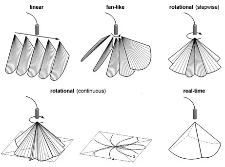
A. Linear acquisition
Parallel imaging can be performed by computer-control-led movement of the ultrasound transducer in a linear direction (Figure 1). Both a transthoracic and tran-soesophageal approach were evaluated for this mode of data acquisition.
B. Fan-like scanning
A pyramidal dataset can be obtained by moving the ultra-sound transducer in a fan-like arc at prescribed angles (Figure 1). This is accomplished by computer-controlled
motors adapted to the transthoracic or transoesophageal probe [3].
C. Stepwise rotational scanning
In this approach, the transducer is rotated around its cen-tral axis, resulting in a conical volume dataset (Figure 1). Different algorithms have been developed for computer-controlled sequential image collection of the heart [4]. The endocardial contours of a series of images obtained with a multiplane (omniplane) precordial transducer can be directly traced and used for volume calculation. In three-dimensional reconstruction, a series of images with rotational intervals between 2° and 16° is acquired and a voxel-based three-dimensional dataset is realised. With volume-rendering and various shading techniques, the reconstructed image accurately portrays the tissue charac-teristics and depth of the cardiac anatomy. Paraplane methods provide multiple equidistant parallel short-axis planes (discs) allowing systematic cross-sectional review of the three-dimensional dataset. It has been shown that slices up to a thickness of 10 mm allow accurate volume determination [5]. By tracing the left ventricular endocar-dial border in each short-axis image, the volume of each slice is calculated by the computer since the slice thickness is known. A summation of the volumes of all slices yields the total volume of the left ventricular cavity at end-dias-tole or end-sysend-dias-tole [4,5]. Subtraction of end-systolic vol-ume from end-diastolic volvol-ume results in stroke volvol-ume of the left ventricle. The percentage of stroke volume over end-diastolic volume represents left ventricular ejection fraction.
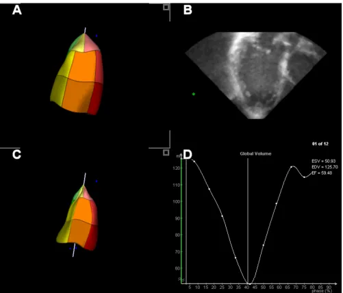
Other algorithms for volume calculation, such as TomTec 4DLV analysis software, are based on the analysis of long-axis views (Figure 2). This software is able to display a dynamic reconstruction of the LV after semi-automated border detection [see additional file 1]. Not only the sizes and shapes of the left ventricle, but also the regional wall motion of the myocardium can be analysed.
D. Continuous rotational scanning
static part of the transducer. The original images are trans-ferred to a workstation for reconstruction of the datasets and semi-automated analysis of the LV endocardial con-tours [10]. This allows rapid calculation of LV volumes, ejection fraction and wall motion analysis. Such a special dedicated system offers advantages for follow-up studies, stress echocardiography and during interventional proce-dures (e.g. resynchronisation therapy).
Initial experience indicates that this near real-time approach is an alternative to real-time volumetric systems for global and regional wall motion analysis of the LV.
Real-time imaging
The ideal way of three-dimensional echocardiography is on-line acquisition of a three-dimensional dataset of the
heart without the need for ECG and respiratory gating avoiding spatial motion artefacts.
The first real-time 3D system has been developed by Von Ramm et al. [11] at Duke University and most experience is with this system (Volumetric Medical Imaging). This system makes use of a sparse matrix phased array trans-ducer of 512 elements to scan a 60° × 60° pyramidal tis-sue volume using parallel processing technology which permits the reception of 16 lines for each transmitted sig-nal (16:1) at a rate of 17 volumes/sec with a depth of 16 cm. Image display for analysis consists of 2 independent B-modes or 3 C-mode scans (these are cross-sections par-allel to the transducer face which are displayed simultane-ously in selected orientations. LV volumes are calculated with dedicated analytic software from either a series of parallel C-scans (short-axis views) or a series of rotated Different methods of data acquisition for transthoracic 3D-echocardiography
Figure 1
apical long-axis views. More recently, Philips Medical Sys-tems has introduced its live 3D system using a matrix phased-array transducer with 3000 transmit-receive ele-ments. In this transducer, multiple recordings are automatically performed to cover the full left ventricle. This is especially useful in dilated ventricles because of the limited sector angle. The multi-directional beam steering capability enables visualization of two views of the heart simultaneously. Experience is still limited, however, promising results have been reported. Movie 2 [see
addi-tional file 2] shows a parasternal long-axis view obtained with the new Philips matrix transducer.
Left ventricular volume measurement by three-dimensional echocardiography
Table 1 and Table 2 provide an overview of studies com-paring left ventricular volumes and function by recon-struction and real-time three-dimensional echocardiography, respectively, in comparison with mag-netic resonance imaging [12–19]. For the various 3D acquisition methods that have been used, a good End-diastolic (A) and End-systolic (C) reconstruction of the left ventricle after semi-automated border analysis of the long-axis views (B)
Figure 2
correlation can be observed. However, most three-dimen-sional echocardiography studies tend to underestimate ESV and EDV. It should also be noted that large differ-ences in study design were present, including number of patients, LV volumes, image quality and analysis meth-ods. In recent studies, new semi-automated endocardial border detection algorithms replace common methods to measure LV volume, as Simpson's rule and algorithms based on manual border tracing.
Conclusion
Three-dimensional echocardiography is a non-invasive technique which can be performed in many clinical sce-narios. It is thus ideal for daily performance and for serial follow-up examinations of left ventricular volume and function.
Regardless of the many improved techniques in three-dimensional echocardiography, time consumption has been the major limitation hampering its routine employ-ment for daily diagnostic echocardiography and for vol-ume and function assessment. Faster data acquisition by
reducing the number of cross-sections for reconstruction of the cavity, using a high-speed rotation transducer or a volumetric real-time three-dimensional echocardio-graphic transducer is being investigated. Data processing and three-dimensional image reconstruction has been accelerated and on-line processing and reconstruction is under investigation. Manual endocardial tracing needed for volume measurement is both laborious and prone for subjective errors. Development of various automatic bor-der detection algorithms along with the improvement of ultrasound spatial resolution and advances in other novel modalities such as harmonic, power-mode Doppler tissue imaging and development of stable intravenous ultra-sound contrast agents that enhance the delineation of endocardium, should be able to avoid the need of manual border tracing and provide automatic, even on-line, vol-ume measurement.
Table 1: Volume and function measurement by reconstruction 3DE in comparison with magnetic resonance imaging
Author/ref. Object N r. SEE Mean Diff. ± SD
Gopal et al.[12,14] LV-EDV 15 0.92 7 ml
LV-ESV -- 0.81 4 ml
Iwase et al.[15] LV-EDV 30 0.93 -17 ± 23 ml
LV-ESV -- 0.96 -4 ± 18 ml
LV-EF -- 0.85 -2 ± 6%
Buck et al.[16] LV-EDV 23 0.97 14.7 ml -10.7 ± 14.5 ml
LV-ESV -- 0.97 12.4 ml -3.4 ± 12.9 ml
LV-EF -- 0.74 5.6% -2.5 ± 6.7%
Altmann et al.[18] LV-EDV 12 0.98 8.7 ml -14.2 ± 8.3 ml
LV-ESV -- 0.98 5.6 ml -3.4 ± 5.5 ml
LV-EF -- 0.85 5.3% -4.4 ± 5.3 %
Nosir et al.[19] LV-EDV 46 0.98 -1.4 ± 13.5 ml
LV-ESV -- 0.98 -1.5 ± 10.5 ml
LV-EF -- 0.98 0.2 ± 2.5%
Kim et al.[20] (patients) LV-EDV 18 6.4 ± 20 ml
LV-ESV -- 0.0 ± 13.3 ml
LV-EF -- 1.4 ± 3.5 %
Kim et al.[20] (volunteers) LV-EDV 10 -3.1 ± 4.9 ml
LV-ESV -- -1.4 ± 2.2 ml
LV-EF -- 0.5 ± 1.8 %
Poutanen et al. [21] (children) LV-EDV 0.80 4.0 ± 19.6 ml
LV-ESV 0.88 0.4 ± 13.0 ml
LV-EF 0.20 1.7 ± 15.1%
Mannaerts et al. [22] LV-EDV 17 0.74 -13.5 ± 13.5 %
LV-ESV -- 0.88 -17.7 ± 23.9 %
LV-EF -- 0.89 -1.8 ± 5.8 %
Krenning et al. (submitted) LV-EDV 15 0.98 13.4 ml -22.7 ± 13.6 ml
LV-ESV -- 0.99 8.7 ml -12.6 ± 9.9 ml
Additional material
References
1. Handschumacher MD, Lethor JP, Siu SC, Mele D, Rivera JM, Picard MH, Weyman AE and Levine RA: A new integrated system for three-dimensional echocardiographic reconstruction: devel-opment and validation for ventricular volume with applica-tion in human subjects.J Am Coll Cardiol 1993, 21:743-753. 2. Gopal AS, King DL, Katz J, Boxt LM, King DL Jr and Shao MY:
Three-dimensional echocardiographic volume computation by pol-yhedral surface reconstruction: in vitro validation and com-parison to magnetic resonance imaging.J Am Soc Echocardiogr
1992, 5:115-124.
3. Delabays A, Pandian NG, Cao QL, Sugeng L, Marx G, Ludomirski A and Schwartz SL: Transthoracic real-time three-dimensional echocardiography using a fan-like scanning approach for data acquisition: methods, strengths, problems, and initial clinical experience.Echocardiography 1995, 12:49-59.
4. Pandian NG, Roelandt J, Nanda NC, Sugeng L, Cao QL, Azevedo J, Schwartz SL, Vannan MA, Ludomirski A and Marx G et al.: Dynamic three-dimensional echocardiography: methods and clinical potential.Echocardiography 1994, 11:237-259.
5. Nosir YF, Fioretti PM, Vletter WB, Boersma E, Salustri A, Postma JT, Reijs AE, Ten Cate FJ and Roelandt JR: Accurate measurement of left ventricular ejection fraction by three-dimensional echocardiography. A comparison with radionuclide angiography.Circulation 1996, 94:460-466.
6. Belohlavek M, Tanabe K, Jakrapanichakul D, Breen JF and Seward JB: Rapid three-dimensional echocardiography : clinically feasi-ble alternative for precise and accurate measurement of left ventricular volumes.Circulation (Online) 2001, 103:2882-2884. 7. Nguyen LD and Leger C: Four-dimensional reconstruction of
the left ventricle using a fast rotating classical phased array scan head: preliminary results. J Am Soc Echocardiogr 2002, 15:593-600.
8. Djoa KK, de Jong N, van Egmond FC, Kasprzak JD, Vletter WB, Lan-cee CT, van der Steen AF, Bom N and Roelandt JR: A fast rotating scanning unit for real-time three-dimensional echo data acquisition.Ultrasound Med Biol 2000, 26:863-869.
9. Voormolen MM, Bouakaz A, Krenning BJ, Lancee CT, ten Cate FJ, Roelandt JRTC, van der Steen AFW and de Jong N: A New Trans-ducer for 3D Harmonic Imaging. In: Proceedings of the IEEE Inter-national Ultrasonics Symposium; Munich 2002:1261-1264.
10. Lancee CT, van Egmond FC, de Jong N, Mastik F, van der Steen AFW, Roelandt JRTC and Bom N: Data Processing for a Fast Rotating Phased Array Real-time 3D Acquisition Unit. In: Proceedings of IEEE Ultrasonics Symposium; San Juan 2000:1597-1600.
11. Sheikh K, Smith SW, von Ramm O and Kisslo J: Real-time, three-dimensional echocardiography: feasibility and initial use.
Echocardiography 1991, 8:119-125.
12. Gopal AS, Keller AM, Rigling R, King DL Jr and King DL: Left ven-tricular volume and endocardial surface area by three-dimensional echocardiography: comparison with two-dimensional echocardiography and nuclear magnetic reso-nance imaging in normal subjects. J Am Coll Cardiol 1993, 22:258-270.
13. Gopal AS, Keller AM, Shen Z, Sapin PM, Schroeder KM, King DL Jr and King DL: Three-dimensional echocardiography: in vitro and in vivo validation of left ventricular mass and compari-son with conventional echocardiographic methods.J Am Coll Cardiol 1994, 24:504-513.
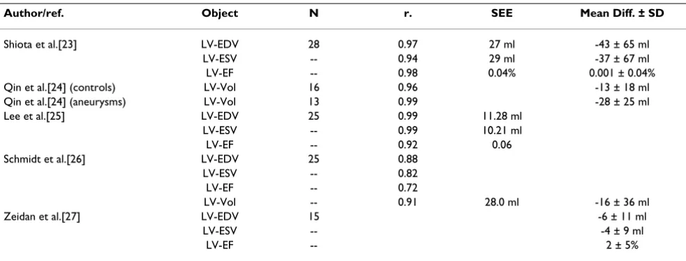
14. King DL, Gopal AS, Keller AM, Sapin PM and Schroder KM: Three-dimensional echocardiography. Advances for measurement of ventricular volume and mass.Hypertension 1994, 23:I172-179. 15. Iwase M, Kondo T, Hasegawa K, Kimura M, Matsuyama H, Watanabe Y and Hishida H: Three-dimensional echocardiography by semi-automatic border detection in assessment of left ven-Table 2: Volume and function measurement by real-time 3DE in comparison with magnetic resonance imaging
Author/ref. Object N r. SEE Mean Diff. ± SD
Shiota et al.[23] LV-EDV 28 0.97 27 ml -43 ± 65 ml
LV-ESV -- 0.94 29 ml -37 ± 67 ml
LV-EF -- 0.98 0.04% 0.001 ± 0.04%
Qin et al.[24] (controls) LV-Vol 16 0.96 -13 ± 18 ml
Qin et al.[24] (aneurysms) LV-Vol 13 0.99 -28 ± 25 ml
Lee et al.[25] LV-EDV 25 0.99 11.28 ml
LV-ESV -- 0.99 10.21 ml
LV-EF -- 0.92 0.06
Schmidt et al.[26] LV-EDV 25 0.88
LV-ESV -- 0.82
LV-EF -- 0.72
LV-Vol -- 0.91 28.0 ml -16 ± 36 ml
Zeidan et al.[27] LV-EDV 15 -6 ± 11 ml
LV-ESV -- -4 ± 9 ml
LV-EF -- 2 ± 5%
3DE = three-dimensional echocardiography; N = number of subjects; LV = left ventricle; r = correlation coefficient; SEE = standard error or regression; Diff. = difference; SD = standard differentiation; EDV = end-diastolic volume; ESV = end-systolic volume; EF = ejection fraction; Vol = volume
Additional File 1
Movie 1: Dynamic LV reconstruction, showing size and shape of the LV during different moments in the heart cycle.
Click here for file
[http://www.biomedcentral.com/content/supplementary/1476-7120-1-12-S1.avi]
Additional File 2
Movie 2: Parasternal long-axis view the LV, using the new Philips Live3D ' matrix transducer (courtesy of Philips Medical Systems).
Click here for file
Publish with BioMed Central and every scientist can read your work free of charge
"BioMed Central will be the most significant development for disseminating the results of biomedical researc h in our lifetime."
Sir Paul Nurse, Cancer Research UK
Your research papers will be:
available free of charge to the entire biomedical community
peer reviewed and published immediately upon acceptance
cited in PubMed and archived on PubMed Central
yours — you keep the copyright
Submit your manuscript here:
http://www.biomedcentral.com/info/publishing_adv.asp
BioMedcentral tricular volume and ejection fraction: comparison with
mag-netic resonance imaging.J Cardiol 1997, 30:97-105.
16. Buck T, Hunold P, Wentz KU, Tkalec W, Nesser HJ and Erbel R: Tomographic three-dimensional echocardiographic deter-mination of chamber size and systolic function in patients with left ventricular aneurysm: comparison to magnetic res-onance imaging, cineventriculography, and two-dimensional echocardiography.Circulation 1997, 96:4286-4297.
17. Vogel M, Gutberlet M, Dittrich S, Hosten N and Lange PE: Compar-ison of transthoracic three dimensional echocardiography with magnetic resonance imaging in the assessment of right ventricular volume and mass.Heart 1997, 78:127-130. 18. Altmann K, Shen Z, Boxt LM, King DL, Gersony WM, Allan LD and
Apfel HD: Comparison of three-dimensional echocardio-graphic assessment of volume, mass, and function in children with functionally single left ventricles with two-dimensional echocardiography and magnetic resonance imaging. Am J Cardiol 1997, 80:1060-1065.
19. Nosir YF, Lequin MH, Kasprzak JD, van Domburg RT, Vletter WB, Yao J, Stoker J, Ten Cate FJ and Roelandt JR: Measurements and day-to-day variabilities of left ventricular volumes and ejec-tion fracejec-tion by three-dimensional echocardiography and comparison with magnetic resonance imaging.Am J Cardiol
1998, 82:209-214.
20. Kim WY, Sogaard P, Kristensen BO and Egeblad H: Measurement of left ventricular volumes by 3-dimensional echocardiogra-phy with tissue harmonic imaging: a comparison with mag-netic resonance imaging.J Am Soc Echocardiogr 2001, 14:169-179. 21. Poutanen T, Ikonen A, Jokinen E, Vainio P and Tikanoja T: Transtho-racic three-dimensional echocardiography is as good as mag-netic resonance imaging in measuring dynamic changes in left ventricular volume during the heart cycle in children.Eur J Echocardiogr 2001, 2:31-39.
22. Mannaerts HF, Van Der Heide JA, Kamp O, Papavassiliu T, Marcus JT, Beek A, Van Rossum AC, Twisk J and Visser CA: Quantification of left ventricular volumes and ejection fraction using freehand transthoracic three-dimensional echocardiography: com-parison with magnetic resonance imaging.J Am Soc Echocardiogr
2003, 16:101-109.
23. Shiota T, McCarthy PM, White RD, Qin JX, Greenberg NL, Flamm SD, Wong J and Thomas JD: Initial clinical experience of real-time three-dimensional echocardiography in patients with ischemic and idiopathic dilated cardiomyopathy.Am J Cardiol
1999, 84:1068-1073.
24. Qin JX, Jones M, Shiota T, Greenberg NL, Tsujino H, Firstenberg MS, Gupta PC, Zetts AD, Xu Y, Ping Sun J, Cardon LA, Odabashian JA, Flamm SD, White RD, Panza JA and Thomas JD: Validation of real-time three-dimensional echocardiography for quantifying left ventricular volumes in the presence of a left ventricular aneurysm: in vitro and in vivo studies.J Am Coll Cardiol 2000, 36:900-907.
25. Lee D, Fuisz AR, Fan PH, Hsu TL, Liu CP and Chiang HT: Real-time 3-dimensional echocardiographic evaluation of left ventricu-lar volume: correlation with magnetic resonance imaging – a validation study.J Am Soc Echocardiogr 2001, 14:1001-1009. 26. Schmidt MA, Ohazama CJ, Agyeman KO, Freidlin RZ, Jones M,
Lau-rienzo JM, Brenneman CL, Arai AE, von Ramm OT and Panza JA: Real-time three-dimensional echocardiography for meas-urement of left ventricular volumes. Am J Cardiol 1999, 84:1434-1439.