R E S E A R C H
Open Access
Management of recurrent unilateral lumbar
disc herniation in a single level: unilateral
versus bilateral pedicle screws fixation with
interbody fusion
Omar El Farouk Ahmed
*, Sherif H. Morad and Ahmed S. Abdelbar
Abstract
Background:Lumbar interbody fusion procedure is a recognized surgical technique in management of a variety of lumbar pathologies including recurrent lumbar disc prolapse. Interbody fusion augmented by pedicle screw fixation has been considered to improve fusion rates and clinical outcomes. Interbody fusion is commonly associated with better fusion potentials through applying the bone graft in the load bearing, vascular position of the anterior, and middle spinal columns. However, it still remains unknown whether interbody fusion with unilateral pedicle screw fixation (UPSF) is superior to that with bilateral pedicle screw fixation (BPSF).
Aim of the work:The aim of this study is to evaluate the efficacy and safety of unilateral versus bilateral pedicle screw fixation associated with interbody fusion for the management of single level unilateral recurrent lumbar disc prolapse as regard the clinical and biomechanical results, operation time, intraoperative blood loss, and
postoperative stay.
Material and methods:This observational prospective comparative study of the two groups who were operated either unilateral (group A /15 patients) or bilateral (group B/15 patients) pedicle screw fixation with interbody fusion was done. Patients were followed up for 1, 6, ad 12 months.
Results:Significant improvement in functional outcome of the two groups was noted compared to preoperatively, except in early postoperative period where the back VAS and ODI in the unilateral group was better than bilateral group. However, on further follow up, no significant difference was noticed. There was no significant difference comparing fusion rate, complication rate, and duration of hospital stay between the two groups at postoperative follow-up. There was significantly less blood loss, and significantly shorter operation time in the unilateral PS fixation group as compared with the bilateral PS fixation group in our study.
Conclusion:Our study suggested that TLIF with unilateral PS fixation was as safe and effective as that with bilateral PS fixation for the management of recurrent single level lumbar disc prolapse; it showed better clinical outcome scores of ODI and back VAS, and a significant reduction of the intraoperative blood loss as well as the operation time, without significant differences considering fusion rate, complication rate, and duration of hospital stay between the two groups at postoperative follow-up. However, BPSF with TLIF likely causes more degeneration at the cranial adjacent segment compared with UPSF techniques. Nevertheless, the long-term follow up is required to demonstrate the impact of these findings.
Keywords:Recurrent Lumbar disc prolapse, Unilateral, Bilateral pedicle screw fixation, Interbody fusion
© The Author(s). 2020Open AccessThis article is distributed under the terms of the Creative Commons Attribution 4.0 International License (http://creativecommons.org/licenses/by/4.0/), which permits unrestricted use, distribution, and reproduction in any medium, provided you give appropriate credit to the original author(s) and the source, provide a link to the Creative Commons license, and indicate if changes were made.
* Correspondence:[email protected]
Introduction
The optimal surgical management for treating recurrent lumbar disc herniation remains controversial [7]. While many authors advocate for repeat discectomy only, particu-larly in the absence of a distinctive evidence of spinal in-stability [28], others support the use of different methods of fusion, stating that the addition of pedicle screw instru-mented fusion might provide added stability that eliminates segmental motion at the involved level, while adding an interbody fusion prosthesis may further improve the clinical results by improving fusion rates, restoring intervertebral height, and maintaining lumbar lordosis [21].
Various surgical techniques of interbody fusion are under discussion, including anterior lumbar interbody fusion (ALIF), posterior lumbar interbody fusion (PLIF), and transforaminal lumbar interbody fusion (TLIF) [10].
Over the last decade, transforaminal lumbar interbody fusion (TLIF), initially described by Harms and Jeszenszky in the 1990s [11], has become increasingly popular as a valuable surgical option to manage a variety of lumbar de-generative diseases. It provides the advantages of reducing the potential surgical risk of excessive neural tissue retrac-tion or epidural fibrosis when compared with the posterior lumbar interbody fusion (PLIF) approach [4, 6], besides, eliminating the potential complications associated with anterior lumbar interbody fusion (ALIF), such as retro-grade ejaculation following presacral sympathetic plexus iatrogenic injury or possible great vessels damage [9,18].
In fact, traditionally practiced bilateral pedicle screw fixation (BPSF) and instrumentation performed together with TLIF can be regarded as a reliable and effective sur-gery for sufficient fusion [16]. On the other hand, it is reported that extreme rigidity caused by bilateral screw fixation may possibly contribute to a higher incidence of adjacent segment disease (ASD) [24]. On the other hand, it is reported in the literature that unilateral pedicle screw fixation (UPSF) with the TLIF procedure has many advantages compared to BPSF including being a less invasive surgical procedure, with shorter operation time, less blood loss, less co-morbidities, less postopera-tive pain, and less cost [26].
In this study, we evaluated the clinical and radiological results and follow up of our patients subjected to fusion procedure through unilateral versus bilateral PSF along with interbody fusion for the management of recurrent single level unilateral prolapsed lumbar disc, aiming at analyzing the results in comparison to the previously published studies.
Material and methods
This is a prospective, randomized, comparative study on 30 patients with first time recurrent lumbar disc hernia-tion, conducted in the period between January 2016 and December 2018.
The patients were divided according to surgical ap-proach into a unilateral group (group A/15 patients) and a bilateral group (group B/15 patients) who were oper-ated either through unilateral or bilateral pedicle screw fixation with interbody fusion respectively. Patients were followed up for 1, 6, and 12 months. This study occurred at Ain shams university hospitals.
Patient selection
Inclusion criteria
1- Patients with a recurrent single level lumbar disc prolapse at the same level of the primary discectomy with at least 6 months of pain relief following initial lumbar disc surgery.
2- The presence of recurrent strictly unilateral radicular pain unresponsive to conservative treatment for at least 6 weeks.
Exclusion criteria
1. Patient with multiple level recurrent lumbar disc prolapse.
2. Patients with conditions requiring bilateral nerve root decompression, such as bilateral radiculopathy, segmental canal stenosis, spondylolisthesis, and those with spinal osteoporosis.
3. The presence of other lumbar spine pathologies such as trauma, tumor, infection, or deformity.
Preoperative data
1. Clinical evaluation
This will include:
Patients’demographics (age, sex).
Relevant past medical and surgical history.
Clinical presentation and its duration including assessment of:
Back pain
radiculopathy
Visual analogue scale (VAS) and the Oswestry Disability Index (ODI) for both back pain and leg pain.
Full general and history and neurological examination.
2. Radiological investigation
Antero-posterior and lateral (standing and dynamic) views to evaluate:
Anatomy of the pedicles, transverse processes, laminae, and facet joints; presence of degenerative changes; neural foraminal dimensions, and bone density.
Measurement of preoperative disc height at the surgical segment, cranial and caudal adjacent segments:
Disc height = (a height + P height)/2.
To assess stability of the spine on dynamic views where lumbar instability is considered as more than 4.5 mm of translation, and/or 15° to 25° of angular motion between adjacent segments.
(B) MRI
Magnetic resonance imaging (MRI) of the lumbosacral spine with Gadolinium enhancement allowing anatom-ical evaluation of the spine and spinal canal, nerve roots, and spinal ligaments complex.
Surgical technique
Our patients were classified into two groups according to the surgical procedure; each consisted of 15 patients. All had revision discectomy and interbody fusion using the same interbody cage type (TLIF PEEK Interbody Cages, Medtronic). While group A consisted of patients who had strictly unilateral pedicle screws fixation (UPSF), group B involved patients who had bilateral ped-icle screws fixation (BPSF) surgery. All surgeons ac-counting for performing the two procedures were efficient with the same skill level. Selection of the pro-cedure was done randomly by alternation method to minimize the risk of selection bias.
Following general anesthesia and endotracheal intub-ation, all patients were positioned prone on Montreal frame or rolls to avoid abdominal compression and sub-sequent venous congestion. Skin incision was performed at the site of the previous surgery through midline pos-terior exposure.
Group A patients had unilateral muscle separation at the symptomatic side exposing the facet joints and the transverse processes, whereas the spinous process, supraspinous and interspinous ligaments and the contra-lateral vertebral plate and facet joints, remained unin-jured. Two transpedicular screws were then placed on the symptomatic side, followed by a unilateral hemilami-nectomy and medial facetectomy. The symptomatic nerve root is then identified and released carefully.
Subsequently, the disc space was to be prepared for cage insertion through discectomy and entire endplate curet-tage using disc shavers, curettes, and rongeurs. The disc space was then distracted and a single cage filled with autogenous bone graft from the removed residual lamina and is inserted into the disc space. A lateral fluoroscopic image was obtained to ensure safe and proper positioning of the cage.
Group B patients had bilateral subperiosteal muscle stripping and the paraspinal muscles were laterally retracted to the outer edge of the facet joint. Bilateral pedicle screws were implanted followed by full laminec-tomy and faceteclaminec-tomy at the symptomatic side. Discec-tomy and cage placement were then carried out.
The fusion level would be moderately compressed by tightening the reduction screws over the rods and hence applying an element of compression over the interbody cage. Intertransverse grafting was also performed.
Finally, closure was performed in a routine fashion over a suction drain. All patients received prophylactic antibiotics perioperatively and were requested to start ambulation on the next day using a lumbar corset for a couple of months. The drains were removed at 48 h.
Evaluation follow up
(A) Clinical outcome
Evaluation of the patient’s neurological outcome dur-ing the hospital stay as regard postoperative VAS and ODI scores of the two groups for back pain and leg pain in comparison to preoperative scores.
(B) Radiological outcome
Was assessed by plain X-ray (AP, lateral standing, flexion, and extension views).
1. Measurement disc height at the surgical segment, and both cranial and caudal adjacent segments.
Disc height = (a height+ P height)/2.
2. Fusion
Assessment of fusion was carried out according to the Bridwell-Lenke grading system and motion on lateral flexion-extension radiographs:
A. (Grade 1) Definite fusion: definitive bony trabecular bridging across the graft/host interface, no
B. (Grade 2) Probable fusion: no definitive bony trabecular crossing, but no detectable motion and no identifiable gap at the interface.
C. (Grade 3) Possible pseudarthrosis: no bony trabecular crossing, no motion, but an identifiable gap at the interface.
D. (Grade 4) Definite pseudarthrosis: no traversing trabecular bone, definitive gap at the interface, and motion on flexion-extension radiographs.
While A and B were considered as successful fusions, C and D were considered as failed fusions.
Statistical analysis
Data were collected, revised, coded, and entered to the Statistical Package for Social Science (IBM SPSS) version 23. The quantitative data were presented as mean, stand-ard deviations, and ranges when parametric. Also qualita-tive variables were presented as number and percentages.
The comparison between groups regarding qualitative data was done by using Chi-square test and/or Fisher exact test only when the expected count in any cell found less than 5.
The comparison between two independent groups re-garding quantitative data with parametric distribution was done by using Independentttest.
The comparison between more than two paired groups regarding quantitative data with parametric distribution was done by using repeated measures ANOVA.
The confidence interval was set to 95% and the margin of error accepted was set to 5%. So, thePvalue was con-sidered significant as follows:
P> 0.05: nonsignificant (NS) P< 0.05: significant (S)
P< 0.01: highly significant (HS)
Results
All the patients’preoperative data was studied and ana-lyzed. There were 21 (70%) males and nine (30%) female patients. Their age ranged from 32 to 55 years in group A with a mean age of 44.21 (± 7.18 STD) years.
In group A subjects, the pathological level was L4-5 in 10 (66.7%) patients, L5-S1 in four (26.7%) patients, and L3–4 in a single case; the left side was the pathological side in 11 (73.3%) patients and the right side accounted for 4 (26.7%) patients. While in group B patients, L4–5 level was involved in eight (53.3%) patients, L5–S1 in five (33.3%) patients, and L3–4 in two (13.4%) cases. The left side was involved in ten (66.7%) cases and five (33.3%) cases had their pathology on the right side.
There was no statistically significant difference found between unilateral group and bilateral group regarding demographic data of the studied patients.
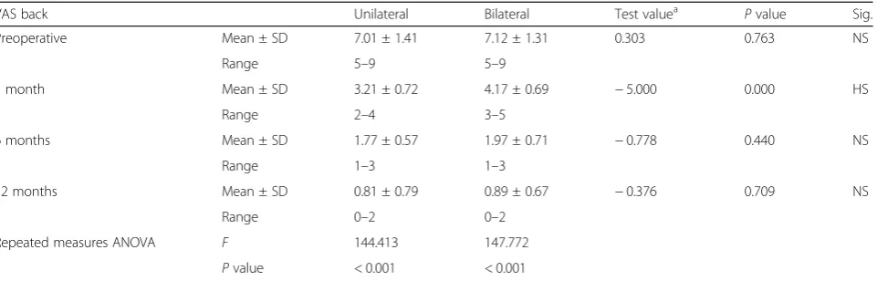
Concerning the VAS score data analysis, there was a highly statistically significant difference between pre-operative and postpre-operative (at 1, 6, and 12 months) VAS of the back pain in both groups; however, on com-paring the two groups, there was no statistically signifi-cant difference regarding VAS of the back pain except during the first postoperative month, which was signifi-cantly lower in unilateral group (3.21 ± 0.72) (Table1).
Similarly, there was a highly statistically significant dif-ference between preoperative and postoperative (at 1, 6, and 12 months) VAS for the leg pain in both groups, but while comparing between the two groups, there was no statistically significant difference regarding VAS for leg pain postoperatively (Table2).
Concerning the subjects ODI evaluation, there was highly statistically significant difference found between preoperative and postoperative (at 1, 6, and 12 months) ODI in both groups; however, on comparing between the two groups, there was no statistically significant dif-ference regarding ODI except during the first postopera-tive month which was significantly lower in unilateral group (Pvalue 0.000) (Table3).
Postoperatively, there was a significant increase in the disc height in both groups. However, there was no statisti-cally difference between the two groups (P= 0.317); while on evaluating the adjacent cranial level mean disc height during the postoperative period, it was reduced by 2.82 ± 0.51, 2.01 ± 0.37, and 1.44 ± 0.32 mm at the first, sixth, and twelfth month respectively in the unilateral group, which was significantly (P< 0.000) less than the values of 3.31 ± 0.37, 2.55 ± 0.33, and 1.89 ± 0.31 mm in the bilat-eral group. The adjacent caudal level mean disc height was reduced by 2.47 ± 0.37, 2.14 ± 0.37, and 1.47 ± 0.35 mm at the first, sixth, and twelfth month respectively in the unilateral group, which was statistically insignificantly in comparison with than the values of 2.41 ± 0.33, 2.07 ± 0.34, and 1.52 ± 0.44 mm in the bilateral group.
Our radiological outcome as regard fusion rate was re-corded in the unilateral group as 72.0% and 88.0% solid fusion at the sixth and twelfth months respectively while in the bilateral group, it was 76.0% and 92.0% solid fusion the same period respectively, without significant difference between the two groups. There was no statistically signifi-cant difference regarding the postoperative complications between the unilateral group and bilateral group.
patient was presented by acute onset of severe sciatic pain; in the first week postoperatively, plain radiographs and CT scan revealed the migrating cage, patient was reoperated and a larger cage was introduced instead; postoperatively, the patient experienced significant relief of the acute pain.
A single patient of the unilateral group developed tem-porary foot drop and sensory affection on the same side of operation immediately after surgery; he received con-servative medical management and had complete recov-ery within 1 month.
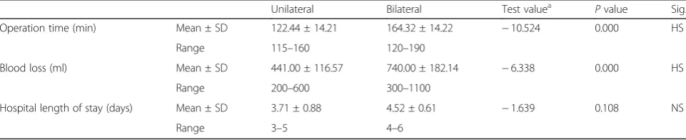
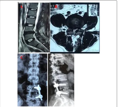
There was a highly statistically significant difference found between the unilateral and the bilateral group concerning operative time and blood loss being signifi-cantly less in the unilateral group, while there was no statistically significant difference regarding the postoper-ative hospital length of stay (Table4) (Fig.1).
Discussion
The use of lumbar fusion together with neural decom-pression for the management of recurrent disc prolapse
cases is a popular surgical option, aiming to maintain disc height, ensure load sharing, and spinal stability [2]; the transforaminal approach for interbody fusion is a recognized practice and remains a sufficient and safe op-tion for fusion particularly in recurrent lumbar disc her-niation cases accompanied by radiculopathy with or without mechanical low back ache [16].
While bilateral PS fixation might provide rigid fix-ation and a consequent satisfactory biomechanical sta-bility and clinical benefits, however, that rigidity may turn the adjacent segment more prone to degener-ation process. To minimize the possible side effects at the adjacent levels and try to achieve optimal bio-mechanical conditions, the use of less rigid fixation systems has been advocated. Hereby, some recent studies questioned the value of unilateral PS fixation as a reliable alternative fusion technique with fewer pedicle screws keeping in mind the distinct nature of revision surgeries with evident perineural scarring and distorted anatomical planes, especially in the presence of strictly unilateral symptoms and the value of
Table 1Comparison between unilateral group and bilateral group regarding the visual analogue scale for back pain
VAS back Unilateral Bilateral Test valuea Pvalue Sig.
Preoperative Mean ± SD 7.01 ± 1.41 7.12 ± 1.31 0.303 0.763 NS
Range 5–9 5–9
1 month Mean ± SD 3.21 ± 0.72 4.17 ± 0.69 −5.000 0.000 HS
Range 2–4 3–5
6 months Mean ± SD 1.77 ± 0.57 1.97 ± 0.71 −0.778 0.440 NS
Range 1–3 1–3
12 months Mean ± SD 0.81 ± 0.79 0.89 ± 0.67 −0.376 0.709 NS
Range 0–2 0–2
Repeated measures ANOVA F 144.413 147.772
Pvalue < 0.001 < 0.001
P> 0.05: nonsignificant (NS),P< 0.05: significant (S),P< 0.01: highly significant a
Independentttest
Table 2Comparison between unilateral group and bilateral group regarding the visual analogue scale for leg pain
VAS back Unilateral Bilateral Test valuea Pvalue Sig.
Preoperative Mean ± SD 7.01 ± 1.32 7.05 ± 1.39 −0.105 0.917 NS
Range 5–9 5–9
1 month Mean ± SD 1.69 ± 0.77 1.65 ± 0.78 0.190 0.850 HS
Range 1–3 1–3
6 months Mean ± SD 1.14 ± 0.71 1.05 ± 0.55 0.872 0.387 NS
Range 0–2 0–2
12 months Mean ± SD 0.88 ± 0.69 0.79 ± 0.73 0.203 0.840 NS
Range 0–2 0–2
Repeated measures ANOVA F 279.159 229.785
Pvalue < 0.001 < 0.001
a
reserving the intact contralateral elements, which may be a contributor for a possible lower complication rate [17].
Harris et al. described the unilateral TLIF procedure with adjunctive pedicular fixation as an interbody fusion technique that minimizes nerve root manipulation and requires less dissection compared with other interbody fusion methods [12].
In a study conducted by Kim et al. with a 10-year follow-up, higher rates of ASD were recorded in the bilateral PSF group [14]. Likewise, Toyone et al. re-ported less adjacent segment degeneration in the unilateral PSF group in their study [20]. Other stud-ies available in the literature supported the same view [8, 15, 23].
Nevertheless, it was suggested by an in vitro study that the use of unilateral PS fixation might be detrimental to the fusion process and the spinal instability [17].
Therefore, it remains debatable whether TLIF with unilateral pedicle screw fixation is superior to that with bilateral pedicle screw fixation for the management of recurrent single level lumbar disc herniation syndromes for patients with strictly unilateral symptoms.
Numerous previous studies attempted to evaluate and compare unilateral and bilateral PS fixation approach and results were inconsistent.
Clinical outcomes
Our study was conducted on 30 patients having a recurrent single level lumbar disc prolapse with strictly unilateral symptoms presented with low back-ache and radicular symptoms who were divided into the unilateral (n = 15) or bilateral (n = 15) pedicle screw fixation groups.
The mean visual analogue score of the back pain for the subjects included in our study showed a highly sta-tistically significant difference between preoperative and postoperative scores at the first, sixth, and twelfth post-operative month in both groups; however, on comparing the two groups, there was no statistically significant dif-ference regarding VAS of the back pain throughout the follow up period except in the first postoperative month, which was significantly lower in unilateral group.
Similarly, our study revealed a highly statistically sig-nificant difference between preoperative and postopera-tive VAS for the leg pain at the first, sixth, and twelfth postoperative month in both groups while there was no statistically significant difference while comparing be-tween the two groups regarding VAS for leg pain.
In agreement with our results, Chen et al. [3] found a highly statistically significant difference between pre-operative and postpre-operative scores for both back pain and leg pain at the first, sixth, and twelfth postoperative
Table 3Comparison between unilateral group and bilateral group regarding the Oswestry disability index ODI
ODI Unilateral Bilateral Test valuea Pvalue Sig.
Preoperative Mean ± SD 59.89 ± 9.41 60.84 ± 9.21 0.000 1.000 NS
Range 45–75 45–75
1 month Mean± SD 22.61 ± 3.96 24.44 ± 3.89 −4.406 0.000 HS
Range 15–25 20–30
6 months Mean± SD 17.55 ± 1.91 16.99 ± 1.87 −0.522 0.604 NS
Range 14–20 14–20
12 months Mean± SD 14.08 ± 1.32 14.27 ± 1.16 −0.323 0.748 NS
Range 12–16 12–16
Repeated measures ANOVA F 277.744 455.119
Pvalue < 0.001 < 0.001
a
Independentttest
Table 4Comparison between unilateral group and bilateral group regarding intraoperative data and hospital length of stay
Unilateral Bilateral Test valuea Pvalue Sig.
Operation time (min) Mean ± SD 122.44 ± 14.21 164.32 ± 14.22 −10.524 0.000 HS
Range 115–160 120–190
Blood loss (ml) Mean ± SD 441.00 ± 116.57 740.00 ± 182.14 −6.338 0.000 HS
Range 200–600 300–1100
Hospital length of stay (days) Mean ± SD 3.71 ± 0.88 4.52 ± 0.61 −1.639 0.108 NS
Range 3–5 4–6
a
month in both groups; and, on comparing the two groups, there was a statistically significant difference re-garding VAS of the back pain in the first postoperative month, which was significantly lower in unilateral group; otherwise, there was no statistically significant difference regarding VAS of the back pain throughout the follow up period.
Numerous authors reported similar results, includ-ing Işik et al. [13], Yang et al. [25], and Villavicencio et al. [22].
Zhang et al. [27] described a statistically significant dif-ference between preoperative and postoperative VAS for both back pain and leg pain at 6, 12, and 24 months
postoperatively in both unilateral and bilateral groups, while on comparing between the two groups, there was no statistically significant differences found throughout the whole follow up period.
In comparison between the two groups included in our study, there was a highly statistically significant difference found between preoperative and postoper-ative at 1, 6, and 12 months concerning the mean ODI score in both groups; however, on comparing between both groups, there was no statistically sig-nificant difference regarding ODI except during the first postoperative month which was significantly lower in unilateral group.
Chen et al. (3) stated no statistically significant differ-ence comparing ODI scores between the two groups at whole postoperative follow-up.
On the other hand, Işik et al. [13] observed a slight in-crease in VAS and ODI scores in both groups as their fol-low up got longer; they theorized that the natural process of degeneration develops by time and this increase may be induced by an adjacent segment degeneration that has de-veloped or may develop in the future in all patients under-going instrumented fusion. Within this framework, the need for longer follow-up is strongly considered.
Concerning the mean disc height at the surgical seg-ment, our study showed there was a significant postop-erative increase in the disc height in both groups while there was no statistically difference between the two groups. However, on evaluating the adjacent cranial level during the postoperative period, there was a reduction in mean disc height in the unilateral group at 1 year post-operatively, which was highly significantly less than the values of the bilateral group, unlike the adjacent caudal level mean disc height that was reduced postoperatively in both groups with statistically insignificant values.
Close to our results, Chen et al. [3] reported that after UPSF with interbody fusion, the mean disc height of the surgical segments has increased postoperatively, which was significantly less than the bilateral group; the mean disc height of adjacent cranial levels was reduced, signifi-cantly less than the values in the bilateral group, whereas the mean disc height of caudal level was reduced with no statistically significant difference between both groups. Similarly, Villavicencio et al. [22] reported a sta-tistically significant mean disc height increase in both groups postoperatively; however, they pointed a finger to a statistically significant foraminal height loss detected in the bilateral group compared to foraminal height loss in the unilateral group that was not statistically signifi-cant. In contrary, Dahdaleh et al. [5] did not demon-strate any radiographic evidence of adjacent segment degeneration during the whole 1 year follow-up period in their study.
In terms of operation time, it is understood that the group subjected to unilateral screw fixation had signifi-cantly shorter surgery time, which is in accordance with the literature.
Our study described a mean operative time for pa-tients of the unilateral group papa-tients to be 122.44 ± 14.21 min, the longest operative time was 160 min, and the shortest was 115 min; while that in the bilateral group was 164.32 ± 14.22 min, the longest operative time was 190 min, and the shortest was 120 min. The mean operative time for the unilateral and the bilateral group according to Işik et al.’s [13] study was 111.7 ± 24.31 min and 158.6 ± 18.30 min respectively, a result close to ours; a lesser mean operative time was noted by
Chen et al. [3], being 89.8 ± 19.7 min in the unilateral group and 109.1 ± 15.3 min in the bilateral group, while Zhang et al. [27] recorded a longer mean operative time of 208 (126–275) minutes and 257 (158–300) respectively.
The use of bilateral PS fixation is associated with an-other drawback which is more blood loss, since it is in-evitable to have more blood loss during neural elements decompression along with screws application on both sides. This is a clear-cut fact reported in all studies [2,8, 19,23,24].
Likewise, in our study, in line with the literature, we noted significantly less blood loss in the unilateral group; the mean intraoperative blood loss for subjects in the unilateral group was 441.00 ± 116.57 ml, the largest amount being 600 ml and the smallest amount was 200 ml, while in bilateral group was 740.00 ± 182.14 ml, the largest operative time being 1100 ml and the smallest was 300 ml.
In our study, the mean hospital length of stay was 3.71 ± 0.88 days for the unilateral group cases ranging from 3 days recorded as the shortest up to 5 days as the longest hospital stay, while it was 4.52 ± 0.61 days in the bilateral group sub-jects and ranging between 4 and 6 days as the shortest and the longest recorded hospital stays respectively. Some stud-ies recorded longer mean hospital stays as both Chen et al. [3] and Zhang et al. [27].
Concerning the radiological outcome, numerous authors reported similar results regarding the fusion rate at 6 and 12 months postoperatively without significant difference between the two group including Chen et al. [3], Işik et al. [13], Villavicencio et al. [22], and Yang et al. [25].
Intraoperative complications included unintended dur-otomy that took place in three patients, two in the bilat-eral group and a single case in the unilatbilat-eral group, where the dural tear was repaired intraoperatively via dural su-tures augmented by a fat graft and no lumbar drain was required. Yang et al. [24] reported also three cases to have a dural tear, again one in unilateral group and two in bilat-eral group. All were managed conservatively as well with no occurrence of any further complications.
Cage migration was recorded in a single case in the bi-lateral group, where the patient was presented by acute onset of severe sciatic pain; in the first week postopera-tively, plain radiographs and CT scan revealed the mi-grating cage, patient was reoperated and a larger cage was introduced instead; postoperatively, the patient ex-perienced significant relief of the acute pain.
There was no statistically significant difference between both groups regarding the postoperative complications.
Conclusion
Our study suggested that unilateral PS fixation with interbody fusion is a safe technique that provides satis-factory short-term and medium-term results in treating single-level recurrent lumbar disc prolapse in selected patients; it is as effective as the bilateral PS fixation tech-niques significantly reducing the operative duration as well as the intraoperative blood loss, provides a satisfac-tory improvement of the clinical outcome scores of ODI and VAS of the back and leg without significant differ-ence between the two groups regarding the fusion rate, complications rate, or the duration of hospital stay. BPSF with interbody fusion likely causes more degeneration at the cranial adjacent segment compared with UPSF tech-niques. However, further randomized controlled trials and long-term evaluation would be necessary to demon-strate the impact of this findings.
Acknowledgments Not applicable.
Authors’contributions
The study design, execution and follow up of the clinical cases, data analysis and results formulation, and writing of the manuscript were all the joint work of all the authors. This work was entirely carried out by the authors without any external contributions. All authors read and approved the final manuscript.
Funding
The authors received no external funding for the design of the study, for the collection, analysis, and interpretation of data or for writing the manuscript.
Availability of data and materials
All the raw data and results of the statistical analysis are available with the authors and ready to be shared with authorized personnel upon request; however, for reasons of patency protection, it was not submitted with the manuscript.
Ethics approval and consent to participate
This research was conducted upon obtaining the approval of the ethical committee of the Faculty of Medicine- Ain Shams University, in July 2015. Since this study involved human subjects, an informed consent was signed and acquired from all the participants or their legal guardians in accordance with the ethical committee recommendations.
Consent for publication Not applicable.
Competing interests
The authors declare that they have no competing interests.
Received: 4 July 2019 Accepted: 3 February 2020
References
1. Aoki Y, Yamagata M, Ikeda Y, Nakajima F, Ohtori S, Nakagawa K, Nakajima A, Toyone T, Orita S, Takahashi K. A prospective randomized controlled study comparing transforaminal lumbar interbody fusion techniques for degenerative spondylolisthesis: unilateral pedicle screw and 1 cage versus bilateral pedicle screws and 2 cages. J Neurosurg Spine. 2012;17(2):153–9.
2. Chen C, Cao X, Zou L, Hao G, Zhou Z, Zhang G. Minimally invasive unilateral versus bilateral technique in performing single-segment pedicle screw fixation and lumbar interbody fusion. J Orthop Surg Res. 2015;10:112. 3. Chen DJ, Yao C, Song Q, Tang B, Liu X, Zhang B, Dai M, Nie T, Wan Z.
Unilateral versus bilateral pedicle screw fixation combined with transforaminal lumbar interbody fusion for the treatment of low lumbar degenerative disc diseases: Analysis of Clinical and Radiographic Results. World Neurosurg. 2018;115:e516–22.
4. Cole CD, McCall TD, Schmidt MH, Dailey AT. Comparison of low back fusion techniques: transforaminal lumbar interbody fusion (TLIF) or posterior lumbar interbody fusion (PLIF) approaches. Curr Rev Musculoskelet Med. 2009;2:118–26.
5. Dahdaleh NS, Nixon AT, Lawton CD, Wong AP, Smith ZA, Fessler RG. Outcome following unilateral versus bilateral instrumentation in patients undergoing minimally invasive transforaminal lumbar interbody fusion: a single-center randomized prospective study. Neurosurg Focus. 2013;35(2): E13.
6. Fleege C, Rickert M, Rauschmann M. The PLIF and TLIF techniques. Indication, technique, advantages, and disadvantages. Orthopade. 2015;44: 114–23.
7. Fu TS, Lai PL, Tsai TT, Niu CC, Chen LH, Chen WJ. Long-term results of disc excision for recurrent lumbar disc herniation with or without posterolateral fusion. Spine (Phila Pa 1976). 2005;30:2830–4.
8. Goel VK, Lim TH, Gwon J, Chen JY, Winterbottom JM, et al. Effects of rigidity of an internal fixation device. A comprehensive biomechanical investigation. Spine. 1991;16:S155–61.
9. Guérin P, Obeid I, Bourghli A, Masquefa T, Luc S, Gille O, Pointillart V, Vital JM. The lumbosacral plexus: anatomic considerations for minimally invasive retroperitoneal transpsoas approach. Surg Radiol Anat. 2012;34:151–7. 10. Hanley EN, David SM. Lumbar arthrodesis for the treatment of back pain. J
Bone Joint Surg Am. 1999;5:716–30.
11. Harms JG, Jeszenszky D. The unilateral transforaminal approach for posterior lumbar interbody fusion. Oper Orthop Traumatol. 1998;10:90–102. 12. Harris BM, Hilibrand AS, Savas PE, et al. Transforaminal lumbar interbody
fusion : the effect of various instrumentation techniques on the flexibility of the lumbar spine. Spine. 2004;29:E65–70.
13. Işik HS, Okutan Ö, Yildirim T, Akpinar E, Yilmaz A. Comparison of unilateral versus bilateral pedicle screw fixation in transforaminal lumbar interbody fusion for single level lumbar degenerative diseases and review of literature. Turk Neurosurg. 2017.https://doi.org/10.5137/1019-5149.JTN.20531-17.1. 14. Kim TH, Lee BH, Moon SH, Lee SH, Lee HM. Comparison of adjacent
segment degeneration after successful posterolateral fusion with unilateral or bilateral pedicle screw instrumentation: a minimum 10-year follow-up. Spine J. 2013;13(10):1208–16.
15. Korovessis P, Papazisis Z, Koureas G, Lambiris E. Rigid, semirigid versus dynamic instrumentation for degenerative lumbar spinal stenosis: a correlative radiological and clinical analysis of short-term results. Spine. 2004;29(7):735–42.
16. Liu F, Feng Z, Zhou X, Liang Y, Jiang C, Li X, Li Z, Jiang X, Dong J. Unilateral versus bilateral pedicle screw fixation in transforaminal lumbar interbody fusion: A monocentric study of 215 patients with a minimum of 4-year follow-up. Clin Spine Surg. 2017;30(6):E776–83.
17. Liu Z, Fei Q, Wang B, Lv P, Chi C, Yang Y, Zhao F, Lin J, Ma Z. A meta-analysis of unilateral versus bilateral pedicle screw fixation in minimally invasive lumbar interbody fusion. PLoS One. 2014;9(11):e111979. 18. Quraishi NA, Konig M, Booker SJ, Shafafy M, Boszczyk BM, Grevitt MP,
Mehdian H, Webb JK. Access related complications in anterior lumbar surgery performed by spinal surgeons. Eur Spine J. 2013;22(Suppl 1):S16–20. 19. Shen X, Zhang H, Gu X, Gu G, Zhou X, He S. Unilateral versus bilateral
pedicle screw instrumentation for single-level minimally invasive transforaminal lumbar interbody fusion. J Clin Neurosci. 2014;21(9):1612–6. 20. Toyone T, Ozawa T, Kamikawa K, Watanabe A, Matsuki K, Yamashita T,
Shiboi R, Takeuchi M, Wada Y, Inada K, Aoki Y, Inoue G, Ohtori S, Tanaka T. Subsequent vertebral fractures following spinal fusion surgery for degenerative lumbar disease: A mean ten-year follow-up. Spine. 2010;35(21): 1915–8.
21. Vaccaro AR. Advances in post lumbar interbody fusion. Ortho Clinc north am. 2002;33(2):367.
World Neurosurg. 2015;83(4):553–9.https://doi.org/10.1016/j.wneu.2014.12. 012Epub 2014 Dec 13. PubMed PMID: 25514613.
23. Xue H, Tu Y, Cai M. Comparison of unilateral versus bilateral instrumented transforaminal lumbar interbody fusion in degenerative lumbar diseases. Spine J. 2012;12(3):209–15.
24. Yang SD, Chen Q, Ding WY, Zhao JQ, Zhang YZ, Shen Y, Yang DL. Unilateral pedicle screw fixation with bone graft vs. bilateral pedicle screw fixation with bone graft or cage: A comparative study. Med Sci Monit. 2016;22:890– 7.
25. Yang X, Wang H, Zhao Q, Xu H, Liu P, Jin Y. A comparison of unilateral and bilateral pedicle screw fixation combined with transforaminal lumbar interbody fusion for lumbar degenerative diseases. Chin Med J (Engl). 2014; 127(20):3592–6 PubMed PMID: 25316235.
26. Zhao CQ, Ding W, Zhang K, Zhao J. Transforaminal lumbar interbody fusion using one diagonal fusion cage with unilateral pedicle screw fixation for treatment of massive lumbar disc herniation. Indian J Orthop. 2016;50(5): 473–8.
27. Zhang K, Sun W, Zhao CQ, Li H, Ding W, Xie YZ, Sun XJ, Zhao J. (2014): Unilateral versus bilateral instrumented transforaminal lumbar interbody fusion in two-level degenerative lumbar disorders: a prospective randomised study. Int Orthop. 2014;38(1):111–6.
28. Zhou Y, Wang J, Chu TW, Li CQ, Zhang ZF, Wang WD, et al. Micro-endoscopic strategies and options for recurrent lumbar disc herniation. Zhonghua Wai Ke Za Zhi. 2008;46:1475–9.
Publisher’s Note