O R I G I N A L A R T I C L E
Open Access
The added value of digital breast
tomosynthesis in improving diagnostic
performance of BI-RADS categorization of
mammographically indeterminate breast
lesions
Mohammad Abd Alkhalik Basha
*, Hadeer K. Safwat, Ahmed M. Alaa Eldin, Hitham A. Dawoud and Ali M. Hassanin
Abstract
Background:Mammographic findings are seen more clearly in tomographic images with consequent
improvement of Breast Imaging Reporting and Data System (BI-RADS) in categorization of indeterminate breast lesions. This study aimed to evaluate the added value of digital breast tomosynthesis (DBT) to BI-RADS classification in categorization of indeterminate breast lesions after digital mammography (DM) as an initial approach.
Methods and results:We prospectively evaluated 296 women with BI-RADS indeterminate breast lesions (BI-RADS 0, 3, and 4) by DM between January 2018 and October 2019. All patients underwent DBT. Two radiologists
evaluated lesions and assigned a BI-RADS category to each lesion according to BI-RADS lexicon 2013 classification using DM, DBT, and combined DM and DBT. The results were compared in terms of main radiological features, diagnostic performance, and BI-RADS classification using histopathology as the reference standard. A total of 355 lesions were detected on DBT and 318 lesions on DM. Thirty-seven lesions were detected by DBT and not seen by DM. The final diagnoses of 355 lesions were 58.3% benign and 41.7% malignant. In comparison to DM, DBT produced 31.5% upgrading and 35.2% downgrading of BI-RADS scoring of breast lesions. DBT reduced number of BI-RADS 3 and 4, compared to DM. All upgraded BI-RADS 4 were malignant. The combination of DBT and DM significantly increased the performance of BI-RADS in the diagnosis of indeterminate breast lesions versus DM or DBT alone (p< 0.001).
Conclusion:Adding DBT to BI-RADS improves its diagnostic performance in detection and characterization of mammography indeterminate breast lesions.
Keywords:Mammography, Tomography, Breast neoplasm
Key points
DBT produced 31.5% upgrading and 35.2% downgrading of BI-RADS scoring
The combination of DBT and DM significantly enhanced the BI-RADS performance
Considering added radiation dose, combined protocol could be limited to suspected lesions
Background
Breast cancer is the most common cancer among women in the world, accounting for about 12% of all
new cancers and 27% of all female cancers [1]. Early
detection becomes a critical job to reduce the
mor-bidity and mortality associated with breast cancer [2].
Digital mammography (DM) is the primary breast im-aging modality for early detection and diagnosis of
breast cancer. However, some limitations persist [3].
One of the substantial limitations of DM is its use in
dense breasts [4]. DM has low sensitivity and
© The Author(s). 2020Open AccessThis article is distributed under the terms of the Creative Commons Attribution 4.0 International License (http://creativecommons.org/licenses/by/4.0/), which permits unrestricted use, distribution, and reproduction in any medium, provided you give appropriate credit to the original author(s) and the source, provide a link to the Creative Commons license, and indicate if changes were made.
* Correspondence:[email protected]
specificity in women with radiographically dense breast due to decrease contrast between a possible tumor and surrounding breast tissue and summation
of tissues may obscure lesions [5]. Digital breast
tomosynthesis (DBT) can be expected to overcome this problem by reducing or eliminating the tissue overlap. DBT technology is a modification of a DM unit to allow the acquisition of a three-dimensional
(3D) volume of thin section data [4]. The role of
DBT for ruling out suspected abnormalities that are identified during screening may be considered an
essential diagnostic application [6]. It also allows
visualization of cancers not apparent by DM [7]. The
more explicit depiction with DBT should allow easier differentiation between benign and malignant lesions [4].
Breast Imaging Reporting and Data System (BI-RADS) was initially developed to allow radiologists to report their level of concern that breast lesions may be missed on DM due to dense tissue but has been widely used in breast cancer research and DM performance research [8,9].
Several previous studies have shown the high bene-fits of the addition of DBT in screening programs
and the diagnostic setting [10–12]. Mammographic
findings are seen more clearly in tomographic images
with the consequent improvement of BI-RADS
categorization. This fact is reflected, among other things, by the upgrade on BI-RADS classification of
malignant lesions not correctly assessed by DM [13],
in a better diagnostic performance in dense breasts
with BI-RADS 0 findings [14] and in the
demonstra-tion of indeterminate lesions (BI-RADS 3 and 4) that
are characterized on DM [15]. Consequently, we
performed this prospective study to evaluate the added value of DBT to BI-RADS classification in categorization of indeterminate breast lesions (BI-RADS 0, 3, and 4) after DM as an initial approach. Additionally, we made a simple comparison between DBT and DM to test their diagnostic performance in this context.
Methods
Study design and population
A prospective study was performed between January 2018 and October 2019. The study was approved by the research and ethical committee, and informed consent was obtained from each patient. Over the 22-month period of the study, three of the authors, who searched in the radiology information system, collected the patients who were categorized as BI-RADS 0, 3, and 4 on DM consecutively to be enrolled in the study and registered the clinical, demographic, and mammographic imaging data of all
patients. Inclusion criteria were (i) female ≥30 years,
(ii) indeterminate breast lesions by DM (BI-RADS 3 and 4), and (iii) dense breast in symptomatic patients (BI-RADS 0). The final cohort of our study included 296 female patients (mean age 46.3 ± 9.4 years, range
32–78 years). The patients’ data are summarized in
Table 1. Once enrolled, all patients were requested
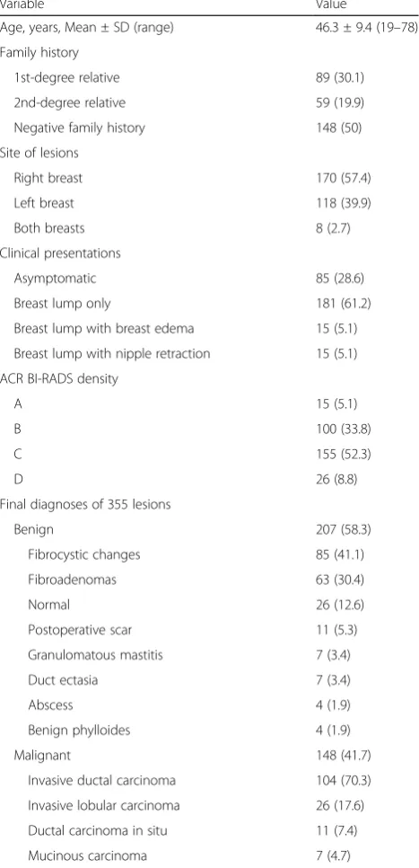
Table 1Patients’data
Variable Value
Age, years, Mean ± SD (range) 46.3 ± 9.4 (19–78)
Family history
1st-degree relative 89 (30.1)
2nd-degree relative 59 (19.9)
Negative family history 148 (50)
Site of lesions
Right breast 170 (57.4)
Left breast 118 (39.9)
Both breasts 8 (2.7)
Clinical presentations
Asymptomatic 85 (28.6)
Breast lump only 181 (61.2)
Breast lump with breast edema 15 (5.1)
Breast lump with nipple retraction 15 (5.1)
ACR BI-RADS density
A 15 (5.1)
B 100 (33.8)
C 155 (52.3)
D 26 (8.8)
Final diagnoses of 355 lesions
Benign 207 (58.3)
Fibrocystic changes 85 (41.1)
Fibroadenomas 63 (30.4)
Normal 26 (12.6)
Postoperative scar 11 (5.3)
Granulomatous mastitis 7 (3.4)
Duct ectasia 7 (3.4)
Abscess 4 (1.9)
Benign phylloides 4 (1.9)
Malignant 148 (41.7)
Invasive ductal carcinoma 104 (70.3)
Invasive lobular carcinoma 26 (17.6)
Ductal carcinoma in situ 11 (7.4)
Mucinous carcinoma 7 (4.7)
Unless otherwise indicated, data are number with the percentage in parenthesis
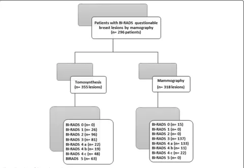
for second attendance to be subjected to DBT exami-nations. The flow chart of our study is illustrated in Fig. 1.
The technique of mammography and tomosynthesis
DBT examinations were performed within 2 weeks after DM examinations. All examinations were per-formed using a full field DM machine with 3D-DBT (Senographe Essential GE healthcare). Each breast was compressed and positioned carefully. Two views for each breast, craniocaudal (CC) and mediolateral ob-lique (MLO), were taken for both techniques. 3D-DBT involved the acquisition of nine projections with 25° scan angle. The 3D volume of the compressed breast was reconstructed from the 2D projections in the form of a series of images (slices) through the en-tire breast. Images from both techniques were sent to liquid-crystal display (LCD) screens for reading. No additional views were needed as further processing can be done while viewing the digital images on LCD panels such as zooming, changing contrast, brightness, darkness, inverting the background, and other pro-cessing to facilitate lesion detection.
Image analysis
The previous DM images and the DBT images were transported to the workstation for assessment. The DM and DBT images were separated for interpret-ation (i.e., the images for DM were interpreted with-out knowledge of the DBT findings). Two radiologists with 10 years of experience in breast imaging inde-pendently reviewed the DM images. After 1 month, the same two radiologists independently reviewed the DBT images. After another month, the same two radiologists independently reviewed DM and DBT im-ages together. Any discrepancies in interpretation were resolved by a third radiologist with over 15 years of experience in breast imaging. The 1-month
interval was to diminish the radiologists’ memory
distribution), and (viii) any other suspicious abnormalities.
The four categories of the American College of Radi-ology (ACR) BI-RADS scale were used to measure
mam-mographic density [16]. The radiologists were requested
to assign the BI-RADS category to all detected lesions in each of the two imaging modalities individually
accord-ing to the BI-RADS lexicon 2013 classification [17].
Fi-nally, each breast lesion had three independent BI-RADS categories (one by DM, one by DBT, and one by com-bined DM and DBT). In the comcom-bined protocol, the ra-diologists assigned a BI-RADS category based on the combined radiological features from each modality. A feature was considered positive when it was seen in at least one of DM or DBT. The results of DM and DBT for each patient were compared in terms of main radio-logical features, BI-RADS classification, and diagnostic performance.
Reference standard
The definitive diagnosis was validated based on
histo-pathologic findings after US-guided biopsy (n = 221
pa-tients), stereotactic biopsy (n= 32 patients), and surgical
mastectomy (n= 43 patients). All specimens were
exam-ined by two experienced pathologists, and the final re-sults were acquired by consensus. Biopsies were performed to determine the lesion type by the request-ing clinician.
Statistical analysis
Statistical analysis was done using SPSS software ver-sion 25 (IBM, 2017). Data were presented in tables and figures. Continuous data were presented as mean and standard deviation. Qualitative data were pre-sented as frequencies and proportions. Pearson’s
chi-square (χ2) test was used to analyze qualitative data.
McNemar test was used to analyze paired qualitative data. The diagnostic performance of DM, DBT, and combined DM and DBT was estimated on a lesion-based analysis. The receiver operating characteristic (ROC) curve analysis was applied to detect the areas
under the curve (AUCs). A p value of ≤0.05 was
ac-cepted as statistically significant.
Results
Study population
We performed our study on 296 female patients. Every enrolled patient had at least one breast lesion catego-rized as BI-RADS 0, 3, and 4 on DM. All patients were submitted to DBT. We detected a total of 318 lesions on DM and 355 lesions on DBT. The final diagnoses of 355 lesions were 207 (58.3%) benign and 148 (41.7%) malig-nant. The most common benign lesion was fibrocystic changes (41.1%), and the most common malignant lesion
was invasive ductal carcinoma (70.3%). According to the ACR BI-RADS lexicon for breast density, our patients were divided into four categories: BI-RADS density A, 15 (5.1%) patients; RADS density B, 100 (33.8%); BI-RADS density C, 155 (52.3%); and BI-BI-RADS density D, 26 (8.8 %).
DM and DBT findings
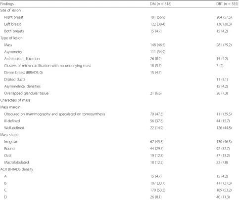
The DM and DBT findings are presented in Table 2.
The DM detected 318 lesions; 148 of them were mass, and 170 were non-mass, while DBT detected 355 lesions; 281 of them were mass, and 74 were non-mass. Of the 26 architectural distortions on DM, 11 revealed under-lying masses on DBT. Of the 18 micro-calcifications on DM, 11 revealed underlying masses on DBT. On DBT, five out of 26 cases revealed superposition of normal glandular tissue which were wrongly diagnosed as masses on DM. Thirty-seven lesions were detected by DBT and could not be detected by DM. These lesions were found in the dense breast (BI-RADS density C and
D) (n= 33) more than non-dense breast (BI-RADS
dens-ity A and B) (n= 4).
Assignment of BI-RADS category of breast lesions by DM and DBT
The BI-RADS scoring of breast lesions is summarized
in Table 3. The change in individual patient breast
le-sion owing to DBT, compared to DM, is presented in
Table 4. In comparison to DM, DBT produced 31.5%
(112/355) upgrading of BI-RADS scoring (4.2% (15/ 355) in BI-RADS 0, 7.3% (26/355) in BI-RADS 3, 18.8% (56/355) in RADS 4a, 3.1% (11/355) in BI-RADS 4 b, and 1.1% (4/355) in BI-BI-RADS 4 c) and 35.2% (125/355) downgrading of BIRADS scoring (18.9% (67/355) in BI-RADS 3, and 16.3% (58/355) in BI-RADS 4 a). Ninety-three (83%) of upgraded were malignant, and 118 (94%) of downgraded were benign. Sixty of BI-RADS 4 (36.1% (60/166)) were upgraded to BI-RADS 5 by DBT. All upgraded BI-RADS 4 were malignant, and seven of downgraded were malignant. DBT reduced the number of RADS 3 and BI-RADS 4 (81 and 89, respectively), compared to DM (137 and 166, respectively).
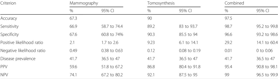
Diagnostic performance of DM and combined DM + DBT
On a lesion-based analysis, the diagnostic perform-ance of DM, DBT, and combined DBT and DM for
breast cancer diagnosis is summarized in Table 5. We
Table 2DM and DBT findings
Findings DM (n= 318) DBT (n= 355)
Site of lesion
Right breast 181 (56.9) 204 (57.5)
Left breast 122 (38.4) 136 (38.3)
Both breasts 15 (4.7) 15 (4.2)
Type of lesion
Mass 148 (46.5) 281 (79.2)
Asymmetry 111 (34.9)
Architecture distortion 26 (8.2) 15 (4.2)
Clusters of micro-calcification with no underlying mass 18 (5.7) 7 (2)
Dense breast (BIRADS 0) 15 (4.7)
Dilated ducts 11 (3.1)
Asymmetrical densities 15 (4.2)
Overlapped glandular tissue 21 (6.6) 26 (7.3)
Characters of mass
Mass margin
Obscured on mammography and speculated on tomosynthesis 70 (47.3) 111 (39.5)
Ill-defined 56 (37.8) 44 (15.7)
Well-defined 22 (14.9) 126 (44.8)
Mass shape
Irregular 67 (45.3) 130 (46.3)
Round 44 (29.7) 92 (32.7)
Oval 19 (12.8) 37 (13.2)
Macrolobulated 18 (12.2) 22 (7.8)
ACR BI-RADS density
A 15 (4.7) 15 (4.2)
B 107 (33.7) 111 (31.3)
C 170 (53.5) 189 (53.2)
D 26 (8.1) 40 (11.3)
The data are represented as numbers with the corresponding percentages given in parentheses
DBTdigital breast tomosynthesis,DMdigital mammography,BI-RADSBreast Imaging Reporting and Data System,ACRAmerican college of radiology
Table 3BI-RADS categories of the 355 breast lesions detected on DBT and DM in relation to the final diagnosis
DBT DM
Malignant Benign Total Malignant Benign Total
Not seen 0 0 0 15 (4.2) 22 (6.2) 37 (10.4)
BI-RADS 0 0 0 0 4 (1.1) 11 (3.1) 15 (4.2)
BI-RADS 1 0 26 (7.3) 26 (7.3) 0 0 0
BI-RADS 2 0 96 (27) 96 (27) 0 0 0
BI-RADS 3 16 (4.5) 65 (18.3) 81 (22.8) 30 (8.5) 107 (30.1) 137 (38.6)
BI-RADS 4 a 16 (4.5) 6 (1.8) 22 (6.3) 66 (18.6) 67 (18.9) 133 (37.5)
BI-RADS 4 b 19 (5.4) 0 19 (5.4) 11 (3.1) 0 11 (3.1)
BI-RADS 4 c 34 (9.6) 14 (3.9) 48 (13.5) 22 (6.2) 0 22 (6.2)
BI-RADS 5 63 (17.7) 0 63 (17.7) 0 0 0
Total 148 (41.7) 207 (58.3) 355 (100) 148 (41.7) 207 (58.3) 355 (100)
The data are represented as numbers with the corresponding percentages given in parentheses
BI-RADS with DBT yielded significantly higher accur-acy, sensitivity, and specificity than BI-RADS with
DM in the diagnosis of breast cancer (p < 0.001). The
combination of DBT and DM significantly increased the performance of BI-RADS in the diagnosis of
breast cancer versus DM or DBT alone (p < 0.001).
DM had more false-positive and false-negative rates than DBT.
ROC analyses
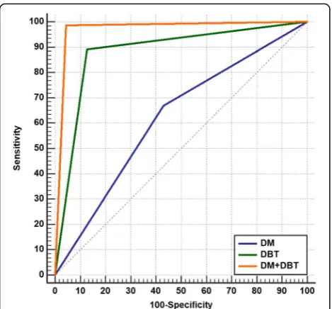
We analyzed the data set of the diagnostic perform-ance of BI-RADS with DM, BI-RADS with DBT, and BI-RADS with combined DM and DBT for breast cancer diagnosis using the ROC curve. When the ROC areas were compared, it was found that RADS with DBT was significantly superior to BI-RADS with DM in breast cancer diagnosis (AUC:
0.883 vs. 0.619; p < 0.0001; 95% CI 0.214 to 0.313),
and the BI-RADS with combined DM and DBT was significantly superior to RADS with DM or
BI-RADS with DBT alone (AUC: 0.971; p < 0.0001; 95%
CI 0.0565 to 0.120) (Fig. 2).
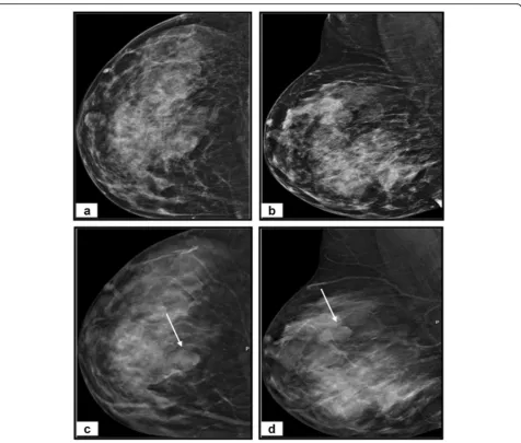
Our study’s representative cases are illustrated in Figs.
3,4, and5.
Discussion
In this study, we mainly focused on indeterminate BI-RADS categories, because reducing BI-BI-RADS 0, 3, and 4 has pivotal implications for patient care. We aimed to display the added value of DBT to the BI-RADS classification. Few studies evaluated this topic; most
of them have investigated category 3 [18, 19], and
some have also included category 0 [20]. However,
this study focused exclusively on BI-RADS 0, 3, and 4 Table 4Change in individual breast lesion grading on account of DBT, compared to DM
The data are represented as numbers with the corresponding percentages given in parentheses
The different colors indicate whether DBT upgraded (yellow), downgraded (blue), or kept the grade the same (green) as DM
DBTdigital breast tomosynthesis,DMdigital mammography,BI-RADSBreast Imaging Reporting and Data System
Table 5The diagnostic performance of BI-RADS with mammography, BI-RADS with tomosynthesis, and BI-RADS with combined tomosynthesis and mammography for confident breast cancer diagnosis considered BI-RADS 4 and 5 as predictive of malignancy
Criterion Mammography Tomosynthesis Combined
% 95% CI % 95% CI % 95% CI
Accuracy 67.3 90 97.5
Sensitivity 66.9 58.7 to 74.4 89.2 83 to 93.7 98.7 95.2 to 99.8
Specificity 67.6 60.8 to 74% 90.3 85.5 to 94 96.6 93.2 to 98.6
Positive likelihood ratio 2.1 1.7 to 2.6 9.23 6.1 to 14.1 29.2 14.1 to 60.4
Negative likelihood ratio 0.49 0.38 to 0.63 0.12 0.08 to 0.19 0.01 0 to 0.06
Disease prevalence 41.7 36.5 to 47 41.7 36.5 to 47 41.7 36.5 to 47
PPV 59.6 51.8 to 67.2 86.8 80.4 to 91.8 95.4 90.8 to 98.1
NPV 74.1 67.2 to 80.2 92.1 87.5 to 95 99 96.5 to 99.9
categories. The overall results of our study confirmed the high diagnostic performance of DBT in the evalu-ation of indeterminate BI-RADS categories. We found that combined DBT and DM in BI-RADS resulted in a superior sensitivity (98.7%), specificity (96.6%), and
accuracy (97.5%) for indeterminate breast lesion
categorization than DM or DBT alone; the sensitivity, specificity, and accuracy declined to 66.9%, 67.6%, and 67.3%, respectively, for the DM assessment and 89.2%, 90.3%, and 90%, respectively, for the DBT assessment. Moreover, we found that DBT had a significantly higher sensitivity, specificity, and accuracy than DM in the diagnosis of indeterminate breast lesions. The above findings are matching with that of many
previ-ous studies [10, 12, 19, 21–25], which have
estab-lished that DBT increases the sensitivity and
specificity of DM. Consequently, in light of our data, and considering the high diagnostic performance of DBT, we recommend the use of DBT as an additional imaging modality to improve diagnostic accuracy in
detecting and characterizing indeterminate breast
lesions.
We found that DBT produced a significant change of BI-RADS category in 66.7% lesions with an up-grade in 31.5% lesions (83% were malignant) and a downgrade in 35.2% lesions (94% were benign) in comparison to the DM. This finding agrees with the
recent study published by Raghu et al. [12] who have
proved that the addition of DBT has been found to
change rates of BI-RADS final assessment over time.
Similarly, Michell et al. [26] showed a reduction of
probably benign cases in 57.8% by an additional DBT.
A remarkable observation of our study was the higher number of lesions identified with DBT than with DM [37 (10.4%)], with BI-RADS 2 lesion repre-senting the greatest number of these missed lesions on DM [19 (51.4%)]. We found that the main cause for missing a lesion on DM was poor visibility due to dense breast parenchyma, tissue overlap, and a radio-graphically non-conspicuous lesion. In contrast, the DBT decreased interference from overlapping breast tissue and improved lesion conspicuity. These missed lesions on DM cause a significant upgrade of the BI-RADS categories between DM and DBT and subse-quently increased diagnostic performance of DBT over DM. This finding indicates that DBT is more ac-curate than DM in the identification of breast lesion,
which is comparable to the previous findings [10, 15,
19]. The increased number of lesions detected on
DBT is most probably due to the use of rebuilt
im-ages in DBT, as stated by Andersson et al. [13].
These images are obtained from different angles from the breast in a short scanning process and allow the assessment of breast parenchyma where lesions may go unnoticed or less evident due to tissue overlap or increased breast density.
The reduction in the number of BI-RADS 3 and 4 le-sions is one of the potential advantages of DBT as some lesions that were categorized as BI-RADS 3 and 4 on DM was upgraded to RADS 5 or downgraded to BI-RADS 1 and 2 based on DBT. This increase in the iden-tification of BI-RADS 3 and 4 lesions by DBT likely result in reduced follow-up of lesions that would not have been identified by DM alone and diminished the requirement for biopsy. These results are comparable to those reported by previous studies [13,27,28].
Although DBT has better diagnostic performance than DM, still some breast lesions could not be
deter-mined on DBT. On DBT images, improved
visualization of a partially or totally smooth boundary in some malignant masses may potentially lead to a
misdiagnosis that is false benign diagnosis [29].
Thirty-six of the breast lesions in this study were misdiagnosed on DBT (20 positives and 16 false-negatives). Sixteen masses were described as probably benign masses on DBT, but histopathology revealed breast cancer. However, the misdiagnosed lesions on DBT were less than that on DM (67 false-positives and 49 false-negatives). The combined DM and DBT decreased misdiagnosed lesions (seven false-positives and two false-negatives) when compared to DM or DBT alone. Accordingly, our study recommends the Fig. 2Comparison of the ROC areas of BI-RADS with DM, BI-RADS
use of BI-RADS with a combined DM and DBT protocol, as it improved the BI-RADS performance for diagnosis of indeterminate breast lesions with sub-sequent potentially better disease management. Simi-lar findings have been seen in various other studies
[30–32], in which the addition of DBT decrease in
the number of false cases.
When comparing the ROC areas, it was found that DBT is significantly superior to DM in breast cancer
diagnosis (AUC = 0.883 vs 0.619; p < 0.0001), and the
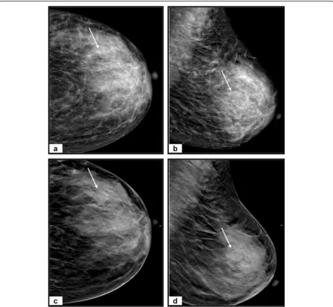
BI-RADS with combined DM and DBT was significantly superior to BI-RADS with DM or BI-RADS with DBT alone (AUC: 0.971; p< 0.0001). Similarly, Cai et al. [33] analyzed 79 cases with pathologic results by using ROC Fig. 3A 35-year-old woman complains of a right breast lump.aCraniocaudal and (b) mediolateral oblique DM images of right breast reveal extremely dense breast (BI-RADS D) with an outer central area of architectural distortion (arrows). No spiculated masses or microcalcifications.c
curve and showed that the AUC of the combined DM and DBT was greater than that of DM alone (0.914 vs.
0.805). Also, Thomassin et al. [34] calculated AUC by
averaging the ROC from four readers; the mean AUC for BI-RADS with combined DM and DBT was higher than that calculated for BI-RADS with DM alone (0.809 vs. 0.685;p< 0.01). In contrast, Gennaro et al. [35] con-cluded that the overall clinical performance with DBT and DM for malignant versus all other cases was not sig-nificantly different (AUCs = 0.851 vs. 0.836,p= 0.645).
diagnosis of being benign or malignant (i.e., BI-RADS 1, 2, or 5), the patients go on to the management without further additional imaging. In cases of indeterminate diagnosis (i.e., BI-RADS 0, 3, or 4), we perform DBT, and the BI-RADS category is determined by the combin-ation of DM and DBT findings. We perform the DM ini-tially and not DBT as the former’s lower cost and wide availability.
There are some limitations to our study. First, we focused on indeterminate breast lesions (BI-RADS 0, 3, and 4) and did not consider other BI-RADS categories (BI-RADS 1, 2, and 5). Second, no trial was conducted to analyze the inter-reader agreement in the classifica-tion of breast lesions. Third, we did not address the
DBT performance in each breast density category. Fourth, the cost-effectiveness and the added radiation dose of combined DM and DBT protocol may be disad-vantages of this protocol. Thus, we suggest that the combined protocol be limited to doubted lesions when there remains uncertainty in the BI-RADS category after conducting DM alone.
Conclusion
Adding DBT to BI-RADS classification improves its
diagnostic performance in the detection and
characterization of lesions categorized as BI-RADS 0, 3, and 4 by DM.
Abbreviations
3D:Three-dimensional; ACR: American College of Radiology; AUC: Area under the curve; BI-RADS: Breast Imaging Reporting and Data System; CI: Confidence interval; DBT: Digital breast tomosynthesis; DM: Digital mammography; LCD: Liquid-crystal display; ROC: Receiver operating characteristic
Acknowledgment
The authors thank all staff members and colleagues in radiology
departments-Zagazig University and Cairo National Cancer Institute for their helpful cooperation and all the study participants for their patience and support.
Ethical approval and consent to participate
Institutional review board approval was obtained. Written informed consent was obtained from all patients.
Authors’contributions
Guarantor of integrity of the entire study—MB. Study concepts and design—MB, AH, and HS. Literature research—HD and AA. Clinical studies—HS, AH, HS, and AA. Experimental studies/data analysis—MB, AH, and HS. Statistical analysis—MB. Manuscript preparation—MB and AH. Manuscript editing—MB and AH. All authors read and approved the final manuscript.
Funding
The authors declare that this work has not received any funding.
Availability of data and materials
The datasets used and/or analyzed during the current study are available from the corresponding author on reasonable request.
Consent for publication
Not applicable.
Competing interests
The authors declare that they have no competing interests.
Received: 19 November 2019 Accepted: 13 January 2020
References
1. Siegel RL, Miller KD, Jemal A (2016) Cancer statistics, 2016. CA Cancer J Clin 66:7–30
2. Dey S (2014) Preventing breast cancer in LMICs via screening and/or early detection: The real and the surreal. World J Clin Oncol 5:509–519 3. van den Biggelaar FJ, Kessels AG, van Engelshoven JM, Flobbe K (2009)
Strategies for digital mammography interpretation in a clinical patient population. Int J Cancer 125:2923–2929
4. Park JM, Franken EA Jr, Garg M, Fajardo LL, Niklason LT (2007) Breast tomosynthesis: Present considerations and future applications. Radiographics 27:S231–S240
5. Fallenberg EM, Dromain C, Diekmann F et al (2014) Contrast-enhanced spectral mammography versus MRI: initial results in the detection of breast cancer and assessment of tumour size. Eur Radiol 24:256–264
6. Gur D (2007) Tomosynthesis: potential clinical role in breast imaging. AJR Am J Roentgenol 189:614–615
7. Helvie M (2010) Digital mammography imaging: breast tomosynthesis and advanced applications. Radiol Clin North Am 48:917–929
8. Yaffe MJ (2008) Mammographic density—measurement of mammographic density. Breast Cancer Res 10:209.
9. Smith-Bindman R, Chu P, Miglioretti DL et al (2005) Physician predictors of mammographic accuracy. J Natl Cancer Inst 97:358–367
10. Skaane P, Bandos AI, Gullien R et al (2013) Comparison of digital mammography alone and digital mammography plus tomosynthesis in a population-based screening program. Radiology 267:47–56
11. Zuley ML, Bandos AI, Ganott MA et al (2013) Digital breast tomosynthesis versus supplemental diagnostic mammographic views for evaluation of noncalcified breast lesions. Radiology 266:89–95
12. Raghu M, Durand MA, Andrejeva L et al (2016) Tomosynthesis in the diagnostic setting: changing rates of BI-RADS final assessment over time. Radiology 281:54–61
13. Andersson I, Ikeda DM, Zackrisson S et al (2008) Breast tomosynthesis and digital mammography: a comparison of breast cancer visibility and BIRADS classification in a population of cancers with subtlemammographic findings. Eur Radiol 18:2817–2825
14. Lee WK, Chung J, Cha ES, Lee JE, Kim JH (2016) Digital breast tomosynthesis and breast ultrasound: Additional roles in dense breasts with category 0 at conventional digital mammography. Eur J Radiol 85:291–296
15. Ray KM, Turner E, Sickles EA, Joe BN (2015) Suspicious findings at digital breast tomosynthesis occult to conventional digital mammography: imaging features and pathology Findings. Breast J 21:538–5342 16. American College of Radiology (2013) Breast imaging reporting and data
system, 5th edn. ACR, Reston, VA
17. D’Orsi CJ, Sickles EA, Mendelson EB et al (2013) ACR BI-RADS® At-las, Breast imaging reporting and data system, 5th edn. ACR, Reston, VA
18. Stepanek T, Constantinou N, Marshall H et al (2019) Changes in the utilization of the BI-RADS Category 3 assessment in recalled patients before and after the implementation of screening digital breast tomosynthesis. Acad Radiol 26:1515–25
19. Bahrs SD, Otto V, Hattermann V et al (2018) Breast tomosynthesis for the clarification of mammographic BI-RADS 3 lesions can decrease follow-up examinations and enables immediate cancer diagnosis. Acta Radiol 59: 1176–1183
20. Emlik GD, Poyraz N, Altunkeser A (2017) Digital breast tomosynthesis and ultrasonography: diagnostic performance and effect on recall rates versus digital mammography in category 0. Int J Clin Exp Med 10:10668–10675 21. Haas BM, Karla V, Geisel J et al (2013) Comparison of tomosynthesis plus digital mammography and digital mammography alone for breast cancer screening. Radiology 269:694–700
22. Waldherr C, Gerny P, Altermatt HJ et al (2013) Value of one-view breast tomosynthesis versus two-view mammography in diagnostic workup of women with clinical signs and symptoms and in women recalled from screening. AJR Am J Roentgenol 200:226–231
23. Poplack SP, Tosteson TD, Kogel CA et al (2007) Digital breast tomosynthesis: initial experience in 98 women with abnormal digital screening
mammography. AJR Am J Roentgenol 189:616–623 24. Gennaro G, Toledano A, Di Maggio C et al (2010) Digital breast
tomosynthesis versus digital mammography: a clinical performance study. Eur Radiol 20:1545–1553
25. Rafferty EA, Park JM, Philpotts LE et al (2013) Assessing radiologist performance using combined digital mammography and breast tomosynthesis compared with digital mammography alone: results of a multicenter, multireader trial. Radiology 266:104–113
26. Michell MJ, Iqbal A, Wasan RK et al (2012) A comparison of the accuracy of film-screen mammography, full-field digital mammography, and digital breast tomosynthesis. Clin Radiol 67:976–981
27. Rose SL, Tidwell AL, Ice MF, Nordmann AS, Sexton R Jr, Song R (2014) A reader study comparing prospective tomosynthesis interpretations with retrospective readings of the corresponding FFDM examinations. Acad Radiol 21:1204–1210
28. Galati F, Marzocca F, Bassetti E et al (2047) (2017) Added value of digital breast tomosynthesis combined with digital mammography according to reader agreement: changes in BI-RADS rate and follow-up management. Breast Care (Basel) 12:218–222
29. Nakashima K, Uematsu T, Itoh T et al (2017) Comparison of visibility of circumscribed masses on Digital Breast Tomosynthesis (DBT) and 2D mammography: are circumscribed masses better visualized and assured of being benign on DBT? Eur Radiol 27:570–577
30. Haas BM, Kalra V, Geisel J, Raghu M, Durand M, Philpotts LE (2013) Comparison of tomosynthesis plus digital mammography and digital mammography alone for breast cancer screening. Radiology 269:694– 700
31. Rose SL, Tidwell AL, Bujnoch LJ, Kushwaha AC, Nordmann AS, Sexton R Jr (2013) Implementation of breast tomosynthesis in a routine screening practice: an observational study. AJR Am J Roentgenol 200:1401–1408 32. Skaane P, Bandos AI, Gullien R et al (2013) Prospective trial comparing
33. Cai SQ, Yan JX, Chen QS, Huang ML, Cai DL (2015) Significance and application of DBT for the BI-RADS classification of breast cancer. Asian Pac J Cancer Prev 16:4109–4114
34. Thomassin-Naggara I, Perrot N, Dechoux S, Ribeiro C, Chopier J, De Bazelaire C (2015) Added value of one-view breast tomosynthesis combined with digital mammography according to reader experience. Eur J Radiol 84:235– 241
35. Gennaro G, Toledano A, Di Maggio C et al (2010) Digital breast tomosynthesis versus digital mammography: a clinical performance study. Eur Radiol 20:1545–1553
Publisher’s Note