M E E T I N G A B S T R A C T S
Open Access
Sepsis 2017 Paris
Paris, France. September 11
–
13, 2017
Published: 11 September 2017
P1
Is Post-Sepsis Syndrome different from Post-Intensive Care (ICU) syndrome? A cohort and propensity score matched analysis Kelly Thompson, Colman Taylor, Stephen Jan, Qiang Li, John Myburgh, Bala Venkatesh, Simon Finfer
The George Institute for Global Health, Newtown, NSW 2042, Australia Correspondence:Kelly Thompson ([email protected]) Intensive Care Medicine Experimental2017,5(Suppl 1):P1
Background
Post-Sepsis Syndrome and Post-ICU Syndrome are terms recently adopted to characterise the health-related quality of life (QoL) issues affecting patients after sepsis or after treatment in an ICU.[1, 2] Whether these syndromes are different is not known.
Objectives
To determine QoL, late mortality, costs and healthcare resource use of ICU patients with sepsis compared to those without sepsis. Methods
After obtaining ethical committee consent we analysed data of ICU patients with sepsis recruited to a randomised controlled trial in New South Wales, Australia. We conducted a cohort and propensity score matched (PSM) analysis using baseline variables including indices of severity of illness compared to ICU patients without sepsis. We assessed QoL using the EQ-5D-3 L at 6 months and mortality, costs (in $AU) and healthcare resource use at 2 years.
Results
QoL was assessed in 4975 patients: 1320 with sepsis and 3655 with-out. There were no significant differences in QoL. For the analysis of mortality, costs and healthcare resource use, 3442 patients were included: 905 patients with sepsis and 2537 without. At 2 years, pa-tients with sepsis had higher mortality: 371/905 (41%) vs 860/2537 (34%) HR1.34, 95%CI 1.18 to1.52, p< 0.01; increased costs: ICU: 47,206 ± 55,121 vs 34,142 ± 43,175; p< 0.0001; Hospital: 73,516 ± 61,100 vs 64,676 ± 56,293;p< 0.0001; extended length of stay: ICU: 10.0 ± 11.9 vs 7.1 ± 9.1 days;p< 0.0001; Hospital: 22.7 ± 21.6 vs 20.4 ± 19.7 days;p= 0.003. Patients with sepsis had fewer hospital read-missions: 506/904 (56%) vs 1597/2563 (63%); p = 0.0002. 1612 pa-tients were included in the PSM analysis: 806 with sepsis and 806 without. 90/905 (10%) patients with sepsis could not be matched due to high illness severity. There were no differences in QoL, late mortality or hospital readmissions. Costs were higher for patients with sepsis: ICU 47,298 ± 53,730 vs 38,952 ± 46,778; p= 0.009; Hos-pital 74,120 ± 60,750 vs 65,806 ± 56,856;p= 0.005, and length of stay: ICU: 10.0 ± 11.9 vs 8.0 ± 9.8 days;p< 0.0001; Hospital: 22.8 ± 21.2 vs 19.1 ± 19.0 days;p= 0.0003.
Conclusions
Compared to patients without sepsis, patients with sepsis report similar QoL but have increased hospital costs, length of stay and fewer hospital readmissions. Aside from cost, these differences are not significant when patients are matched for severity of illness and other baseline variables, notwithstanding the limitation of our inabil-ity to match 10% of patients in the sepsis cohort.
References
1. Winters, B.D., Eberlein, M., Leung, J., Needham, D., Pronovost, P., Sevransky, J. Long-term mortality and quality of life in sepsis: a systematic review. Crit Care Med, 2010. 38(5): p. 1276-1283.
2. Elliott, D., et al., Exploring the scope of post-intensive care syndrome therapy and care: engagement of non-critical care providers and survivors in a second stakeholders meeting. Crit Care Med, 2014. 42(12): p. 2518-2526.
P2
Extracellular histones induce erythrocyte fragility and anaemia and identification of compounds that prevent this process
Farzaneh Kordbacheh1, Connor H. O
’Meara1, Lucy A. Coupland1, Patrick M. Lelliott2, Chris R. Parish1
1Cancer and Vascular Biology Group, ACRF Department of Cancer
Biology and Therapeutics, The John Curtin School of Medical Research, Australian National University, Canberra, Australia;2Department of Immunology and Infectious Disease, John Curtin School of Medical Research, Australian National University, Canberra, ACT, 2601, Australia Correspondence:Farzaneh Kordbacheh
Intensive Care Medicine Experimental2017,5(Suppl 1):P2
Background
Sepsis is a life-threatening systemic inflammatory response to infection or trauma associated with, and mediated by, the activation of a number of host defence mechanisms. Neutrophils play an essential role in sep-sis by releasing neutrophil extracellular traps (NETs) which exhibit very effective anti-microbial activity. Uncontrolled formation of NETs can, however, become pathogenic particularly via their associated histones that can be cytotoxic in the vasculature causing endothelial cell death, systemic vascular obstruction and multiple organ failure. They can also initiate coagulation by both activating platelets and damaging erythro-cytes such that they become pro-thrombotic. Recently we discovered an additional effect of histones on erythrocyte, which is to enhance their fragility when they are subjected to sheer stress associated with blood flow through the vascular system and spleen in particular. In parallel studies our laboratory has synthesised a family of small polya-nions (SPAs) which have been screened for their ability to neutralise the pathogenic effects of histones and NETs.
Methods
We have used a novel mechanically-induced sheer stress method as well as anin vitrospleen filtration model to investigate the effects of mam-malian histones ± SPAs, over a wide concentration range, on RBC fragility under both isotonic and hypotonic conditions. Thein vitrofindings were mirroredin vivowith the injection of histones and SPAs in mice. Results
Our results revealed that free histones bind to RBCs and render RBCs sus-ceptible to lysis by shear stress in a dose-dependent manner bothin vitro andin vivomodels as well as being retained by the spleen. Some SPAs have been identified that can totally prevent the ability of histones to induce RBC fragilityin vitroand anaemiain vivo, with the compounds also preventing the rapid and acute histone-induced thrombocytopenia. Remarkably we have also discovered that the active SPAs can rapidly rescue histone dam-aged erythrocytes, the fragility of the restored RBCs returning to normal levels within minutes, even following prolonged exposure to histones. Conclusion
Based on ourin vitroandin vivodata we propose that erythrocytes can capture histones in the circulation and transport them to the spleen for disposal where overload of this disposal system could
cause the unexplained anaemia associated with sepsis, cancer and other pathological conditions and our SPAs can both prevent and re-verse these effects.
P3
Innate immune receptor gene polymorphisms and its association with the risk of neonatal sepsis
Snehal L Martin1, Saumil Desai2, Roshan B Colah1, Kanjaksha Ghosh1, Ruchi Nanavati2, Malay B Mukherjee1
1Department of Haematogenetics, National Institute of
Immunohaematology (ICMR), KEM Hospital campus, Parel, Mumbai, India;2Department of Neonatology, KEM Hospital, Parel, Mumbai, India Correspondence:Snehal Martin ([email protected]) Intensive Care Medicine Experimental2017,5(Suppl 1):P3
Background
Neonatal sepsis is one of the major causes of neonatal death in devel-oping countries. Identifying single nucleotide polymorphisms (SNPs) in the genes involved in sepsis may help to understand the pathophysi-ology of neonatal sepsis. The aim of the present study to look for asso-ciation between the sepsis in neonates and SNPs in different immune receptor genes involved in host responses to bacterial infection. Methods
The study involved 151 preterm neonates with signs and symptoms of clinical sepsis and had a blood culture that was positive for a bacterial or fungal pathogen, 136 preterm neonates with signs and symptoms of sepsis along with a positive septic screen but negative blood culture and 146 healthy neonates born between June 2013 to June 2016. A total of 9 SNPs in 6 innate immune related genes were genotyped using PCR- RFLP and Real time SNP genotyping technique.
Results
Birth weight, gestational age, duration of rupture of membranes, respira-tory distress, apnea, abdominal distension, sclerema and anemia were observed in sepsis cases. Incidence of early onset sepsis (EOS) in the blood culture positive neonates was significantly higher as compared to late onset sepsis (p= 0.01). Infections caused due to Gram negative or-ganisms (67.92%) were more common than Gram positive (18.86%) and fungal infections (13.20%). An overall mortality rate among the sepsis cases was found to be 26.82%, however, a higher prevalence of mortal-ity (27.94%) was observed in clinical sepsis as compared to in the proven sepsis (23.17%). Although polymorphisms in innate immune related genes did not show significant association with overall risk of develop-ing sepsis, nevertheless, genotype CT of rs5744168 (TLR5 gene) was as-sociated with a significantly reduced risk of developing sepsis (p=0.02). Genotypes TT of rs2569190 (CD14 gene) and CT of rs5744168 (TLR5 gene) were significantly associated with a reduced risk of developing sepsis in the neonates with bacteriologically confirmed sepsis (p= 0.02) and clinical sepsis (p= 0.03) respectively. Among the patients with bac-teriologically confirmed sepsis, genotype CC of rs3804100 (TLR2 gene) was associated with a significantly increased risk of developing severe sepsis (p= 0.02) while same genotype was found to be reduced the risk of developing sepsis (p= 0.04) in the neonates with clinical sepsis. Conclusions
This study demonstrates that polymorphisms in the innate immune related genes seems to play a role in sepsis in preterm neonates by influencing susceptibility and the severity of the disease.
Acknowledgement
We would like to thank Indian council of Medical research (ICMR) for providing fellowship to Ms. Martin S.
P4
Association of Red cell Distribution Width with sepsis and sepsis related mortality in newborns
Snehal L Martin1, Saumil Desai2, Roshan B Colah1, Kanjaksha Ghosh1, Ruchi Nanavati2, Malay B Mukherjee1
1Department of Haematogenetics, National Institute of
Immunohaematology (ICMR), KEM Hospital campus, Parel, Mumbai, India;2Department of Neonatology, KEM Hospital, Parel, Mumbai, India Correspondence:Snehal Martin ([email protected]) Intensive Care Medicine Experimental2017,5(Suppl 1):P4
Background
Red Cell Distribution Width (RDW) is derived from the routine Complete Blood Count and provides a simple means to stratify patients by sever-ity of illness to facilitate focused interventions without any additional costs. The rise in RDW among neonatal sepsis cases may therefore reflect the degree of the underlying inflammatory state and provide useful prognostic information about the risk of mortality. The present study was undertaken to look for association of Red cell distribution width (RDW) with neonatal sepsis and sepsis related mortality. Methods
We performed a prospective observational study on 251 sepsis neo-nates along with 284 controls. RDW along with other RBC (Red blood cell) indices was performed on an automated cell counter (Sysmex, Japan). We categorized RDW at admission into tertiles of≤16.9%, 17-20% and≥20.1%. The cumulative survival rate across the RDW levels during the neonatal period (≤30 days) was assessed using the Kaplan–Meier curve and the log rank test.
Results
Of the RBC indices studied, the median (IQR) RDW levels were signifi-cantly higher among the neonatal sepsis cases (19.90%) as compared to the controls (18.90%) (p= 0.001). The rate of mortality due to sepsis was 28.28% and RDW was significantly higher amongst the non-survivors (median: 20.30, IQR: 18.3-23.5) than the survivors (median: 19.80, IQR: 17.5-22) (p= 0.003). Kaplan-Meier curve with log rank test showed that increased RDW tertiles are significantly associ-ated with an increased mortality (p= 0.03) during the 28-day neo-natal period.
Conclusions
The present study suggests that increased RDW is associated with neonatal sepsis and could be an independent outcome predictor for mortality associated with neonatal sepsis. The results of this study might help in the effective management of sepsis in neonates.
Acknowledgement
We would like to thank Indian council of Medical research (ICMR) for providing fellowship to Ms. Martin S.
P5
Effects of symbiotics on systemic inflammation in animal model of pediatric sepsis
Pricila Romão Marcondes Ávila, Monique Michels, Cleonice Maria Michelon, Maria Vitória Meller, Rafaela Bilésimo, Mariane Abatti, Felipe Dal Pizzol Universidade do Extremo Sul Catarinense- UNESC, Criciúma/SC, Brazil Correspondence:Pricila Romão Marcondes Avila
Intensive Care Medicine Experimental2017,5(Suppl 1):P5
Background
Sepsis is a very common clinical syndrome, also affecting pediatric population. The main characteristic of this condition is the imbalance of inflammatory response. A relation between immune system and microbiome has been proposed. This study aimed to investigate the effects of symbiotics on systemic inflammation induced in animal models of pediatric sepsis.
Methods
Results
In general, results showed that FOS and probiotics treatment was protective against LPS. In addition, strains had different behaviors on each tissue and in each marker, but their protective effect was more pronounced in the gut tissue. When it was evaluated the role of the intestinal microbiota through FMT, it was observed that FMT was protective, independently of previous administration of probiotics to fecal donor groups.
Conclusion
In conclusion, symbiotics had protective effect upon endotoxemia that was independent of change in intestinal microbiota. However, FMT may offer an additional immunomodulatory benefit that was in-dependent on symbiotic modulation.
Keywords: Pediatric sepsis. Probiotics. Symbiotic. Fecal microbiota transplant.
P6
Der p 2 from Dermatophagoides pteronissinus potentiates the endotoxic activity of lipopolysaccharide fromEscherichia coli Isabella Prokhorenko1, Anastasia Morozova1, Dmitry Kabanov1, Sergey Grachev1,2
1FSBSI Institute of Basic Biological Problems RAS, Pushchino, Moscow
Region, Russia;2SIEI HE I.M. Sechenov’s First Moscow State Medical University of Russian’s Ministry of Healthcare, Moscow, Russia Correspondence:Dmitry Kabanov ([email protected]) Intensive Care Medicine Experimental2017,5(Suppl 1):P6
Background
CD14, Toll-like receptor 4 (TLR4) and TLR4-associated adaptor protein MD-2 are essential receptors involved in lipopolysaccharide (LPS) rec-ognition leading to the synthesis and releasing of pro-inflammatory cytokines among them TNF-α playing a fundamental role [1]. It is well known that p38 MAP kinase pathway is involved in TLR4-dependent TNF-αproduction triggered by LPS•MD-2 complexes [2]. Recently, it has been shown that the main component from house dust mite Dermatophagoides pteronissinus extract (DpE)–Der p 2 mimics to MD-2 and acts as LPS•MD-2 complex inducing synthesis of pro-inflammatory cytokines [3, 4]. However, LPS•Der p 2 induced Fig. 1 (abstract P5).Effect of cronic administration of symbiotics
treatment on damage oxidative and proinflammatory cytokines in the lung of rats submitted to endotoxemia. twelve hours after submitted to endotoxemia, TBARS (A), Carbonyl (B), MPO(C), IL-1b (D), IL-6 (E) and TNFa (E), were measured in the brain. Data are expressed as the mean ± standard deviation for five to seven animals per group (mean ± SD). *Different from Sham + water; #Different from LPS+ water; % Different from LPS + FOS; & Different from LPS + bifidus; $ Different from LPS + rhamnosus;βDifferent from LPS + casei; @ Different from LPS + acidophilus; p <0.0001, ANOVA on one, followed the Tukey's multiple comparisons test
Fig. 2 (abstract P5).Effect of cronic administration of symbiotics treatment on damage oxidative and proinflammatory cytokines in the gut of rats submitted to endotoxemia. twelve hours after submitted to endotoxemia, TBARS (A), Carbonyl (B), MPO(C), IL-1b (D), IL-6 (E) and TNFa (E), were measured in the brain. Data are expressed as the mean ± standard deviation for five to seven animals per group (mean ± SD). *Different from Sham + water; #Different from LPS+ water; % Different from LPS + FOS; & Different from LPS + bifidus; $ Different from LPS + rhamnosus;βDifferent from LPS + casei; @ Different from LPS + acidophilus; p <0.0001, ANOVA on one, followed the Tukey's multiple comparisons test
Fig. 3 (abstract P5).Effect of fecal microbiota transplant treatment on damage oxidative and proinflammatory cytokines in the lung of rats submitted to endotoxemia, TBARS (A), Carbonyl (B), MPO(C), IL-1b (D), IL-6 (E) and TNFa (E), were measured. Data are expressed as the mean ± standard deviation for five to seven animals per group (mean ± SD). *Different from Sham, #Different from LPS, & Different from control transplant, % Different from transplant l casei;p<0.0001
intracellular signaling mechanism is not yet known. In this connec-tion the effect of p38 MAP kinase inhibiconnec-tion on TNF-α production induced by LPS or DpE as well as by their combined action has been elucidated in whole blood modelex vivo. It is necessary to note that an inflammatory mechanism in allergic patients with suspected sep-sis has not also fully explained.
Methods
Whole blood from healthy volunteers was collected into heparin-ized tubes (5%), diluted with RPMI-1640 medium (1:5 v/v), and transferred into CellStar 24-well tissue culture plates. Blood samples were pre-incubated with 10 μM of p38 MAP kinase inhibitor SB203580 for 15 min at 37 °C in a highly humidified atmosphere containing 5% CO2. Then whole blood was stimulated by LPS Escherichia coliO55:B5 (40 ng/ml), DpE (5 000 PNU/ml) or in their combination during 6 h. When incubation period had been fin-ished, the cell culture plates were centrifuged for 10 min at 15 °C and 1000 rpm. The supernatants were stored at -20 C until pending analysis. Concentrations of TNF-α in the supernatants were mea-sured by ELISA Kit according to the manufacturer’s instructions. Statistical significance was determined by Mann-Whitney U-test. Results
The incubation of whole blood with LPSE. colicaused cell activation as determined by TNF-αproduction, whereas combined applying of LPS E. coli and DpE further increased synthesis of TNF-α(Fig.5). Very low amounts of TNF-αwere detected in the response to DpE alone. Without LPS E. coli or DpE the TNF-α production was almost un-detectable. Pre-incubation of whole blood with SB203580 followed by LPS and DpE, either alone or together, resulted in 43% and 48% inhibition of LPS-induced TNF-α production as compared to LPSE. coli or LPS plus DpE, respectively. DpE triggered TNF-α production was inhibited by SB203580 completely (Fig. 5). These results indi-cated that the effect of LPSE. colior DpE on the induction of TNF-α synthesis at least in part depends on p38 MAPK activation. It is ne-cessary to note that there were some variations in TNF-αproduction triggered by DpE or DpE and LPSE. colitogether. These variations may be related to the use of primary whole blood cells rather than cell lines, with different donors having different sensitivities to the applying drugs.
Conclusions
In this study we have found that the synthesis of TNF-α was ampli-fied during combined action of LPSE. coliand DpE. The similar sup-pressive effect of p38 MAP kinase inhibition on LPS- as well as on LPS and DpE-induced synthesis of TNF-αhas been revealed. We have supposed that at least two signaling pathways (NF-kB and AP-1) may be involved in the observed TNF-αproduction [5, 6]. It seems likely that the DpE leading to TNF-α production does not involve any additional intracellular signaling pathways but probably its effect is realized on the cell surface receptor level [3, 4]. Our study may result in a better understanding of intracellular signaling mechanisms trig-gered by combined action of LPSE. coliand Der p 2 from Dermato-phagoides pteronissinus extract.
References
1. Christiansen D, Brekke O, Stenvik J, et al: Differential effect of inhibiting MD-2 and CD14 on LPS- versus whole E. coli bacteria-induced cytokine responses in human blood. Adv Exp Med Biol 2012, 946: 237–251. 2. Fehr S, Unger A, Schaeffeler E, et al: Impact of p38 MAP kinase inhibitors
on LPS-induced release of TNF-αin whole blood and primary cells from different species. Cell Physiol Biochem 2015, 36: 2237–2249.
3. Park B, Lee N, Kim M, et al: Interaction of Der p 2 with Toll-like receptor 4 and its effect on cytokine secretion. Biomed Sci Lett 2015, 21: 152–159. 4. Trompette A, Divanovic S, Visintin A, et al: Allergenicity resulting from
functional mimicry of a Toll-like receptor complex protein. Nature 2009. 457: 585–588.
5. Takashiba S, Shapira L, Amar S, Van Dyke T: Cloning and characterization of human TNF alpha promoter region. Gene 1993, 131: 307–308. 6. Karin M: The regulation of AP-1 activity by mitogen-activated protein
ki-nases. J Biol Chem 1995, 270: 16483–16486.
P7
Chemiluminescent analysis of the neutrophil function: from bench to bedside
Igor V. Obraztsov1,3, Marina A. Sukhina1, Vadim N. Sinyutin2, Sergey I. Achkasov1, Oleg I. Sushkov1, Anton L. Safin1 1
A. N. Ryzhikh State Scientific Centre for Coloproctology, Moscow, Russia; 2M. V. Lomonosov Moscow State University, Moscow, Russia;3D.
Rogachev National Scientific and Clinical Centre for Pediatric Haematology, Oncology and Immunology, Moscow, Russia Correspondence:Igor Obraztsov ([email protected]) Intensive Care Medicine Experimental2017,5(Suppl 1):P7
Background
Neutrophils play a crucial role in antimicrobial resistance; evaluation of reactive oxygen species (ROS) production by the neutrophils could provide valuable clinical data on neutrophils’ microbicidal capacity. Our aim was to develop a standardized methodology for the neutro-phil's oxidative output assessment and to implement it in a clinical practice for management of surgical patients.
Methods
Oxidative metabolism was evaluated in the whole blood by means of luminol-enhaced chemiluminescence (CL) induced by priming with 50 ng/ml 4-phorbol-12-myristate-13-acetate (PMA) prior to 10μM N-for-myl-methionyl-leucyl-phenylalanine (fMLP) stimulation. We defined an experiment design in order to obtain a signal of high intensity and re-producible pattern. We assessed the time-dependent neutrophils’ phenotypic shift by means of CD64, CD16, CD62L, CD11b, CD15 and CD33 expression evaluation after PMA stimulation.
Results
We revealed changes of CD62L (p< 0,05), CD11b (p< 0,01) and CD15 (p< 0,05) expression as well as CD11b/CD15(high) and CD11b/ CD15(bright) subsets formation (p= 0,001) under PMA priming of the neutrophils. Surface markers expression under PMA stimulation has various impacts on oxidative output kinetics. PMA priming of the neutrophils also leads to a dramatic increase of fMLP-induced ROS production (p< 0,001) that is accompanied by reduction of relative intracellular ROS production (p< 0,001). We built reference CL param-eters in a control group of 95 healthy individuals. Investigation of 17 patients with severe burns in dynamics revealed association between intracellular CL insufficiency and development of severe septic complications. We built a fitting linear-logarithmic function to create an unbiased approach to intracellular CL quantification and substanti-ated clinically relevant kinetic parameters (Fig.6). We used prognostic coefficient C = I2/I3 to perform a precise quantification of intracellular oxidative output in 21 colorectal cancer patients after surgery in
dynamics. CD16, CD64 as well as CD11b expression by the CD11b/ CD15(bright) subset drive C to the higher levels (p< 0,05), while CD62L and CD11b expression by the CD11b/CD15(high) subset tend to low C (p< 0,05). Assessment of colorectal cancer patients on the first four days after surgery has shown an increase of C (p< 0,05) as well as CD64+ neutrophils (p< 0,01) and procalcitonin (p< 0,01) levels in patients with longer hospitalization duration due to subsequent in-flammatory complications.
Conclusion
Thus kinetic CL assay being high-sensitive, cost-effective, easy-to-standardize and easy-to-perform, is promising for use in a clinical routine of a surgical hospital.
Acknowledgement
Authors express their deepest gratitude to prof. Yu. A. Vladimirov and all the stuff of the department for medical biophysics at M. V. Lomonosov Moscow State University for the encouragement, advice and help.
P8 Withdrawn
P9
Preventive diagnosis of sepsis in newborns with respiratory diseases on mechanical ventilation
Marina Puchtinskaya, Viadimir Estrin
Department of Anesthesiology and Critical Care Medicine, Rostov-on-Don State Medical University, Rostov-on-Don, Russia Correspondence:Marina Puchtinskaya ([email protected]) Intensive Care Medicine Experimental2017,5(Suppl 1):P9
Background
The decision on the confirmation of the diagnosis of sepsis remains a challenge of neonatology[1,2]. The purpose of this study was to im-prove the efficiency of preventive diagnosis of sepsis in newborns with respiratory diseases on mechanical ventilation.
Methods
The presentation of the monitored, randomized clinical trials was ap-proved by the local committee on ethical issues and bioethics of the Research Institute of Obstetrics and Pediatrics, Rostov-on-Don, Russia (protocol№8/1 of 26.11.2008 and№20/1 of 14.01.2009). Patients were included in the study with the written permission of their parents or
other legal representatives. The text of the authorization and the research protocol are formulated in accordance with the Declaration of Helsinki, Federal Law N323-F of the Zot on November 21, 2011 "On the fundamentals of protecting the health of citizens in the Russian Feder-ation"; the basis of the legislation of the Russian Federation "On the protection of public health, the rules for clinical practice in the Russian Federation" (Order of the Ministry of Health of the Russian Federation N266 of 19.07.03, order of Roszdravnadzor N2325-Pr/06 of 17.10.06). 200 full-term newborns with respiratory pathology, admitted to the neonatal intensive care unit on a ventilator in 1 day of life, without clin-ical signs of bacterial infection on admission, 3-5, 20 days was deter-mined by ELISA (Victor Multilabel Coulter-21420, Finland) plasma concentration of IL-1ß, IL-6, IL-8, TNF-α, G-CSF, s-Fas, FGF, NO; method of immunophenotyping (Beckman Coulter Epics XL-II, USA) CD3 + CD19-, CD3-CD19+, CD3 + CD4+, CD3 + CD8+, CD69+, CD71+, CD95+, HLA-DR+, CD34+, CD14+, CD3-CD56+; lymphocytes with the expression of AnnexinV-FITC + PI- and AnnexinV-FITC + PI+. Retrospectively the pa-tients were divided into two groups: with sepsis and without sepsis. Cluster analysis formed the base of the immunological data of prevent-ive diagnosis of sepsis, the assessment of the value of the correlation coefficient by the method of "decision Trees" was performed in the statistical environment "R".
Results
Of the 200 patients develop sepsis confirmed at 45. Statistical clus-ter analysis of a set of immunological criclus-teria of diagnosis of sepsis at admission to ICU confirmed the presence of two clusters (the presence or absence of sepsis: these two components explain 60.81% of the point variability). The method of "decision Trees" for-mulated the diagnostic rule for preventive diagnosis of sepsis: if CD95≥16.8% NO≤9.6 mkmol/l or CD95≤16.8% and CD34≤0.2% and CD69≥4.12% or CD95≤16.8% and CD34≤0.2% CD69≤4.12% and lymphocytes with the expression of AnnexinV-FITC + PI-≥12.3 per cent - is the development of sepsis. Diagnostic accuracy 95.41%; sensitivity 97.06%; specificity 94.67%; false-positive diagno-ses, the share of 5.33%; the proportion of false-negative diagnoses of 2.94%, the accuracy of a positive result 89.19%; the accuracy of a negative result up 98.61%.
Conclusions
Determination of the relative content of lymphocyte expression of CD95+, CD69+, AnnexinV-FITC + PI-; stem cells CD34+ and plasma NO concentration to diagnose sepsis in newborns with respiratory path-ology on a ventilator at admission to the NICU. A significant role in the development of sepsis belongs to prevalence of process of alteration of immunocompetent cells on the proliferation and endogenous synthesis of nitric oxide.
References
1. Puchtinskaya M., Estrin V. Prevention of sepsis by correcting apoptosis// Critical Care.–2013.–Vol. 17, Suppl 2.–P. 8.
2. Puchtinskaya M. Effect of inhaled nitric oxide on apoptosis of lymphocytes in newborns in critical states//Critical Care.–2014.–Vol. 18, Suppl 1.–P. 288.
P10
The mortality attributable to sepsis in adult patients in intensive care Marinelle Schout1,2,3, Naomi Hammond2,3,4, Frances Bass2,3, Anthony Delaney2,3,4, Simon Finfer2,3,4
1Radboud University, Nijmegen, The Netherlands;2Intensive Care Unit,
Royal North Shore Hospital, Sydney, Australia;3University of Sydney, Sydney, Australia;4The George Institute for Global Health, Sydney, Australia Correspondence:Marinelle Schout ([email protected]) Intensive Care Medicine Experimental2017,5(Suppl 1):P10
Background
Chronic comorbid conditions are present in the majority of sepsis pa-tients and these papa-tients have a significantly worse outcome than patients without comorbidities [1,2]. It is thought that sepsis occurs as a terminal event in a proportion of sepsis patients dying from a disease process other than sepsis [1,3].
Methods
We conducted an inception cohort study including all patients admitted to the ICU at a tertiary hospital in Sydney, Australia be-tween October 1st 2016 and December 31st 2016. In all patients diagnosed with sepsis using clinical criteria[4] we used an adjudi-cation system where clinicians estimated the pre-sepsis life ex-pectancy of patients who died in-hospital within 60 days. Patients who had an estimated life expectancy of less than 90 days were adjudicated to have died with sepsis rather than from sepsis.
Results
From 864 patients admitted, 146 (16.9%) patients were prospect-ively diagnosed with sepsis and included (Fig. 7). Comorbidities were present in 117/146 (80.1%) of patients (Table 1). 39/146 (26.7%) died in-hospital within 60 days. 30/39 (76.9%) of de-ceased patients were adjudicated not to be in a terminal phase of an underlying disease process before onset of sepsis. These deaths were therefore adjudicated to be directly attributable to sepsis.
Conclusions
Despite the presence of comorbidities in the majority of patients, most patients who died were not in a terminal stage of an underlying disease process when sepsis occurred. Therefore, sep-sis did result in an appreciable loss of life years. Further research in this area is warranted.
References
1. Angus DC, Linde-Zwirble WT, Lidicker J, Clermont G, Carcillo J, Pinsky MR. Epidemiology of severe sepsis in the United States: analysis of incidence, outcome, and associated costs of care. Crit Care Med. 2001;29(7):1303-1310.
2. Kaukonen KM, Bailey M, Suzuki S, Pilcher D, Bellomo R. Mortality related to severe sepsis and septic shock among critically ill patients in Australia and New Zealand, 2000-2012. JAMA. 2014;311(13):1308-1316.
3. Daviaud F, Grimaldi D, Dechartres A, et al. Timing and causes of death in septic shock.Ann Intensive Care.2015;5(1):16.
4. Finfer S, Bellomo R, Lipman J, French C, Dobb G, Myburgh J. Adult-population incidence of severe sepsis in Australian and New Zealand intensive care units.Intensive Care Med. 2004;30(4):589-596.
P11
Automated screening system for sepsis in hospital wards (Serial Patient Screen for Infectious Source, SePSIS)
Rosemary Grant1, Natalie Kress2, Benjamin Wolpaw2, David Carlbom3 1
Clinical Education, Harborview Medical Center, Seattle, WA, USA; 2University of Washington School of Medicine, Seattle, WA, USA; 3
Pulmonary and Critical Care Medicine, Harborview Medical Center, Seattle, WA, USA
Correspondence:Rosemary Grant ([email protected]) Intensive Care Medicine Experimental2017,5(Suppl 1):P11
Background
Early identification and timely intervention are essential in the manage-ment of patients with sepsis. Numerous clinical studies have shown that patients developing sepsis on acute care hospital wards often Fig. 7 (abstract P10).Inclusion and adjudication process
Table 1 (abstract P10).Patient characteristics of all included patients and per adjudication group
All patients N = 146
Group 1* N = 107
Group 2** N = 30
Group 3*** N = 9
p-value ****
Age, mean (SD) 64.5
(15.4) 64.4 (15.7)
65.6 (14.0)
62.0 (17.6)
0.53
Male sex,n (%) 90 (61.6) 70 (65.4) 13 (43.3) 6 (66.7) 0.27 Comorbidities,n(%)
Myocardial infarction 20 (13.7) 16 (15.0) 4 (13.3) 0 (0.0) 0.56
Congestive heart failure 14 (9.6) 10 (9.3) 3 (10.0) 1 (11.1) 0.99
Peripheral vascular disease 8 (5.5) 8 (7.5) 0 (0.0) 0 (0.0) N/A
Cerebrovascular disease 13 (8.9) 10 (9.3) 3 (10.0) 0 (0.0) 0.99
Dementia 7 (4.8) 4 (3.7) 3 10.0) 0 (0.0) 0.99
Chronic pulmonary disease 31 (21.2) 19 (17.8) 11 (36.7) 1 (11.1) 0.23
Connective tissue disease 14 (9.6) 7 (6.5) 5 (16.7) 2 (22.2) 0.65
Ulcer disease 6 (4.1) 4 (3.7) 2 (6.7) 0 (0.0) 0.99
Mild liver disease 12 (8.2) 8 (7.5) 2 (6.7) 2 (22.2) 0.22
Diabetes 33 (22.6) 21 (19.6) 8 (26.7) 4 (44.4) 0.42
Hemiplegia 10 (6.8) 9 (8.4) 1 (3.3) 0 (0.0) 0.99
Moderate/severe renal disease 5 (3.4) 4 (3.7) 0 (0.0) 1 (11.1) 0.23
Diabetes with end organ damage
3 (2.1) 3 (2.8) 0 (0.0) 0 (0.0) N/A
Any (solid) tumour 9 (6.2) 7 (6.5) 2 (6.7) 0 (0.0) 0.99
Leukaemia 8 (5.5) 5 (4.7) 3 (10.0) 0 (0.0) 0.99
Lymphoma 5 (3.4) 3 (2.8) 0 (0.0) 2 (22.2) 0.05
Moderate/severe liver disease 5 (3.4) 2 (1.9) 2 (6.7) 1 (11.1) 0.56
Metastatic solid tumour 13 (8.9) 7 (6.5) 3 (10.0) 3 (33.3) 0.12
Comorbidities per patient,
mean (SD)
1.5 (1.1) 1.4 (1.1) 1.8 (0.9) 1.9 (1.1)
-Patients with≥1 comorbidity,n (%)
117 (80.1) 82 (76.6) 27 (90.0) 8 (88.9)
-CCS,median (IQR) 2 (1-3) 2 (1-3) 2 (1-3) 4 (2-6) 0.14
None (0),n (%) 29 (19.9) 25 (23.4) 3 (10.0) 1 (11.1)
-Low (1-2),n (%) 70 (47.9) 53 (49.5) 15 (50.0) 2 (22.2)
-Moderate (3-4),n (%) 28 (19.2) 19 (17.8) 7 (23.3) 2 (22.2)
-High (≥5),n (%) 19 (13.0) 10 (9.3) 5 (16.7) 4 (44.4)
experience delays in diagnosis and treatment resulting in in-creased length of stay, ICU transfer, and higher mortality. At Har-borview Medical Center, hospital-acquired sepsis is associated with a 26% mortality rate, which is 70% worse than expected and markedly elevated compared just 5% for sepsis present on admission.
Our sepsis early detection and warning system (Serial Patient Screen for Infectious Source, SePSIS) was implemented in 2012 to address this disparity. SePSIS makes use of vital sign and la-boratory data present in the electronic health record (EHR) to gener-ate an automgener-ated screening form to query bedside nurses for suspicion of infection. Concern for infection is subsequently communicated to providers to prompt additional diagnostic tests and treatment interven-tions. The SePSIS warning system is very sensitive (89%), but not very specific (51%), leading to a high number of false positive screens and subsequent alert fatigue.
Using a Rapid Process Improvement (RPI), we assembled a multi-disciplinary team of nurses and physicians. With senior physician and process improvement specialist mentorship, we re-designed and implemented a successful overhaul of our current screening system, focusing on improving workflow delays, communication and user satisfaction.
Methods
We identified three areas for improvement: 1) Our low specificity al-gorithm, 2) Delays in response times to automated alerts, and 3) Lack of closed-loop communication. We simplified the algorithm, im-proved workflow, created a new“Sepsis Order Set,”and trained our teams. Clinical informatics staff helped develop refinements to our algorithm.
We will track several outcomes in admitted patients with a discharge diagnosis of sepsis: Mortality, Length of Stay, ICU Transfer Rate, Time to Completion of 3 Hour Bundle.
Results
In February 2017 we implemented the workflow changes. We will implement our updated algorithm and expect improved specificity.
Ten weeks after implementation, rates of compliance with our order set reached 54%, markedly higher than previously implemented order sets at our institution. Nurses now complete screening tasks within two hours in 90% of alerts.
We believe that systematic changes to our EHR-based sepsis screen-ing will improve time to delivery and reduce mortality.
Conclusions
Automated screening for sepsis in acute care hospital wards may be helpful in the early identification and treatment of sepsis.
P12
The incidence of sepsis in an Australian ICU: a prospective clinical diagnosis versus a retrospective database diagnosis
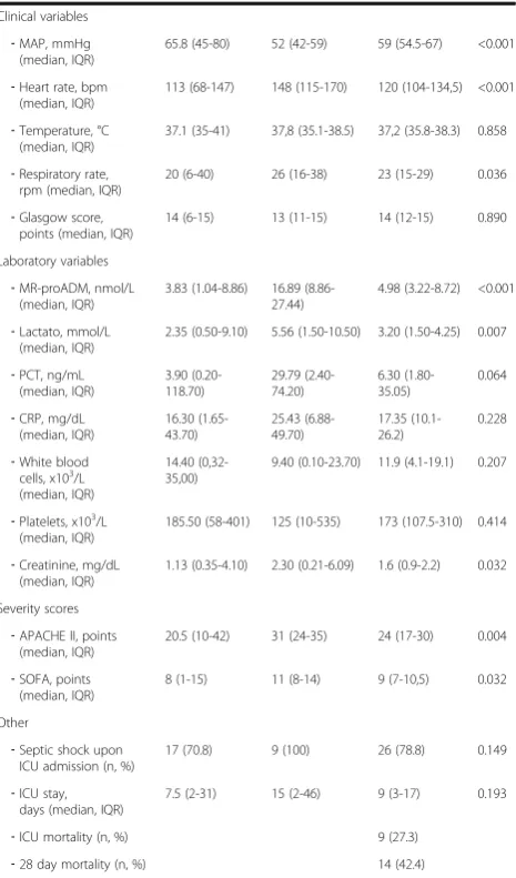
Manon Heldens1,2,3, Naomi Hammond2,3,4, Frances Bass2,3,4, Anthony Delaney2,3, Simon Finfer2,3,4
1
Radboud University, Nijmegen, The Netherlands;2Intensive Care Unit, Royal North Shore Hospital, Sydney, Australia;3University of Sydney, Sydney, Australia;4The George Institute for Global Health, Sydney, Australia
Correspondence:Manon Heldens ([email protected]) Intensive Care Medicine Experimental2017,5(Suppl 1):P12
Background
Estimates of the incidence of sepsis are often based on retrospective database or coding studies and the accuracy of these estimates in comparison with prospective cohort studies is not clear. The object-ive of this study is to compare the incidence of sepsis derobject-ived from a prospective inception cohort study (clinically diagnosed sepsis) and that derived from retrospective database (Australian and New Zealand Intensive Care Society Evaluation Centre for Outcome and Resource Evaluation (ANZICS CORE)) coding in ICU patients. Methods
Design, setting, patients: This study is an inception cohort study in-cluding all patients admitted between the 1st of October and the 31st of December 2016 at the ICU of The Royal North Shore Hospital,
St Leonards, Australia. Main outcome measures: Incidence of sepsis and septic shock using a clinical and a database definition.
Results
A total of 864 patients were admitted during the study period (Table2), of these 146 patients (16.9%) met the clinical definition of sepsis, of which 49/146 (33.6%) met the clinical definition for septic shock. A total of 98/864 patients (11.3%) met the database definition of sepsis, with 83/98 (84.7%) diagnosed with septic shock (Figs. 8 and 9). Hospital mortality was higher in patients who met the clinical definition of sepsis (39/146, 26.7%), com-pared to patients that met the database definition of sepsis (17/ 98, 17.3%).
Conclusions
Our study showed that the incidence of sepsis using the ANZICS CORE database definition is lower and the incidence of septic shock higher when compared with a prospective clinical definition. The database definition also provides a falsely low estimate of sepsis mortality.
Acknowledgement
Radboud Honours Programme 'Beyond the Frontiers'
Table 2 (abstract P12).Patient characteristics
Groupsa All
patients Clinical Sepsis No clinical sepsis Database sepsis No database sepsis Neither P valuesd
n = 864 n = 146 n = 718 n = 98 n = 766 n =
696
Male, n (%) 484
(56.0) 86 (58.9)
398 (55.2)
50 (51.0) 434 (56.7) 385 (55.3) 0.24 APACHE III score, mean (SD)b 45.0 (23.5) 62.2 (24.7) 41.4 (21.7) 59.8 (25.6) 43.1 (22.5) 41.4 (21.7) 0.44
Age in years, mean (SD) 62.1 (17.6) 64.4 (15.4) 61.6 (18.0) 64.7 (15.6) 61.8 (17.8) 61.6 (18.2) 0.89 Postoperative admissions, n (%) 380 (44.0) 19 (13.0) 361 (50.3)
12 (12.2) 368 (48.0) 354 (50.9)
0.99
Length of ICU stay in days, median (IQR)
2 (1-4) 4.5 (2-8)
2 (1-3) 3 (2-6.8) 2 (1-3) 2 (1-3) 0.02
Died in ICU, n (%)
64 (7.4) 32 (21.9)
32 (4.5) 16 (16.3) 48 (6.3) 31 (4.5) 0.33
Died in
hospital, n (%)c 83 (9.6) 39(26.7) 44 (6.1) 17 (17.3) 66 (8.6) 42 (6.0) 0.12
Died within
28 days, n (%)c 76 (8.8) 34(23.3) 42 (5.8) 14 (14.3) 62 (8.1) 41 (5.9) 0.10
Septic shock, n (%)
49 (33.6)
83 (84.7) <0.0005
•
Died in ICU, n (%)
●18
(36.7) ●
12 (14.5) 0.005
• Died in hospital, n (%)c
●18 (36.7)
●13 (15.7) 0.01
• Died within 28 days, n (%)c
●16 (32.7)
●11 (13.3) 0.013
No septic shock, n (%)
97 (66.4)
15 (15.3) <0.0005
aPatients admitted more than once were classified as sepsis if they had sepsis during any
admission and classified as not having sepsis if they did not have sepsis during any admission
bTo calculate the APACHE III score and length of stay for those who did not have
sepsis, data from the first admission is included in this table. For those who did have sepsis, data from the first admission with sepsis is included in this table
cFor mortality data all patients were followed until hospital discharge
dP values refer to the difference between the clinical sepsis group and the database
P13
Antibiotic administration and lactate level measurement timing in septic shock patients admitted in the medical ICU: hospital ward versus emergency department
Veerapong Vattanavanit1, Theerapat Buppodom2 1
Department of Internal Medicine, Prince of Songkla University, Hat Yai, Songkhla, Thailand;2Faculty of Medicine, Prince of Songkla University, Hat Yai, Songkhla, Thailand
Correspondence:Theerapat Buppodom ([email protected]) Intensive Care Medicine Experimental2017,5(Suppl 1):P13
Background
The timing of lactate level measurement and the timing of intraven-ous antibiotics administration are associated with survival of septic shock patients. Septic shock patients were admitted to medical in-tensive care unit (MICU) from 2 major sources, hospital ward and
emergency department. The differences in management on each sources will affect the lactate and intravenous antibiotics timing. Methods
The medical data of adult patients with septic shock from hospital wards and emergency department, admitted to the MICU, Songkla-nagarind hospital, during January 2015 to December 2016, were collected. Timing of antibiotic administration and lactate level meas-urement from “time zero” (time diagnosis sepsis) were recorded. The association between timing and risk-adjusted 28-day mortality are assessed.
Results
Among 150 septic shock patients admitted to medical ICU, 58.7% were from hospital ward. The median time (min) from time zero to antibiotic administration time was higher in hospital ward compared with the emergency department (290.5, interquartile range (IQR), 210 to 486 versus 122, IQR 82 to 212;P< 0.10). With the same trend of lactate level measurement timing (min) (97, IQR, 42 to 164 versus 95, IQR, 57 to 183;P= 0.37). A longer timing of antibiotic administration was associated with higher risk-adjusted 28-day mortality (odds ratio 0.99; 95% confidence interval, 0.99-0.99;P< 0.05).
Conclusions
Septic shock patients from hospital wards had longer timing of anti-biotic administration and lactate level measurement.
P14
Antibacterial and antibiofilm activity of silver nanoparticles with reducing agent extracted from Eucalyptus coating on endotracheal tube surface: phase I study in the 'PSU endotracheal tube' innovation project
Bodin Anantravanit1, Veerapong Vattanavanit1, Chalongrat Daengngam2, Supayang Voravuthikunchai3
1Department of Internal Medicine, Faculty of Medicine, Prince of Songkla
University, Hat Yai, Songkhla, Thailand;2Department of Physics, Faculty of Science, Prince of Songkla University, Hat Yai, Songkhla, Thailand; 3
Department of Microbiology, Faculty of Science, Prince of Songkla University, Hat Yai, Songkhla, Thailand
Correspondence:Bodin Anatravanit ([email protected]) Intensive Care Medicine Experimental2017,5(Suppl 1):P14
Background
Ventilator associated pneumonia (VAP) is a serious complication of mechanical ventilation that has been shown to be associated with increased medical costs and mortality. Bacterial biofilms contribute significantly to the recalcitrant nature of VAP. Commercial silver-coated endotracheal tube (Commercial ETT) had been developed but limit using due to highly cost and using metallic nanoparticles. To create and investigate antibacterial and antibiofilm efficacy of silver-coated green synthesis endotracheal tube with reducing agent ex-tracted from Eucalyptus (AgNPs ETT).
Methods
Silver nanoparticles and Eucalyptus reducing agents were pre-pared and coated on endotracheal tubes by electrostatic self-assembly technique. Three types of ETT were compared antibac-terial and antibiofilm activity; AgNPs ETT, commercial ETTs and uncoated ETTs. Staphylococcus aureus, Pseudomonas aeruginosa and Actinobacter baumanni were inoculated to form biofilm. We used modified Kirby-Bauer method to identify the diameter of growth inhibition zone (mm), optical density and biofilm forma-tion method to detect antibacterial activity and bacterial adher-ence respectively.
Fig. 8 (abstract P12).Outcome of 909 ICU admissions. The difference between the number of admissions and patients is due to readmissions
Results
AgNPs ETT was found to be most effective in inhibition ofP.aeruginosa, followed by commercial ETT and uncoated ETT (4.0 (interquartile range (IQR) 2.2 to 5.0 versus 3.5, IQR 2.6 to 4.4, P = 0.02). This effect also found onS.aureus (3.5, IQR 3.0 to 4.6 versus 3.5, IQR 2.3 to 4.0, P = 0.02) andA.baumanii(4.0, IQR 4.0 to 4.4 versus 3.0, IQR 2.6 to 3.0, P < 0.01). In biofilm formation testing, AgNPs ETT had insignificant bacterial logarithm colony forming unit (log CFU/ ml) when compared with commercial ETT but better than uncoated ETT. Conclusions
Silver-coated green synthesis ETT could inhibit bacterial growth and biofilm formation ofP. aeruginosa, S.aureusandA.baumanii in vitro. Keywords: Antibiofilm, silver nanoparticle, endotracheal tube, Eucalyptus
Acknowledgements
This study shows no conflict of interest.
P15
Ferritin heavy chain controls glucose metabolism and affords disease tolerance in sepsis
Sebastian Weis1,2, Ana Rita Carlos1, Maria Raquel Moita1, Sumnima Singh1, Birte Blankenhaus1, Silvia Cardoso1, Rasmus Larsen1, Sofia Rebelo1, Sascha Schäuble3, Laura Del Barrio1, Gilles Mithieux4, Fabienne Rajas4, Sandro Lindig2,5, Michael Bauer2,5, Miguel P. Soares1
1Instituto Gulbenkian de Ciência, Oeiras, Portugal;2Department of
Anesthesiology and Intensive Care Medicine, Jena University Hospital, Jena, Germany3Language & Information Engineering Laboratory, Friedrich-Schiller-University, Jena, Germany;4INSERM U1213, Université Claude Bernard Lyon, France, 5Center for Sepsis Control and Care, Jena University Hospital, Jena, Germany
Correspondence:Sebastian Weis ([email protected]) Intensive Care Medicine Experimental2017,5(Suppl 1):P15
Background
Sepsis results from maladaptive immune and metabolic responses to infection compromising host homeostasis[1, 2]. Infected organisms use 2 distinct defense mechanisms against infections, i.e. resistance and dis-ease tolerance [3]. Resistance mechanisms aim at reducing the number of pathogens. Disease tolerance is a preserves host homeostasis, with-out exerting a direct negative impact on pathogens[3]. Instead, it relies on tissue damage control that provide metabolic adaptation and pre-serves the functional output of parenchymal tissues, maintaining homeostatic parameters within a dynamic range compatible with host survival[4]. Previously, it was shown that expression of the gene encod-ing the heavy/heart subunit of the iron sequesterencod-ing molecule ferritin (Fth) is essential for the establishment of disease tolerance in malaria [5] as well as to viral and fungal infections in plants[6]. We hypothe-sized that expression of Fth protects against the deleterious outcome of bacterial sepsis via affording disease tolerance to infection. Methods
We applied genetic loss-of function studies in different knock-out vs. control mice (Mx1CreFthΔ/Δ, AlbCreFthΔ/Δ,LyzMCreFthΔ/Δ, AlbCreERT2G6Pc1Δ/Δ, Tlr4-/- and Ifng-/-) as well as adenovirus-mediated gene overexpresion in C57B6 mice. Mice underwent low- and high grade cecal ligation and punc-ture (CLP) or peritoneal contamination and infection (PCI) or were adminis-tered heme. Survival, disease severity, gene expression, cytokine content, cardio-vascular function, organ damage/dysfunction were assessed. Tran-scriptomics and hepatic metabolomics performed and integrated in a flux-variance analysis.
Results
Here, we provide experimental evidence that deletion of Fth in sepsis is deleterious and that expression of Fth in the liver is critical to con-trol glucose metabolism and establish disease tolerance to sepsis. Deletion of Fth in sepsis does not cause overt organ damage but in-stead results in failed metabolic adaptation and increased glycolysis, i.e. a parenchymal Warburg effect. The protective effect acts via a mechanism that counters iron-driven oxidative inhibition of the liver Glucose-6-Phosphatase (G6Pase). This is required to prevent the de-velopment of septic hypoglycemia that would compromise disease tolerance to sepsis, hence overcoming its lethal outcome. In addition, application of apoferritin reduces sepsis mortality via enhancing dis-ease tolerance infection even when given 6 hours after infection.
Conclusions
Disease tolerance to sepsis relies on a cross talk between host iron and glucose adaptive metabolic responses. Ferritin sustains endogen-ous hepatic gluconeogenesis. This maintains blood glucose levels in response to infection. The described pathways can be targeted pharmacologically providing a novel therapeutic approach against sepsis that acts independently of pathogens.
Acknowledgements
Grant: DFG WE 4971/3 to SW. SFRH/BPD/101608/2014 to ARC. GlioPATH (grant FKZ 01ZX1402C) to SAS. BMBF; grant 01EO1002 and Meta-ZIK (03Z2J52) to MB. e:Med initiative ERC-2011-AdG 294709-DAMAGECONTROL, PTDC/SAU-TOX/116627/2010, HMSP-ICT/0022/2010 to MPS.
The full manuscript has been published: Metabolic Adaptation Establishes Disease Tolerance to Sepsis.
Weis S, Carlos AR, Moita MR, Singh S, Blankenhaus B, Cardoso S, Larsen R, Rebelo S, Schäuble S, Del Barrio L, Mithieux G, Rajas F, Lindig S, Bauer M, Soares MP.
Cell. 2017 Jun 15;169(7):1263-1275.e14.
References
1. Singer M, Deutschman CS, Seymour CW, Shankar-Hari M, Annane D, Bauer M, Bellomo R, Bernard GR, Chiche JD, Coopersmith CM et al: The Third International Consensus Definitions for Sepsis and Septic Shock (Sepsis-3). Jama 2016, 315(8):801-810.
2. Kotas ME, Medzhitov R: Homeostasis, Inflammation, and Disease Susceptibility. Cell 2015, 160(5):816-827.
3. Medzhitov R, Schneider DS, Soares MP: Disease Tolerance as a Defense Strategy. Science 2012, 335:936-941.
4. Soares MP, Gozzelino R, Weis S: Tissue damage control in disease tolerance. Trends in immunology 2014, 35(10):483-494.
5. Gozzelino R, Soares MP: Coupling heme and iron metabolism via ferritin H chain. Antioxid Redox Signal 2013, 20(11):1754-1769.
6. Deak M, Horvath GV, Davletova S, Torok K, Sass L, Vass I, Barna B, Kiraly Z, Dudits D: Plants ectopically expressing the iron-binding protein, ferritin, are tolerant to oxidative damage and pathogens. Nature biotechnology 1999, 17(2):192-196.
P16
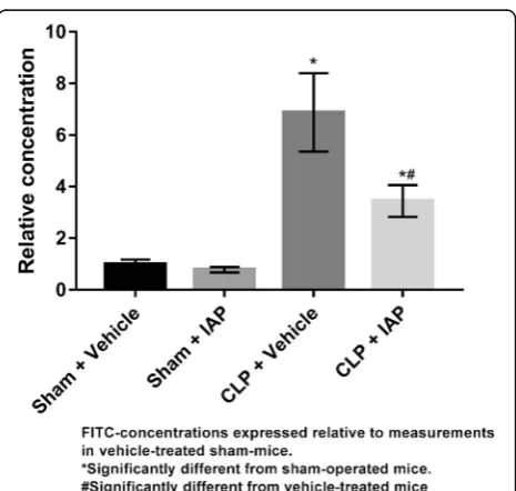
Intestinal Alkaline Phosphatase (IAP) enhances the gastrointestinal barrier function and modulates tight junction expression in an experimental model of polymicrobial sepsis
Philip Plaeke1, Joris G De Man1, Annemieke Smet1, Hanne Van Spaendonk1, Sara Nullens1, Philippe G Jorens1,2, Guy Hubens3,4, Benedicte Y De Winter1
1Laboratory of Experimental Medicine and Paediatrics, University of
Antwerp, Wilrijk, Belgium;2Department of Intensive Care Medicine, Antwerp University Hospital, Edegem, Belgium;3Antwerp Surgical Training, Anatomy and Research Centre, University of Antwerp, Wilrijk, Belgium;4Department of Abdominal Surgery, Antwerp University Hospital, Edegem, Belgium
Correspondence:Philip Plaeke ([email protected]) Intensive Care Medicine Experimental2017,5(Suppl 1):P16
Background
Sepsis is a severe condition characterized by the activation of a sys-temic and intestinal inflammatory cascade and mucosal barrier dys-function. Intestinal alkaline phosphatase (IAP), an endogenously expressed, brush-border-bound enzyme is ubiquitously produced in the mucosa of the small intestine. IAP was demonstrated to detoxify several proinflammatory molecules (e.g. LPS, UDP) and is presumed to play a role in the maintenance of the healthy intestinal microbiome. Additionally, recent clinical trials established a nephroprotective func-tion of IAP when administered systemically in septic patients. This study aimed to investigate the effects of intraperitoneally administered IAP on the intestinal barrier function of the small intestine in a cecal ligation and puncture (CLP)-model for sepsis.
Methods
first day, mice received an intraperitoneal (ip) injection of calf IAP (1 IU/g mouse, New England Biolabs) or saline (vehicle) 5 min. prior to a CLP-procedure (50% ligation, single 21G punc-ture) or a sham-laparotomy. Subsequently, ip injections of IAP or the vehicle were repeated twice a day. Analgesia and fluid resuscitation were regularly provided. A validated clinical dis-ease score [1] and the body weight were monitored daily. On the 2nd postoperative day, the abdomen was reopened, 100μl of 4 kDa FITC-Dextran was injected directly in the distally li-gated ileum and quantified one hour later by fluorescence-spectrophotometry to measure intestinal permeability. Total RNA was extracted from an ileal bowel specimen and the expression of cytokine and tight junction genes was determined by RT-PCR.
Results
Septic mice had significantly worse clinical disease scores, lost more weight and had increased intestinal permeability com-pared to their sham-operated counterparts. After the CLP-procedure, gene expression of IL1β, IL6, Tnfα and Cldn14 were significantly elevated in both vehicle- and IAP-treated mice (Table3).
Similarly, Occludin and Cldn2 gene expression decreased signifi-cantly. IAP-treatment did not result in better clinical outcomes. Nevertheless, IAP did significantly reduce the observed disturbances in intestinal permeability in septic animals (Fig.10). Additionally, IAP-therapy significantly elevated the expression levels of Cldn1 and Cldn14.
Conclusion
In mice, CLP-induced sepsis is associated with enhanced intestinal permeability, modified expression of intestinal tight junctions and increased expression of ileal inflammatory markers. Treatment of septic mice with IAP significantly lowered the permeability of the intestinal barrier at the small bowel and elevated expression levels of Cldn1 and Cldn14. We expect this increase to be linked to the observed improvements of the intestinal barrier function after IAP-treatment.
Reference
1. Heylen M.: Colonoscopy andμPET/CT are Valid Techniques to Monitor Inflammation in the Adoptive Transfer Colitis Model in Mice. Inflamm Bowel Dis. 2013, 19:967-97.
P17
Possible mechanisms of dying of septic patients associated with aromatic microbial metabolites
NV Beloborodova1, NI Fedotcheva2, VV Teplova2, AK Pautova1 1Negovsky Research Institute of General Reanimatology, Federal
Research and Clinical Center of Intensive Care Medicine and Rehabilitology, Petrovka, 25/2, Moscow, 107031, Russia;2Institute of Theoretical and Experimental Biophysics, Russian Academy of Sciences, Institutskaya 3, Pushchino, Moscow region, 142290, Russia
Correspondence:NV Beloborodova ([email protected]) Intensive Care Medicine Experimental2017,5(Suppl 1):P17
Background
The main causes of death in ICU are multiple organ failure (MOF) associated with sepsis and refractory septic shock[1]. Concept of mitochondrial dysfunction during sepsis is not new[2], but it remains unclear what role bacteria play. Our experimental approaches have revealed the influence of aromatic microbial metabolites (AMM) on some mitochondrial function[3]. We want to show how AMM is asso-ciated with mortality rates through the development of MOF and septic shock.
Methods
The study included adult non-survivors patients (N= 91), from which serum samples were taken and frozen for one or two days before the death. Serum samples from 20 donors and 24 survivors of sepsis were as comparison groups. The main equipment: GH-MS (Thermo Scientific) and GH-FID. All chemicals were from Sigma-Aldrich (US). Results
One day before death in 100% of cases only three AMM were increased, while other biochemical parameters of organs dysfunction in 9-40% of cases did not exceed normal values (Table4). In the dynamics the closer the moment of death the increase of PhLA, HPhAA, p-HPhLA were more significant than the growth of other routine parame-ters and severity scales (Table5).
The serum levels of the mitochondrial metabolites - tricarboxylic acid cycle (TCA) were measured simultaneously with AMM. More than half of septic patients showed a multiple increase in succinic and fumaric acids, also the appearance in the blood of the alpha-ketoglutarate. This can be interpreted as a inhibition of the TCA. Correlations of mitochondrial metabolites (fumaric acid) with AMM (r= 0.68,p<0.01) and among themselves (fumaric with succinic) (r= 0.74, p <0.01) Table 3 (abstract P16).See text for description
Sham + Vehicle Sham + IAP CLP + Vehicle CLP + IAP
Clinical follow-up
Clinical disease score 0.00 ± 0.00 0.08 ± 0.08 5.17 ± 0.32* 4.25 ± 0.36*
Baseline Weight (g) 36.24 ± 1.15 35.04 ± 1.40 36.92 ± 1.41 36.39 ± 1.32
Weight loss (%) day 2 5.35 ± 1.01 2.65 ± 1.43 10.37 ± 0.70* 10.55 ± 0.92*
Permeability
FITC-Dextran Concentration (1)
1.00 ± 0.17 0.79 ± 0.11 6.88 ± 1.51* 3.45 ± 0.62*#
PCR-results
IL1β 1.19 ± 0.19 1.15 ± 0.23 10.28 ± 3.73* 7.45 ± 2.35*
IL6 1.27 ± 0.27 1.36 ± 0.29 8.84 ± 3.26* 5.68 ± 1.56*
TNFα 1.17 ± 0.19 1.17 ± 0.13 2.03 ± 0.30* 2.85 ± 0.47*
Claudin-1 (Cldn1) 1.27 ± 0.16 1.55 ± 0.23# 1.34 ± 0.28 2.42 ± 0.43#
Claudin-2 (Cldn2) 1.14 ± 0.14 1.17 ± 0.12 0.79 ± 0.08* 0.84 ± 0.12*
Claudin-14 (Cldn14) 1.13 ± 0.24 2.37 ± 0.84# 2.53 ± 0.43* 5.28 ± 1.56*#
Occludin 1.22 ± 0.17 1.26 ± 0.19 0.68 ± 0.10* 0.69 ± 0.10*
Results are expressed as mean ± SEM (Standard error of mean) (1) Relative FITC-concentration to the FITC-levels measured in vehicle-treated sham-mice
* Significantly different from sham-operated mice # Significantly different from vehicle-treated mice
Statistics–Two Way Analysis of Variance with LSD Post-Hoc analysis when appropriate, a p-value of 0.05 was considered statistically significant
were noted. Direct correlation (r= 0.59, p< 0.01) between ∑3AMM and presence of arterial hypotension was found. Only in septic shock patients but never in comparison groups the intermediate (DOPAC) and final (HVA) product of a minor pathway of tyrosine were detected in serum (Fig. 11). The microbial origin of these metabolites[4, 5] and their ability to inhibit the tyrosine hydroxy-lase are known[6, 7]. The correlation between ∑3AMM and HVA reached a high rate, r = 0.76.
Conclusions
AMM can be involved in dying of sepsis: 1) due the development of the MOF via the inhibition of TCA metabolism in the mitochondria and 2) through the mechanism of septic shock by inhibiting of the metabolic pathway of catecholamine synthesis, particularly tyrosine hydroxylase. The search in this direction will help to identify new therapeutic targets to improve the survival in sepsis.
Acknowledgements
Supported by Russian Science Foundation Grant№15-15-00110.
References
1. Vincent JL, Nelson DR, Williams MD. Is worsening multiple organ failure the cause of death in patients with severe sepsis? Crit Care Med. 2011;39(5):1050–1055. doi:10.1097/CCM.0b013e31820eda29
2. Singer M. The role of mitochondrial dysfunction in sepsis-induced multi-organ failure. Virulence. 2014 Jan 1; 5(1): 66–72. PMCID: PMC3916385 DOI: 10.4161/viru.26907
3. Beloborodova N.V., Teplova V.V., Fedotcheva N.I. Monograph. “The role of microbial metabolites in mitochondrial dysfunction in sepsis” LAP Lambert Academic Publishing, Germany, 2013, 89 p. ISBN: 978-3-659-43111-1
4. Martin M, Gibello A, Fernández J, Ferrer E, Garrido-Pertierra A. Catabolism of 3- and 4-hydroxyphenylacetic acid by Klebsiella pneumoniae. 1991 Mar;137(3):621-8.
5. Sparnins VL, Chapman PJ, Dagley S. Bacterial degradation of 4-hydroxyphenylacetic acid and homoprotocatechuic acid. J Bacteriol. 1974 Oct; 120(1):159-67
6. Laschinski, G., Kittner, B., Brautigam, M., 1986. Direct inhibition of tyrosine hydroxylase from PC12 cells by catechol derivaties.
Naunyn-Schmiedeberg’s Arch. Pharmacol. 3324, 346–350
7. Rong-Sen Shen. Potent inhibitory effects of tyrosine metabolites on dihy-dropteridine reductase from human and sheep liver. 1984, Biochimica et Biophysica Acta (BBA) - Protein Structure and Molecular Enzymology, 785, 3, 181-185
P18
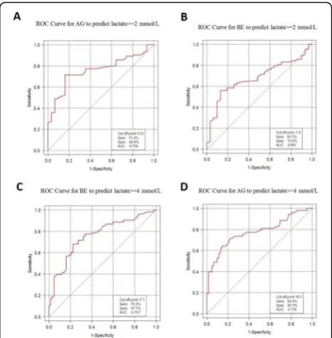
Can base excess and anion gap predict lactate level in diagnosis of septic shock?
Werapon Pongmanee1, Veerapong Vattanavanit1 1
Department of Internal Medicine, Faculty of Medicine, Prince of Songkla University, Hat Yai, Songkhla, Thailand
Correspondence:Werapon Pongmanee ([email protected])
Intensive Care Medicine Experimental2017,5(Suppl 1):P18
Background
Lactate measurement is the key component in septic shock identifica-tion and resuscitaidentifica-tion[1]. However, point-of-care lactate testing is not widely used due to lack of access. Biomarkers such as serum lactate, anion gap (AG), and base excess (BE) have been shown to be of use in determining shock in patients with seemingly normal vital signs. Objective
To determine if these biomarkers can be used interchangeably in pa-tients with septic shock in the emergency setting based on their test characteristics and correlation to each other.
Methods
A prospective observational cohort study was undertaken at a ter-tiary hospital in southern Thailand. Baseline point-of-care BE, AG, and serum lactate were recorded in all patients who presented with septic shock at the emergency department. Overall correlations in-cluding area under the receiver operating characteristic curve (AUROC) for both BE and AG to predict serum lactate level were calculated.
Table 4 (abstract P17).The frequency and severity of changes in the levels of aromatic microbial metabolites (p-HPhAA, PhLA, p-HPhLA) in comparison with other biochemical parameters of organ dysfunctions in septic patients in point of «the last day before death», median (IR 25-75%)
Parameter Donors, n = 20
Pts, the last day before death, n = 91
The incidence of abnormalities, %
a
p-HPhAA,μM 0.87–2.53 24.11 (14.16–53.77) 100
b
PhLA,μM 0.27–1.04 7.23 (5.10–17.00) 99
c
p-HPhLA,μM 0.50–2.27 23.22 (10.62–65.30) 98
∑3AMM,μM 1.80–5.15 80.41 (39.78–143.67) 100 lactate,мM 0.5–2.2 5.6 (3.3–10.0) 91 urea,мM 2.5–8.3 22.3 (14.7–32.2) 90 creatinine,μM 44–110 257 (173–348) 88 total bilirubin,мM <18.8 22.1 (14,0–32.1) 60
a
p-HPhAAp-hydroxyphenylacetic acid, b
PhLAphenyllactic acid, c
p-HPhLAp-hydroxyphenyllactic acid
Table 5 (abstract P17).The increase in MODS score and some metabolites (median, IR 25-75%) in septic patients during the last 48 hours before death (n = 34)
Parameter Time to death (retrospective analysis) the multiplicity of changes, Me
p=
48-25 hours 24-0 hours MODS1
5 (3–7) 9 (6–10) 1,8 <0.001 Lactate, mM 3.6 (2.4–5.1) 4.2 (3.0–10.6) 1,2 0.03 PhLA,μM 4.27 (2.14–9.33) 7.97 (5.07–17.90) 1,9 <0.001 p-HPhAA,μM 10.45 (1.99–25.81) 20.14 (7.27–65.49) 1,9 <0.001 p-HPhLA,μM 9.14 (5.73–25.51) 22.06 (13.64–67.04) 2,4 <0.001
∑3 AMM,μM 33.47 (12.40–63.98) 78.02 (38.92–156.26) 2,3 <0.001
1
MODS - Multiple Organ Dysfunction Score
Results
One hundred fifteen patients were enrolled. The median age was 66 years (interquartile range, 52-80 years), and 61% were male. About thirty-five percent of septic shock were pneumonia. Pearson correlation of serum lactate to BE was -0.59 (r2 = 0.35; 95% confi-dence interval [CI], -0.69 to -0.44;P< .001), that of BE to AG was -0.67 (r2 = 0.49; 95% CI, -0.76 to -0.55;P< .001), and that for serum lactate to AG was 0.64 (r2 = 0.41; 95% CI, 0.52-0.74;P< .001) (Fig.12). A cut-off point of AG 15.8 identified lactate level≥2 mmol/L (sensitivity 71.4%, specificity 80.7%, AUROC 0.76) and the best cutoff values to predict lactate level≥4 mmol/L were 18.5 (sensitivity 64.2%, specifi-city 85.5%, AUROC 0.78) (Fig.13).
Conclusions
In septic shock patients, lactate and AG showed strong correl-ation with each other; while, lactate and BE showed moderate correlation with each other. Thus, these biomarkers can be used interchangeably to help earlier determination of patients with septic shock.
Keywords: base excess, anion gap, lactate, septic shock
Acknowledgements
This study was fully funded by the Faculty of Medicine, Prince of Songkla University.
The authors report no conflicts of interest in this work.
Reference
1. Singer M, Deutschman CS, Seymour CW, Shankar-Hari M, Annane D, Bauer M, et al. The Third International Consensus Definitions for Sepsis and Septic Shock (Sepsis-3). Jama. 2016;315(8):801-10.
P19
Cholecystokinin modulates the colonic inflammatory response and prevents epithelial barrier dysfunction in endotoxemic rats Aline B. Ribeiro1, Humberto Giusti1, Rafael S. Saia1
1
Department of Physiology, Ribeirão Preto Medical School, University of São Paulo, Ribeirão Preto, São Paulo, Brazil
Correspondence:Rafael Simone Saia ([email protected]) Intensive Care Medicine Experimental2017,5(Suppl 1):P19
Background
The acute phase of sepsis is characterized by the exacerbated production of inflammatory mediators (chemokines, cytokines and nitric oxide). Especially the cytokines may contribute to the intestinal permeability, bacterial translocation and tight junction rearrange-ment. Cholecystokinin (CCK) was firstly described as a gastrointestinal hormone; however our research group has demonstrated an anti-inflammatory involvement of this peptide in experimental models of endotoxemia and bacterial infection. Our aim was to determine the possible role of CCK on lipopolysaccharide (LPS)-induced intestinal epithelial barrier dysfunction. Furthermore, we evaluated its ability to modulate the mucosal inflammatory response and also the expres-sion of proteins of the intestinal tight junctions.
Methods
To conduct these experiments, male rats had their jugular vein cannulated for endotoxin administration, one day before the experiment. Rats were pre-treated with CCK (0.4 or 40μg/kg, intravenously) before LPS administration (1.5 mg/kg, intravenously). At 4 h after endotoxemia induction, the intestinal permeability was evaluated by injecting FITC-dextran 4 kDa in the ileum and colon; mesenteric lymph nodes were col-lected for microbiological analysis and also; cytokines were quantified in the plasma and intestinal mucosa by ELISA technique. Additionally, the integrity of the tight junctions was determined by the expression of its constituents proteins (occludin, claudin-1, junctional adhesion molecule-A) and the histological damages were evaluated in tissue sections. Results
Our results demonstrated that CCK administration reduces the permeabil-ity only in the colon and prevented the bacterial translocation to the mesenteric lymph nodes. The levels of pro-inflammatory mediators TNF-α, IL-1β, IL-6 and IFN-γwere reduced in CCK-treated rats, being in the co-lonic mucosa the most significant effect. Furthermore, treatment with Fig. 12 (abstract P18).A-E, Correlation of (serum lactate to BE, AG,
correct AG) and (BE to AG, correct AG)