REVIEW
The nanomaterial toolkit
for neuroengineering
Shreyas Shah
*Abstract
There is a growing interest in developing effective tools to better probe the central nervous system (CNS), to under-stand how it works and to treat neural diseases, injuries and cancer. The intrinsic complexity of the CNS has made this a challenging task for decades. Yet, with the extraordinary recent advances in nanotechnology and nanoscience, there is a general consensus on the immense value and potential of nanoscale tools for engineering neural systems. In this review, an overview of specialized nanomaterials which have proven to be the most effective tools in neuroscience is provided. After a brief background on the prominent challenges in the field, a variety of organic and inorganic-based nanomaterials are described, with particular emphasis on the distinctive properties that make them versatile and highly suitable in the context of the CNS. Building on this robust nano-inspired foundation, the rational design and application of nanomaterials can enable the generation of new methodologies to greatly advance the neuroscience frontier.
Keywords: Nanomaterials, Nanoparticles, Neuroscience, Neuroengineering, Stem cells, Regenerative medicine, Drug delivery, Optogenetics
© 2016 The Author(s). This article is distributed under the terms of the Creative Commons Attribution 4.0 International License (http://creativecommons.org/licenses/by/4.0/), which permits unrestricted use, distribution, and reproduction in any medium, provided you give appropriate credit to the original author(s) and the source, provide a link to the Creative Commons license, and indicate if changes were made.
1 Background
The fields of biology and medicine have heavily relied on advances in technology to better understand how the human body works. These advances range from the creation of simple tools to conduct surgery (e.g. scalpel), devices to measure physiological levels (e.g. electrocar-diograph) and instrumentation to image the body in real time (e.g. fMRI). Besides studying the human physiol-ogy, these types of technological advances have further enhanced our capabilities to diagnose, prevent and even treat medical ailments such as disease, cancer and trau-matic injuries. In general, the continual development of precision tools has enabled scientists and clinicians to acquire a remarkable breadth of knowledge about bio-logical systems.
Among other disciplines, the field of neuroscience has greatly benefited from such advances. Neuroscientists have long strived to acquire a complete understanding of how the nervous system works. Early work involved
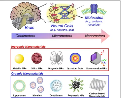
investigating the bulk anatomical makeup of the brain, primarily through dissecting human cadavers. Taking the human brain as an example, it is organized into dis-tinct lobes within the centimeter range (Fig. 1). The lobes were recognized to correspond to specific physiological functions, whether it be processing sensations of touch (parietal lobe) or controlling body movement (frontal lobe). Thereafter, the discovery of the role of electric-ity in nerve signaling, along with the development of microscopy, allowed scientists to go even deeper to the micrometer scale of neurons and glia cells. Millions of neurons in the distinct regions of the brain, are organ-ized into ensembles or circuits, which serve to process and carry information throughout the nervous system. Going even deeper to the nanometer scale, the distinct neural cells are composed of numerous biomolecules and receptors on the surface membrane, which enable multi-directional interactions with the surrounding microenvi-ronment. There is growing interest in the miniaturization of tools to better control and understand neural systems at this fundamental scale [1]. Biological systems function with extraordinary fidelity at the molecular level, which permits robust structure and function at the cellular,
Open Access
*Correspondence: [email protected]
tissue and organ level. The ability to probe systems like the nervous system at the fundamental resolution that they have naturally evolved to function is revolutionary. Nanotechnology has enabled the design of materials and devices to do just that. It has enhanced our understand-ing of how biological systems work at the nanoscale, and further allowed the development of nanoscale tools to improve the quality of life after disease or injury [2]. The integration of engineered materials with intact biologi-cal systems has proven to be highly transformative and is possible due to the growing understanding of nanosci-ence and nanotechnology.
To this end, nanoscale materials hold immense poten-tial for engineering neural systems. The ability to pre-cisely tailor the properties of nanomaterials permits scientists and clinicians to effectively employ them for a
wide variety of applications. In this review, we will first explore the current challenges and existing approaches in the field of neuroscience. We will then see how nano-materials can be utilized to address these challenges, with a focus on the distinctive properties that make them highly suitable tools for advancing neuroscience research (Fig. 1).
2 Current challenges and approaches in neuroscience
of two key cell types: neurons and glia. Neurons serve to process changes in the environment, communicate the changes throughout the system and direct the body’s response to such changes. Glia are the supportive and most abundant cells of the nervous system which help support neighboring neurons and maintain homeosta-sis. The intricate network of billions of these neural cells, presumably organized in defined arrangements to impart specific neural activity, gives rise to thoughts, feelings, memories and life as we know it. In this section, we will look at three key areas of active CNS research: (a) neuro-regenerative therapies, (b) delivery of therapeutics, and (c) neuromodulation.
2.1 Neuro‑regenerative therapies
The CNS is very sensitive to damage, including infection, hypoxia, stroke, neurodegenerative diseases, and injury. The inevitable loss of neural cells makes this particu-larly devastating, since it leads to debilitating motor and cognitive impairment. For example, Parkinson’s disease (PD) results in the gradual loss of midbrain neurons in the substantia nigra which synthesize the neurotrans-mitter dopamine (DA), leading to rigidity and tremors [3]. Like neurodegenerative diseases, traumatic injuries can cause the loss of neural cells, in addition to complex microenvironments as the injury progresses from acute to chronic stages [4]. If kept untreated, a series of dam-aging conditions continue to accumulate, resulting in continued degeneration and dysfunction [5]. Due to the limited regenerative capabilities of the CNS, the loss of nervous tissue is extremely detrimental. To this end, cel-lular-based therapies have emerged as a promising route of therapy for CNS-related diseases and injuries [6]. The rationale is simple: replace the lost cells with new cells, in order to restore function. Cell transplantation started with clinical trials in patients with PD, in which trans-plantation of human fetal mesencephalic tissue rich in dopaminergic neurons was found to normalize dopamine release and reverse impairment in cortical activation [7, 8]. However, such an approach relies on the availabil-ity of donor tissue, making it an impractical long-term solution.
In this context, stem cell-based therapies have gained tremendous attention for neural disorders [9]. Stem cells are particularly suitable since they have the innate capability to self-renew, serving as a renewable source of transplantable cells that can be routinely expanded. At the same time, stem cells can differentiate into vari-ous cellular lineages, allowing for the generation of spe-cific neural cell types of interest. Various types of stem cells, including embryonic stem cells (ESCs), induced pluripotent stem cells (iPSCs), fetal neural stem cells and mesenchymal stem cells (MSCs), have proven to be
therapeutically beneficial after transplanting into dam-aged neural systems [10–12]. While cell transplantation initially started as an approach for cell replacement, the transplanted stem cells have been observed in recent years to imbue a number of favorable therapeutic effects in the CNS recovery process. This includes a decrease in inflammation, neuro-protection, remyelination, produc-tion of neurotrophic factors to enhance axonal regen-eration and the enhancement of endogenous recovery processes [13].
Nevertheless, an active area of current research lies in achieving reproducible control of stem cell differentia-tion towards a pure, defined neural cell populadifferentia-tions. The uncontrolled stem cell growth or differentiation after transplantation (e.g. teratoma formation from pluripo-tent cells [14]) is clearly unacceptable for clinical appli-cations. Moreover, in vitro differentiation protocols tend to be fairly lengthy and complex; the general biologist’s approach tends to require supplementing a number of chemical compounds, biological factors or viral gene vec-tors, which can lead to high variability between experi-ments. Another common problem is the limited survival of transplanted cells and poor interaction with host tis-sue. For this reason, long-term viability and integration are critical factors to consider when it comes to re-estab-lishing the damaged neuronal circuitry [13]. At the same time, the in vivo CNS microenvironment can be highly heterogeneous, with major fluctuations at the molecular and cellular level especially in the damaged site. In turn, achieving spatiotemporal control of stem cell behavior and differentiation after transplantation is quite challeng-ing [15]. Engineering how the cell interacts with the sur-rounding environment is therefore critical when it comes to advancing stem cell-based neuro-regenerative thera-pies [16].
2.2 Delivery of therapeutics to the CNS
including paclitaxel and temozolomide [20]. Biopharma-ceutics have also become attractive for CNS therapies. These include peptides, recombinant proteins, enzymes, monoclonal antibodies, and gene vectors. Compared to small molecule drugs, this class of therapeutics tends to have higher specificity and potency [21]. For instance, genomic sequencing and bioinformatics approaches have identified therapeutic targets for GBM that can be tar-geted with viral vectors and microRNAs [22]. In another example, in vivo administration of antibody inhibitors targeting β-secretase and α-synuclein were found to reduce amyloid-β concentrations [23] and α-synuclein aggregates [24], respectively, for treating dementia.
While multitudes of therapeutics exist for treatment, delivery to the CNS has proven to be challenging. Intrac-erebroventricular injection is one direct delivery option, wherein therapeutics are injected directly into the cer-ebral lateral ventricles [25]. However, such a strategy is highly invasive and not a feasible option for therapies requiring frequent injections. Intrathecal administra-tion via cerebrospinal routes is also popular and gener-ally favorable, but the restricted diffusion in the brain compared to the blood is a limiting factor [21]. These challenges arise from the fact that the CNS is highly-protected and dynamically-regulated by key physical bar-riers, which prevent the invasion of foreign or unwanted substances. While favorable for maintaining homeo-stasis, it is a critical obstacle for the systemic delivery of therapeutic agents. The blood–brain barrier (BBB) is the primary barrier protecting the CNS, consisting of a layered structure composed of endothelial cells, the cap-illary basement membrane, pericytes and astrocyte foot processes [26]. The tight junctions formed between the endothelial cells permits the free diffusion of small mol-ecules, such as oxygen, carbon dioxide and water, but highly limits the movement of large molecules including most therapeutics [27].
Even though systemic delivery is limited by the BBB, targeted therapies have been developed to enhance the permeability across the BBB by: modifications of the drug, temporary disruption of the BBB using chemical or physical perturbation, catheter-based interstitial deliv-ery or drug-eluting reservoir systems [28]. Depending on the approach, there are multiple design considerations to take into account. The first is the type of therapeutic that is to be delivered. Given that different compounds have varying chemical, physical and biological proper-ties (e.g. small molecules versus antibody versus RNA molecule), the stability and formulation must be main-tained for maximum efficacy. Second, sufficient dosing must be achieved to stay within the therapeutic window. The amount of drug administered versus the actual drug that reaches the target can significantly differ due to
fluctuations in pharmacokinetics. This may thus require higher effective doses, which can be lethal, expensive, and compromise patient compliance. Balancing these considerations based on the neurological disorder to be treated is therefore essential for enhancing therapeutic delivery to the CNS.
2.3 Neuromodulation
Mapping the neural circuitry of the brain is currently a major initiative for neuroscientists worldwide [29]. Determining the specific organization of billions of neu-rons, interconnected via trillions of synapses, is funda-mental to unlocking how the CNS processes information to coordinate neural activity, cognition and behavior [30]. In this regard, there is a general consensus on the need for tools to better interface with the nervous system to enable the measurement and manipulation of neural signaling.
Electrodes are commonly used to record and stimulate neural activity. The most basic system is an electrolyte-filled micropipette, which is still employed for in vitro electrophysiology experiments to measure changes in current and/or potential of neurons [31]. By further modifying the physical dimensions, electrodes have been placed into mammalian brains for local neural stimu-lation and recording as well. One such example is deep brain stimulation, in which electrodes are implanted and stimulated near the internal globus pallidus and sub-thalamic nucleus to treat PD patients [32]. In order to acquire multipoint readings, microfabrication techniques and MicroElectroMechanical Systems (MEMS) have also been widely used to generate micron-scale multielec-trode arrays [33]. For electrode compositions in general, a number of different metals have been explored, includ-ing gold, platinum, steel and iridium oxide [34]. However, metallic electrodes tend to be mechanically hard (50– 500 GPa) compared to soft nervous tissue (0.1–1 kPa), which causes neural damage and incurs an inflammatory response after insertion [35]. Yet, electrodes must also be brought in close proximity to the target region for both effective stimulation and measurement. At the same time, long-term implantation further causes a chronic inflammatory response, leading to gliosis near the surface of the electrode and thus reducing signal transduction due to the increase in the impedance [36].
the expression of visible light-activated cation chan-nels from algal species, such as the 470-nm blue-light responsive channelrhodopsin-2 from Chlamydomonas reinhardtii (ChR2) or the 535-nm green-light responsive channelrhodopsin-1 from Volvox carteri (VChR1), into mammalian neurons were found to transduce trains of millisecond-duration light flashes into time-locked depo-larizations [37]. On the other hand, chloride-pumping halorhodopsin from Natronomonas pharaonis (NpHR) can hyperpolarize and thus inhibit neuronal firing using yellow-light (589 nm) [38]. Molecular engineering tech-niques to modify these microbial proteins and encode them in viral vectors has allowed for the introduction of these opsin genes into mammalian cells, with the first in vitro demonstration using mammalian neurons in 2005 [39]. By 2007, the first in vivo demonstration which linked optically-manipulated neural activity with spe-cific behavioral changes in freely-moving mammals was reported [40]. By combining the spatiotemporal resolu-tion of optical hardware with the genetic manipularesolu-tion of specific cell types, optogenetics has allowed for the pre-cise control of neural activity in select regions of the brain [41]. In other words, exposure of genetically-manipulated neuronal cells to light has facilitated the ability to modu-late neural activity at a timescale relevant to brain func-tion. Over the last decade, optogenetic techniques have elucidated neuronal circuits of numerous neural-related states and disorders including fear and anxiety, addiction, depression, reward-seeking, schizophrenia and PD [42]. While optogenetics is continuing to enable novel studies that were previously impossible, a number of fundamen-tal limitations exist, including lack of deep tissue penetra-tion using convenpenetra-tional visible light sources, the need for invasive surgeries to deliver light, and difficulties in tar-geting deeper brain regions [43]. A completely non-inva-sive approach for neural modulation would be ideal, but it may prove to be difficult due the lack of precise control in mapping or stimulating specific regions of the brain without intervention.
Regardless of the approach employed to modulate neu-ral activity, a clear consideration for future development is to reduce invasiveness while achieving maximal qual-ity of signal recording or stimulation. The key will be to design materials that offer optimal interfacing with intact nervous tissue, both in regard to structural (i.e. mechani-cal) and surface (i.e. chemical, physimechani-cal) properties.
3 Nanomaterials for neuroengineering
Nanomaterials have a number of unique properties that make them attractive for addressing the abovemen-tioned challenges. For instance, the small size (below 1 micron) enables facile delivery throughout the body and into cells by crossing the plasma membrane [44]. While
different cell types may have a different composition of lipids and proteins in the plasma membrane, nanoma-terials cross the plasma membrane and are internalized in a size-dependent manner via endocytosis pathways, such as clathrin-mediated endocytosis, caveolae-medi-ated endocytosis, or phagocytosis [45]. Moreover, the surface chemistry of the nanomaterial also plays a defin-ing role, wherein it can be adjusted to selectively bind biomolecules found on the cell membrane, in specific normal/diseased tissues, or in bodily fluids (e.g. blood, interstitial fluids, etc.). This can be achieved by conju-gating cell-specific targeting ligands or antibodies to the surface. As a result, nanomaterials can be preferentially targeted to specific tissues (e.g. cancerous tissue) upon injection into the blood stream. At the same time, the nanomaterial surface can be chemically-functionalized (e.g. PEGylation) to improve circulation time in the body and evade clearance by the liver or kidney [46]. In addi-tion, nanomaterials with a variety of different composi-tions, both inorganic and organic, can be synthesized. This is especially advantageous since different composi-tions impart specific physicochemical, thermal, electrical, magnetic, mechanical, and/or optical properties of the nanomaterial. In this section, we will explore the vari-ous types of inorganic and organic nanomaterials which have been used for to address the prominent challenges in neuroscience.
3.1 Inorganic nanomaterials
The following inorganic nanomaterials will be described in this section: metallic nanoparticles, silica nanoparti-cles, magnetic nanopartinanoparti-cles, quantum dots and upcon-version nanoparticles.
3.1.1 Metallic nanoparticles
These properties have been exploited for numerous neural applications. In one study, 10-nm AuNPs were employed to dissolve amyloid beta (Aβ) aggregates linked with Alzheimer’s disease (AD) [50]. A specialized PEP peptide (sequence H-Leu-Pro-Phe-Phe-Asp-NH2) was
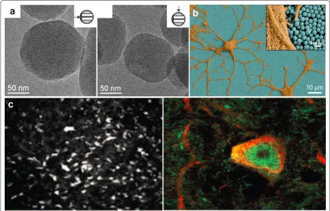
then attached to the AuNP, which can facilitate selec-tive binding to Aβ aggregates. Thereafter, exposure to a low gigahertz electromagnetic field prompted local heat dissipation from the AuNPs, which then dissolved the aggregates. In another study, AgNPs were employed to study the interaction of amyloid β-derived diffusible ligand (ADDL) and the anti-ADDL antibody for an opti-cal biosensor to diagnose AD (Fig. 2a) [51].
In order to make effective electrodes, AuNPs assembled using a layer-by-layer approach to form electrodes which were shown to yield low impedance and high charge stor-age capacity (Fig. 2b) [52]. This initial demonstration of using AuNPs for neural interfaces showed improve-ments in the signal-to-noise ratio, long-term recording, and delivery of a higher charge per area of electrode to the surrounding tissue. In recent work, AuNPs have been
used to further target neurons for neuromodulation [53]. Spherical 20-nm AuNPs were attached to high-avidity ligands targeting different membrane proteins of dorsal root ganglion (DRG) neurons (Fig. 2c). After binding to the neuron, the particles transduce millisecond pulses of light into heat, which sufficiently altered the membrane capacitance to elicit action potentials. This AuNP-based strategy worked well for all tested ligands to induce selective stimulation, providing an alternative to optoge-netic techniques. These diverse studies highlight the potential of metallic nanoparticles for a diverse array of neuroapplications.
3.1.2 Silica nanoparticles
Silica is categorizes as a “Generally Recognized As Safe” material by the FDA, and is widely used for food addi-tives and cosmetics [54]. Besides the favorable biocom-patibility features, nanoparticles composed of silica are promising due to their robust structural stability and high drug loading [54]. It is also a highly transparent, dielectric material that does not absorb light nor conduct electrons
[55]. As an inert host, silica can further serve as a matrix for the construction of well-ordered particles capable of containing small molecule drugs and biomolecules.
Silica NPs are generally categorized as nonporous or mesoporous. While both are derived from an amor-phous silica structure, mesoporous silica NPs have a porous structure (2–50 nm pore size), which can allow for enhanced drug loading [56]. Mesoporous silica can therefore deliver a payload (e.g. drugs, proteins, genes) by entrapping it within the pores and releasing it through passive diffusion or the controlled opening of a chemi-cal/biological cap covering the pores (Fig. 3a) [57]. For instance, a recent study released nerve growth factors (NGF) using mesoporous silica nanoparticles, which not only prevented clearance and degradation of NGF, but improved delivery to promote nerve cell proliferation and neurite outgrowth [58]. Others have loaded agents, like
111 In radiolabeling, to enable multimodal in vivo imaging
and tracking [59].
Recent studies have explored the response of differ-ent neural tissue-type cells, like neural stem cells, neu-rons, astrocytes and microglia, to silica NP treatment in order to assess optimal surface modifications that ensure minimal cytotoxicity [60]. These silica-cell interactions have further been exploited to provide nanotopographi-cal features on interfacial surfaces. For instance, a self-assembled silica nanoparticle monolayer was employed to deliver negatively-charged RNA-based molecules (e.g. siRNA, miRNA) into neural stem cells to control neu-ronal differentiation (Fig. 3b) [61]. This substrate-medi-ated delivery for the nanoparticle film was non-toxic, highly effective, and achieved in the absence of cationic polymers.
The biocompatibility of silica has made it attractive for brain delivery. It is often used as an inert shell layer to coat other types of nanoparticles, as seen with mag-netic nanoparticles delivered to track neural progeni-tor cells in ischemic mice [62]. Among the multitude of
reports utilizing silica NPs for delivery to cells, Bharali et al. provided the first demonstration for in vivo deliv-ery using silica NPs as a nonviral vector [63]. Stable aque-ous dispersions of organically-modified silica NPs, bound with DNA-encoding EGFP, were prepared. Interestingly, this report described stereotaxic injection of the com-plexes into the mouse ventral midbrain and lateral ven-tricle. In addition to exhibiting no toxicity four weeks after transfection, the green fluorescence was visualized in the substantia nigra along with localization in tyros-ine hydroxylase (TH)-positive dopamtyros-inergic neurons (Fig. 3c). This initial study gave promise for using silica NPs for in vivo delivery and brain-targeting therapies. These features of silica NPs make them an attractive option for future neural studies.
3.1.3 Magnetic nanoparticles
Magnetic nanoparticles (MNPs) are attractive due to the superior contrast enhancement they offer for in vivo imaging. Magnetic resonance imaging (MRI) is one of the most widely used medical imaging techniques, which relies on measuring the relaxation times of excit-able hydrogens in the tissue to acquire high-resolution images. Since such intrinsic differences tend to be insuf-ficient for obtaining a detectable signal, contrast agents bearing paramagnetic or superparamagnetic properties are often used. MNPs, such as iron oxide-based parti-cles (Fe2O3 and Fe3O4), are excellent MRI contrast agents
for improved sensitivity in T-2-weighted imaging [64].
MNPs composed of iron oxide are clinically approved as MRI contrast agents, in which Feridex and Resovist well-known commercial products [65].
MNPs tend to be most effective when the size is typi-cally around 10–20 nm [66]. Over the years, synthetic procedures have been optimized to: (a) facilitate the incorporation of metals into the magnetic core that offer enhanced magnetic properties (e.g. zinc, cobalt, nickel), (b) coat with organic species (e.g. surfactants, polymers) to prevent degradation, and (c) deposit inorganic shell layers (e.g. silica, gold) for greater stability and additional surface functionalization [66].
The inherent magnetic properties of MNPs enable these particles to serve as useful multifunctional neu-ral platforms. For instance, MNPs have been designed to selectively cross the BBB by minimizing the size and coating the surface with biocompatible polymer layers and chemical functionalities [67]. Further complexing with tumor-specific peptides (e.g. chlorotoxin, CTX) allowed the MNPs to be targeted to highly invasive gli-oma brain tumors in mice models [67]. Moreover, this entire process could be monitored using MRI, making MNPs a versatile nanoparticle platform.
The high saturation magnetization properties of MNPs has also facilitated magnetic-based targeting. In this case, the dragging force of a permanent magnet is used to deliver MNPs to a target site. This has been demon-strated for both in vitro gene delivery to neural stem cells [68] and in vivo for drug/gene delivery to brain tumors of 9L-gliosarcoma-bearing rats [69]. Along with magnetic targeting, MNPs are becoming attractive for neuromodu-lation as well. In one study, 30-nm CoFe2O4-BaTiO3
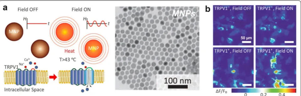
nan-oparticles were injected through the mouse’s tail vein and forced to cross the BBB via a d.c. field, and then used to modulate the electric waveforms in the brain upon expo-sure to an external a.c. field [70]. In another recent dem-onstration, MNPs were even used to stimulate deep brain structures in vivo through magnetic heating (Fig. 4a) [71]. The heat-sensitive capsaicin receptor TRPV1 was expressed with lentiviruses in the ventral tegmental area (VTA), followed by MNP injection into the same region. Subsequent exposure of the mice to alternating magnetic fields induced heat dissipation by hysteresis from the MNPs, permitting neuronal excitation by TRPV1-activa-tion for up to one month after MNP injecTRPV1-activa-tion (Fig. 4b). These diverse properties of MNPs offer unique applica-tions for neural research.
3.1.4 Quantum dots
Quantum dots (QDs), also referred to as semiconductor nanocrystals, have emerged as an exciting class of fluo-rescent probes. Conventional organic dyes readily pho-tobleach upon prolonged irradiation, and tuning their optical properties is a general synthetic challenge [72]. In contrast, QDs are resistant to photobleaching, extremely bright, and exhibit size-dependent emission wavelengths. In particular, QDs tend to have a broad absorption spec-trum and a narrow emission specspec-trum. This allows for the generation of QDs that can be excited with a single wavelength yet emit at different wavelengths [73]. Along with the small size range (~ 2-10 nm), this enables unique applications of QDs in multiplex imaging and biosensing.
Early on, a major limitation of QDs was the possibility of cytotoxic side effects. The most prominent QDs are composed of heavy metal ions, such as cadmium, sele-nide and tellurium, which exhibit adverse effects upon exposure to cells and tissues [74]. Strategies have been devised to overcome these issues, which include coating the core QD with inert shells (e.g. ZnS, silica) to prevent leaching of toxic elements [75], or using biocompatible elements to generate non-toxic QDs (e.g. CuInS2,
ZnS-AgInS2) [76].
functionalization strategies to complex biomolecules like siRNA to QDs for the delivery into U87-glioblastoma brain tumor cells [77]. In such a case, the innate fluores-cence property allows QDs to serve as a single vehicle for drug/biomolecule delivery, visualization and monitor-ing. For instance, the QDs can be targeted to specific cell types to enable cellular tracking within the body. A recent study showed that the conjugation of QDs with cell-penetrating lipopeptides and the subsequent injection into intact embryonic chick brains helped to identify and monitor neural stem cells as they migrate in the develop-ing brain [78].
The long-term stability and robust fluorescence prop-erties of QDs make them useful for mechanistic studies as well. For instance, the movement of QD-labeled nerve growth factor (NGF) was tracked in cultures of rat dorsal root ganglion (DRG) neurons to understand the mecha-nism of retrograde axonal transport [79]. Preliminary studies have also exploited the optoelectronic proper-ties of QDs to activate ion channels on neurons [80]. In this case, remote-controlled membrane depolarization or hyperpolarization was achieved in cortical neurons by activating K+ and Na+ channels using QD films and
QD-coated micropipettes. The combination of the innate optical properties with the additional bioconjugation capabilities make QDs a favorable option for advanced studies.
Carbon-based quantum dots (CQDs or C-dots) have also recently emerged as a new category of fluorescent probes, with sizes below 10 nm. CQDs were first dis-covered as a major impurity during the purification of single-walled carbon nanotubes using preparative electrophoresis [81]. Thereafter, a number of synthetic strategies were devised to generate CQDs, including
electrochemical carbonization, laser ablation and hydro-thermal treatment [82]. Compared to the traditional semiconductor-based quantum dots described above, CQDs have enhanced photoluminescence, solubility in water and biocompatibility [82]. These remarkable prop-erties have made CQDs particularly useful for applica-tions in bioimaging and biosensing [83]. In the context of neuroscience, CQDs were recently employed to tar-get brain cancer glioma in mice [84]. Synthesized using a simple thermolysis route with d-glucose and l-aspar-tic acid as starting materials, the as-prepared CQDs not only showed tunable emission spectra, but also intrinsic targeting to brain C6 glioma cells. While there are still limited investigations using CQDs for neuroapplications, such favorable properties makes this class of QDs attrac-tive for future studies.
3.1.5 Upconversion nanoparticles
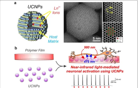
Upconversion nanoparticles (UCNPs) have attracted significant biomedical interest due to their ability to absorb low-energy photons and emit high-energy pho-tons. In other words, UCNPs convert long-wavelength near-infrared light (NIR; >800 nm) to short-wavelength visible light (300–700 nm), known as an anti-Stokes process [85]. This phenomenon is possible due to the unique optical properties of the lanthanide-series ele-ments, which are used as dopants within a crystalline host matrix, such as NaYF4 (Fig. 5a). Two different types
of lanthanide-dopants are usually employed: a sensitizer and an activator. Upon NIR irradiation, the sensitizer (e.g. Yb3+) harvests NIR energy and transfers it through
a non-radiative process to the activator (e.g. Er3+, Tm3+).
The ladder-like arrangements of energy levels in trivalent lanthanide ions (Ln3+) thus allows for visible light
emis-sions through various energy transfer pathways, depend-ing on the pre-selected ion pairdepend-ing [87]. As a result, while conventional organic dyes are sensitive to their chemical surroundings, the shielded 4f–4f intra-configurational transitions in UCNPs permit emissions that are independ-ent of the particle size and environmindepend-ent [86]. The fixed energy levels, high resistance to photoblinking and pho-tobleaching, long luminescence lifetimes (micro/milli-sec-ond) and the upconversion process using deep-penetrating NIR light makes UCNPs ideal as in vivo probes [88].
UCNPs first emerged in neuroscience research for imaging and cancer application. In one study, the conju-gation of the RGD peptide to the surface of NaYF4:Yb3+/
Tm3+ UCNPs allowed for targeted imaging of nude mice
bearing human glioblastoma U87MG tumors [89]. Oth-ers have conjugated neurotoxins such as chlorotoxin, which has the ability to target primary brain tumors, for imaging of xenograft glioma tumors in Balb-c nude mice
in vivo and ex vivo [90]. In a recent study, a UCNP-based sensor was also designed for the detection of Zn2+ in
brain slices from mice bearing Alzheimer’s disease [91]. The application of UCNPs has recently been extended to optogenetically modulate neuronal activity using near-infrared light. Optogenetic approaches are contingent upon delivering a sufficient dose of visible light to the target opsin-expressing cells. Since visible light is highly scattered in tissue, invasive procedures are required to precisely implant optical fibers at the target site. In an alternative strategy, UCNPs have been shown for the first time to serve as mediators for converting deep-penetrat-ing NIR light into visible light (e.g. blue light) to facili-tate optogenetic neuronal control [92]. Embedding the UCNPs in a biodegradable polymer can further ensure a sufficient neural interface for repeated NIR-stimula-tion (Fig. 5b). There is continued efforts to enhance the upconversion efficiency in order to improve UCNP-based optogenetic control [93], as well as open new appli-cations of UCNPs for neural research.
3.2 Organic nanomaterials
The following organic nanomaterials will be discussed in this section: liposomes/micelles, dendrimers, polymeric NPs and carbon-based nanomaterials.
3.2.1 Liposomes/micelles
Amphiphiles, which contain both hydrophilic and hydro-phobic domains, are powerful building blocks in biology [94]. They self-assemble in order to minimize the ener-getically unfavorable interaction of hydrophobic moieties with the surrounding water molecules, leading to the for-mation of well-defined nanoassemblies [95]. An example of this phenomena is the cellular membrane, which is the dynamic assembly of phospholipids. The chemical con-trol of this assembly process has enabled the generation
of various types of amphiphilic nanocarriers, in which liposomes and micelles are two well-known categories (Fig. 6a) [94].
Liposomes are spherical-shaped vesicles comprised of one or more vesicular bilayers (lamellae) [96]. This unique bilayer formation into a spherical assembly gen-erally results in a core of aqueous solution, which can be loaded with hydrophilic compounds. At the same time, hydrophobic compounds can be loaded within the lipid bilayer itself. Liposomes are usually composed of natural phospholipids such as sphingomyelin and glyc-erophospholipids, or synthetic polymers such as block copolymers [97]. Micelles, on the other hand, are spher-ical-shaped assemblies that have a hydrophilic exterior and a hydrophobic interior [98]. In contrast to molecules
used to form liposomes, micelles consist of amphiphilic molecules (also known as surfactants) with one hydro-phobic tail linked to one hydrophilic head. In turn, dis-persion in water leads to the spontaneous formation of micelles, wherein the tail portion of the molecules sequester away from the water molecules into a highly hydrophobic core [99]. Pluronic block copolymers, such as ethylene oxide and propylene oxide, are wide-used for micelle formation [100].
Amphiphilic-based nanoassemblies like liposomes and micelles have been utilized for several decades now in neuroscience research, particularly for the gene/drug delivery to the CNS. Early work demonstrated lipo-some-based drug vehicles capable of delivering antican-cer agents like daunomycin across the BBB into the rat brain [101]. In such cases, modification with targeting moieties (e.g. antibodies) and stabilization with polyeth-ylene glycol (PEG) conjugation to increase in vivo circu-lation times proved to be essential. Recent studies have advanced the application of multifunctional liposomes to address a variety of neurological ailments in vivo, includ-ing neuroprotection after cerebral ischemia [102], tar-geting gliomas [103], treating brain metastasis [104] and reducing β-amyloid plaques in Alzheimer’s disease [105]. Polymeric micelles have been similarly applied. In one study, the cell-penetrating peptide TAT was anchored to micelles in order to deliver the antibiotic ciprofloxacin across the BBB to treat brain infections [106].
These amphiphilic nanoassemblies have also grown popular for other specialized neural applications. For example, liposomes have been used to mediate neural regeneration, wherein genes coding for neurotrophic growth factors (e.g. GDNF, NGF) were delivered after neuronal injury [107, 108]. The in vivo expression of these growth factors was seen to promote axonal regen-eration and improve locomotive function in adult rats [107]. Besides serving as a drug/gene carrier, liposomes and micelles have been uniquely used as externally-trig-gerable agents. For example, a hetero-assembly of siRNA-complexed polymeric micelles and gas-cored liposomes was recently synthesized to develop an ultrasound-based nanobubble therapy (Fig. 6b) [109]. In this case, the ultra-sound-sensitive gas-cored liposomes carried the siRNA-loaded micelles, resulting in enhanced delivery efficiency and gene silencing in a mouse glioma model. In general, the biocompatibility, facile surface functionalization, and lack of immune response have made these amphiphile-based nanocarriers invaluable.
3.2.2 Dendrimers
Dendrimers are synthesized by the cross-linking of repeating monomer subunits [110]. This regular arrange-ment of the monomers results in a highly-branched and
well-defined hierarchical structure. Emanating from an initiator core, the layer-by-layer expansive growth allows for the synthesis of varying ‘generations’ of dendrimers with different hydrodynamic sizes, branching points and surface functionality (Fig. 7a) [111]. Further modification of the surface to introduce chemical functionalities (e.g. positive-charged amine groups) can render dendrim-ers useful for complexation with drugs and gene vectors [112, 113]. Various types of dendrimer systems have been used for biological studies, including poly(propylene imine) (PPI) and poly(amidoamine) (PAMAM) [111].
Dendrimers are promising for targeted delivery to the brain. Attaching different classes of drugs for CNS thera-pies, including anticancer, inflammatory, and anti-microbial agents, is facilitated by either encapsulation within the dendrimer or through chemical bonding [114]. Studies have shown the hydroxy-functionalized PAMAM dendrimers to be non-toxic, yet only minimal uptake was observed in both healthy and tumor-bearing animals [115]. However, enhanced PAMAM dendrimer uptake was observed into the brain following neuroinflamma-tion, possibly due to impairment in the BBB [116]. The localization of dendrimers to activated microglia after systemic administration was observed in rabbit models of cerebral palsy, which in turn allowed for the targeted delivery of N-acetyl-l-cysteine (NAC), an antioxidant and anti-inflammatory agent [116]. Recent work further advanced these findings from the small animal rabbit injury model to a larger canine model [117]. After sys-temic administration, PAMAM localized to the injured neurons and microglia in the brain of canines (Fig. 7b), allowing for the delivery of both NAC and valproic acid for enhanced neuroprotection [117]. Such seemingly inherent targeting, in addition to further modification of the dendrimer to incorporate therapeutics, makes the dendrimer class of nanomaterials clinically-relevant.
3.2.3 Polymeric nanoparticles
a protective coating for the therapeutic, which ensures enhanced stability and efficacy after in vivo adminis-tration compared to the free form [119]. Moreover, the pharmacokinetic properties of the therapeutic-loaded polymeric nanoparticles can be further enhanced by functionalizing the surface, which can enable targeted delivery as well as increased permeability through the BBB [120].
The CNS delivery of drugs or biomolecules is a key application of polymeric nanoparticles. Early studies
to the CNS, including doxorubicin, methotrexate, lopera-mide and temozololopera-mide [123].
Polyester-based nanoparticles like PLGA have been observed to be safer alternatives for brain delivery since the degradation products are mainly water and carbon dioxide [124]. Besides drugs, polymeric nanoparticles have been useful for the sustained release of growth fac-tors to treat neurodegenerative disorders. In a Hunting-ton’s disease rat model, the local administration of nerve growth factor (NGF)-loaded PLGA enabled neuropro-tection after excitotoxin quinolinic acid injections [125]. Similar loading with other tropic factors and neurotrans-mitters has led to significant results for neuroprotection and repair [18, 126]. Polymeric nanoparticles have also been used to direct neural stem cell behavior in vivo. For example, PEI-based nanoparticles were complexed with retinoic acid to control neural differentiation in the sub-ventricular zone (neural stem cell niche) [127] and after ischemia [128]. In this way, polymeric nanoparticles will continue to provide utility in advancing studies.
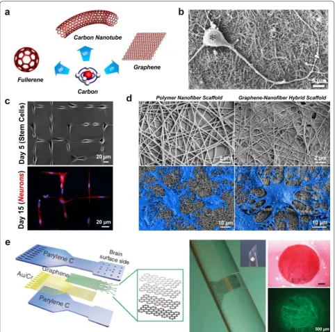
3.2.4 Carbon‑based nanomaterials
Carbon-based nanomaterials are becoming attractive due to their unique optical, thermal, mechanical, electrical and chemical properties. Composed of sp2-bonded
gra-phitic carbon, these nanomaterials are categorized into zero-dimensional, one-dimensional and two-dimensional structures (Fig. 8a) [129]. Laser ablation of graphite was used to isolate the well-known C60 buckyball in 1985, a
zero-dimensional fullerene derivative, which was the first carbon nanomaterial to be isolated [130]. Soon after, one-dimensional carbon nanotubes (CNTs) were pre-pared using arc discharge techniques in 1991 [131]. The cylindrical carbon structure has an extended sp2 carbon
with physical properties that can be tuned, such as the diameter, length, number of walls/cylindrical layers and chirality [132]. CNTs are in fact made of graphene sheets wrapped onto themselves, but two-dimensional graphene was not isolated until 2004, using mechanical exfoliation [133]. As a single-atom thick sheet, graphene exhibits a number of remarkable properties, including: high planar surface area, superior mechanical strength, unparalleled thermal conductivity, and favorable electronic properties and optical properties [134]. Further modifications to the graphene surface, like oxidation to create graphene oxide, has resulted in derivatives with complementary proper-ties for biological studies (e.g. enhanced water solubility, facile functionalization for biomolecule conjugation, etc.) [135].
Besides serving as a remarkable material for the bio-medical field in general, carbon-based nanomateri-als have become especially useful in neuroengineering, where the focus is the development of devices or surfaces
to effectively interface with the CNS [136]. The first report using these materials for neural research was the growth of embryonic rat brain neurons on multi-walled CNTs (Fig. 8b) [137]. This early work highlighted the importance of modifying the CNT surface for enhanced neurite outgrowth, a critical feature for enhanced in vivo performance in terms of biocompatibility, neuron growth and neurite/axonal elongation. While graphene is similar to CNTs in many ways, the two-dimensional structure and flexibility of graphene allows for facile coating on numerous types of cell culture surfaces. The early dem-onstration of the biocompatible interaction of neurons with graphene showed favorable long-term outcomes, in which mouse hippocampal neurons had enhanced neu-rite sprouting and outgrowth on graphene-coated tissue culture polystyrene (TCPS) compared to bare TCPS sub-strates [138].
The promising results from neuronal cultures led to the examination of carbon-based nanomaterials for stem cell cultures. One of the earliest studies showed the success-ful differentiation of mouse NSCs on single-walled CNT-polyelectrolyte multilayer thin films into neural cells [139]. The viability, neurite outgrowth and neural marker expression was found to be comparable between the con-ventional poly-l-ornithine (PLO) surface and the CNT surface. Further modifications of CNTs has also resulted in enhanced neuronal differentiation, both by using dif-ferent types of stem cells, like hESCs [140] and hMSCs [141], and by combining with other biomaterials, like col-lagen [142] and silk [143]. Graphene-coated surfaces have also shown similar enhancements in neuronal formation [144], axonal alignment [145], neuronal patterning [146] and oligodendrocyte differentiation [147] (Fig. 8c, d).
neurophysiological recording and optogenetic activa-tion in the rodent brain (Fig. 8e) [152]. Considering these exceptional features, carbon-based nanomaterials have great promise for improving neural interfaces. With more and more studies verifying the compatibility of such materials, there is immense scope for using these robust materials in translational studies.
4 Conclusions
From both a fundamental and an applied science point-of-view, nanotechnology and nanoscience has greatly advanced in a relatively short period of time. Nanomedi-cine in particular has seen a steady progress in the last two decades, with tremendous efforts being placed in translating these advances to the field of neuroscience. A wide array of nanomaterials show promise for enhancing our understanding of the CNS, moreover offering thera-peutic opportunities in CNS-related treatment. Many of the inorganic-based nanomaterials, such as metal-lic nanoparticles, magnetic nanoparticles and quantum dots, are being extensively employed as imaging agents. Other inorganic nanomaterials provide unique advan-tages, such as enhanced small molecule/biomolecule loading with silica nanoparticles and improved optical penetration with upconversion nanoparticles. This makes them quite versatile and adaptable to crucial neuroappli-cations such as CNS drug delivery and deep tissue imag-ing, respectively. In contrast, organic nanomaterials such as micelles, liposomes dendrimers and polymeric nano-particles are generally biocompatible and biodegradable right from the start. In addition, carbon-based nanoma-terials offer superior material properties, making this class of nanomaterials attractive candidates for neural interfaces. The availability of such a diverse nano-toolkit has changed the way scientists approach challenges in neuroscience.
Nevertheless, a growing need exists to create nano-based platforms that bear multiple functionalities on a single platform. This is mainly due to highly complex nature of the CNS, and furthermore it’s sensitivity to slight damage and the consequent limited capability for autonomous repair. As a result, approaches that enable maximal effectiveness with minimal perturbation of the intact tissue would be ideal. In developing the next generation of nanoscale CNS platforms, critical design criteria consist of: attachment of multiple types of thera-peutic agents, spatiotemporal control within the body, built-in modalities for long-term tracking, and capabili-ties to record and modulate neural activity. Integrating these features on a single nanoplatform holds remark-able potential for utilizing the nanomaterial toolkit for advanced neuroengineering applications.
Acknowledgements
I would like to acknowledge Dr. Randy Giles (Director; Bell Labs) for his help in reviewing and revising this manuscript. I would also like to acknowledge Dr. Sanjay Patel (Program Leader; Bell Labs) and Dr. Marcus Weldon (President of Bell Labs and CTO at Nokia) for their guidance and support.
Competing interests
The author declares that he has no competing interests.
Received: 30 July 2016 Accepted: 29 September 2016
References
1. A.P. Alivisatos, A.M. Andrews, E.S. Boyden, M. Chun, G.M. Church, K. Deisseroth, J.P. Donoghue, S.E. Fraser, J. Lippincott-Schwartz, L.L. Looger, S. Masmanidis, P.L. McEuen, A.V. Nurmikko, H. Park, D.S. Peterka, C. Reid, M.L. Roukes, A. Scherer, M. Schnitzer, T.J. Sejnowski, K.L. Shepard, D. Tsao, G. Turrigiano, P.S. Weiss, C. Xu, R. Yuste, X. Zhuang, Nanotools for neuro-science and brain activity mapping. ACS Nano 7, 1850–1866 (2013) 2. L.Y. Rizzo, B. Theek, G. Storm, F. Kiessling, T. Lammers, Recent progress
in nanomedicine: therapeutic, diagnostic and theranostic applications. Curr Opin Biotechnol 24, 1159–1166 (2013)
3. J. Jankovic, Parkinson’s disease: clinical features and diagnosis. J Neurol Neurosurg Psychiatry 79, 368–376 (2008)
4. X.Z. Liu, X.M. Xu, R. Hu, C. Du, S.X. Zhang, J.W. McDonald, H.X. Dong, Y.J. Wu, G.S. Fan, M.F. Jacquin, C.Y. Hsu, D.W. Choi, Neuronal and glial apop-tosis after traumatic spinal cord injury. J Neurosci 17, 5395–5406 (1997) 5. A.J. Mothe, C.H. Tator, Advances in stem cell therapy for spinal cord
injury. J Clin Invest 122, 3824–3834 (2012)
6. M.A. Fischbach, J.A. Bluestone, W.A. Lim, Cell-based therapeutics: the next pillar of medicine. Sci Transl Med 5, 179ps7 (2013)
7. P. Piccini, O. Lindvall, A. Bjorklund, P. Brundin, P. Hagell, R. Ceravolo, W. Oertel, N. Quinn, M. Samuel, S. Rehncrona, H. Widner, D.J. Brooks, Delayed recovery of movement-related cortical function in Parkinson’s disease after striatal dopaminergic grafts. Ann Neurol 48, 689–695 (2000)
8. P. Piccini, D.J. Brooks, A. Bjorklund, R.N. Gunn, P.M. Grasby, O. Rimoldi, P. Brundin, P. Hagell, S. Rehncrona, H. Widner, O. Lindvall, Dopamine release from nigral transplants visualized in vivo in a Parkinson’s patient. Nat Neurosci 2, 1137–1140 (1999)
9. J. Yoo, H.S. Kim, D.Y. Hwang, Stem cells as promising therapeutic options for neurological disorders. J Cell Biochem 114, 743–753 (2013) 10. E.N. Momin, A. Mohyeldin, H.A. Zaidi, G. Vela, A. Quinones-Hinojosa,
Mesenchymal stem cells: new approaches for the treatment of neuro-logical diseases. Curr Stem Cell Res Ther 5, 326–344 (2010)
11. O. Einstein, T. Ben-Hur, The changing face of neural stem cell therapy in neurologic diseases. Arch Neurol 65, 452–456 (2008)
12. D. Lukovic, V. Moreno Manzano, M. Stojkovic, S.S. Bhattacharya, S. Erceg, Concise review: human pluripotent stem cells in the treatment of spinal cord injury. Stem Cells 30, 1787–1792 (2012)
13. T. Ben-Hur, Reconstructing neural circuits using transplanted neural stem cells in the injured spinal cord. J Clin Invest 120, 3096–3098 (2010)
14. R.V. Nelakanti, N.G. Kooreman, J.C. Wu, Teratoma formation: a tool for monitoring pluripotency in stem cell research. Curr Protoc Stem Cell Biol 32, 4a.8.1–4a.8.17 (2015)
15. O. Lindvall, A. Björklund, Cell replacement therapy: helping the brain to repair itself. NeuroRx 1, 379–381 (2004)
16. S. Shah, A. Solanki, K.-B. Lee, Nanotechnology-based approaches for guiding neural regeneration. Acc Chem Res 49, 17–26 (2016) 17. D.J. Begley, Delivery of therapeutic agents to the central nervous
system: the problems and the possibilities. Pharmacol Ther 104, 29–45 (2004)
19. Y. Ramirez, J. Weatherbee, R. Wheelhouse, A. Ross, Glioblastoma multiforme therapy and mechanisms of resistance. Pharmaceuticals 6, 1475–1506 (2013)
20. J. Zhou, K.B. Atsina, B.T. Himes, G.W. Strohbehn, W.M. Saltzman, Novel delivery strategies for glioblastoma. Cancer J (Sudbury, Mass) 18, 89–99 (2012)
21. S. Mitragotri, P.A. Burke, R. Langer, Overcoming the challenges in admin-istering biopharmaceuticals: formulation and delivery strategies. Nat Rev Drug Discov 13, 655–672 (2014)
22. A. Mohyeldin, E.A. Chiocca, Gene and viral therapy for glioblastoma: a review of clinical trials and future directions. Cancer J 18, 82–88 (2012) 23. J.K. Atwal, Y. Chen, C. Chiu, D.L. Mortensen, W.J. Meilandt, Y. Liu, C.E.
Heise, K. Hoyte, W. Luk, Y. Lu, K. Peng, P. Wu, L. Rouge, Y. Zhang, R.A. Lazarus, K. Scearce-Levie, W. Wang, Y. Wu, M. Tessier-Lavigne, R.J. Watts, A therapeutic antibody targeting BACE1 inhibits amyloid-beta produc-tion in vivo. Sci Transl Med 3, 84ra43 (2011)
24. E. Masliah, E. Rockenstein, M. Mante, L. Crews, B. Spencer, A. Adame, C. Patrick, M. Trejo, K. Ubhi, T.T. Rohn, S. Mueller-Steiner, P. Seubert, R. Bar-bour, L. McConlogue, M. Buttini, D. Games, D. Schenk, Passive immuniza-tion reduces behavioral and neuropathological deficits in an alpha-synu-clein transgenic model of lewy body disease. PLoS One 6, e19338 (2011) 25. S.E. Laursen, J.K. Belknap, Intracerebroventricular injections in mice:
some methodological refinements. J Pharmacol Methods 16, 355–357 (1986)
26. J.T. Hansen, B.M. Koeppen, Netter’s atlas of human physiology (Saunders,
Philadelphia, 2002)
27. J. Stockwell, N. Abdi, X. Lu, O. Maheshwari, C. Taghibiglou, Novel central nervous system drug delivery systems. Chem Biol Drug Des 83, 507–520 (2014)
28. G.F. Woodworth, G.P. Dunn, E.A. Nance, J. Hanes, H. Brem, Emerging insights into barriers to effective brain tumor therapeutics. Front Oncol
4, 126 (2014)
29. L.A. Jorgenson, W.T. Newsome, D.J. Anderson, C.I. Bargmann, E.N. Brown, K. Deisseroth, J.P. Donoghue, K.L. Hudson, G.S. Ling, P.R. MacLeish, E. Marder, R.A. Normann, J.R. Sanes, M.J. Schnitzer, T.J. Sejnowski, D.W. Tank, R.Y. Tsien, K. Ugurbil, J.C. Wingfield, The BRAIN initiative: developing technology to catalyse neuroscience discovery. Philos Trans R Soc Lond B Biol Sci 370, 20140164 (2015)
30. L. Luo, E.M. Callaway, K. Svoboda, Genetic dissection of neural circuits. Neuron 57, 634–660 (2008)
31. M. Karmazinova, L. Lacinova, Measurement of cellular excitability by whole cell patch clamp technique. Physiol Res 59, S1–S7 (2010) 32. M.L. Kringelbach, N. Jenkinson, S.L.F. Owen, T.Z. Aziz, Translational
princi-ples of deep brain stimulation. Nat Rev Neurosci 8, 623–635 (2007) 33. L.R. Hochberg, M.D. Serruya, G.M. Friehs, J.A. Mukand, M. Saleh, A.H.
Caplan, A. Branner, D. Chen, R.D. Penn, J.P. Donoghue, Neuronal ensem-ble control of prosthetic devices by a human with tetraplegia. Nature
442, 164–171 (2006)
34. D.R. Merrill, Materials considerations of implantable neuroengineering devices for clinical use. Curr Opin Solid State Mater Sci 18, 329–336 (2014) 35. P. Fattahi, G. Yang, G. Kim, M.R. Abidian, A review of organic and
inorganic biomaterials for neural interfaces. Adv Mater 26, 1846–1885 (2014)
36. C. Marin, E. Fernandez, Biocompatibility of intracortical microelectrodes: current status and future prospects. Front Neuroeng 3, 8 (2010) 37. F. Zhang, V. Gradinaru, A.R. Adamantidis, R. Durand, R.D. Airan, L. de
Lecea, K. Deisseroth, Optogenetic interrogation of neural circuits: tech-nology for probing mammalian brain structures. Nat Protoc 5, 439–456 (2010)
38. F. Zhang, L.P. Wang, M. Brauner, J.F. Liewald, K. Kay, N. Watzke, P.G. Wood, E. Bamberg, G. Nagel, A. Gottschalk, K. Deisseroth, Multimodal fast opti-cal interrogation of neural circuitry. Nature 446, 633–639 (2007) 39. E.S. Boyden, F. Zhang, E. Bamberg, G. Nagel, K. Deisseroth,
Millisecond-timescale, genetically targeted optical control of neural activity. Nat Neurosci 8, 1263–1268 (2005)
40. A.R. Adamantidis, F. Zhang, A.M. Aravanis, K. Deisseroth, L. de Lecea, Neural substrates of awakening probed with optogenetic control of hypocretin neurons. Nature 450, 420–424 (2007)
41. F. Zhang, A.M. Aravanis, A. Adamantidis, L. de Lecea, K. Deisseroth, Circuit-breakers: optical technologies for probing neural signals and systems. Nat Rev Neurosci 8, 577–581 (2007)
42. K.M. Tye, K. Deisseroth, Optogenetic investigation of neural circuits underlying brain disease in animal models. Nat Rev Neurosci 13, 251–266 (2012)
43. A.M. Packer, B. Roska, M. Hausser, Targeting neurons and photons for optogenetics. Nat Neurosci 16, 805–815 (2013)
44. W. Jiang, Y.S. KimBetty, J.T. Rutka, C.W. ChanWarren, Nanoparticle-medi-ated cellular response is size-dependent. Nat Nano 3, 145–150 (2008) 45. L. Shang, K. Nienhaus, G.U. Nienhaus, Engineered nanoparticles
inter-acting with cells: size matters. J Nanobiotechnol 12, 1–11 (2014) 46. J.V. Jokerst, T. Lobovkina, R.N. Zare, S.S. Gambhir, Nanoparticle
PEGyla-tion for imaging and therapy. Nanomedicine (London, England) 6, 715–728 (2011)
47. P.K. Jain, X. Huang, I.H. El-Sayed, M.A. El-Sayed, Noble metals on the nanoscale: optical and photothermal properties and some applica-tions in imaging, sensing, biology, and medicine. Acc Chem Res 41, 1578–1586 (2008)
48. P.K. Jain, K.S. Lee, I.H. El-Sayed, M.A. El-Sayed, Calculated absorption and scattering properties of gold nanoparticles of different size, shape, and composition: applications in biological imaging and biomedicine. J Phys Chem B 110, 7238–7248 (2006)
49. D.A. Giljohann, D.S. Seferos, W.L. Daniel, M.D. Massich, P.C. Patel, C.A. Mirkin, Gold nanoparticles for biology and medicine. Angew Chem Int Ed 49, 3280–3294 (2010)
50. M.J. Kogan, N.G. Bastus, R. Amigo, D. Grillo-Bosch, E. Araya, A. Turiel, A. Labarta, E. Giralt, V.F. Puntes, Nanoparticle-mediated local and remote manipulation of protein aggregation. Nano Lett 6, 110–115 (2006) 51. A.J. Haes, L. Chang, W.L. Klein, R.P. Van Duyne, Detection of a biomarker
for Alzheimer’s disease from synthetic and clinical samples using a nanoscale optical biosensor. J Am Chem Soc 127, 2264–2271 (2005) 52. H. Zhang, J. Shih, J. Zhu, N.A. Kotov, Layered nanocomposites from gold
nanoparticles for neural prosthetic devices. Nano Lett 12, 3391–3398 (2012)
53. J.L. Carvalho-de-Souza, J.S. Treger, B. Dang, S.B. Kent, D.R. Pepperberg, F. Bezanilla, Photosensitivity of neurons enabled by cell-targeted gold nanoparticles. Neuron 86, 207–217 (2015)
54. F. Tang, L. Li, D. Chen, Mesoporous silica nanoparticles: synthesis, bio-compatibility and drug delivery. Adv Mater 24, 1504–1534 (2012) 55. N.J. Halas, Nanoscience under Glass: the versatile chemistry of silica
nanostructures. ACS Nano 2, 179–183 (2008)
56. L. Tang, J. Cheng, Nonporous silica nanoparticles for nanomedicine application. Nano Today 8, 290–312 (2013)
57. I.I. Slowing, J.L. Vivero-Escoto, C.W. Wu, V.S.Y. Lin, Mesoporous silica nanoparticles as controlled release drug delivery and gene transfection carriers. Adv Drug Deliv Rev 60, 1278–1288 (2008)
58. B. Sun, A. Taing, H. Liu, G. Nie, J. Wang, Y. Fang, L. Liu, Y. Xue, J. Shi, Y.P. Liao, J. Ku, T. Xia, Y. Liu, Nerve growth factor-conjugated mesoporous silica nanoparticles promote neuron-like PC12 cell proliferation and neurite growth. J Nanosci Nanotechnol 16, 2390–2393 (2016) 59. S.H. Cheng, D. Yu, H.M. Tsai, R.A. Morshed, D. Kanojia, L.W. Lo, L. Leoni, Y.
Govind, L. Zhang, K.S. Aboody, M.S. Lesniak, C.T. Chen, I.V. Balyasnikova, Dynamic in vivo SPECT imaging of neural stem cells functionalized with radiolabeled nanoparticles for tracking of glioblastoma. J Nucl Med 57, 279–284 (2016)
60. E. Izak-Nau, K. Kenesei, K. Murali, M. Voetz, S. Eiden, V.F. Puntes, A. Duschl, E. Madarasz, Interaction of differently functionalized fluorescent silica nanoparticles with neural stem- and tissue-type cells. Nanotoxicology
8(Suppl 1), 138–148 (2014)
61. A. Solanki, S. Shah, P.T. Yin, K.-B. Lee, Nanotopography-mediated reverse uptake for siRNA delivery into neural stem cells to enhance neuronal differentiation. Sci Rep 3, 1553–1559 (2013)
62. L. Zhang, Y. Wang, Y. Tang, Z. Jiao, C. Xie, H. Zhang, P. Gu, X. Wei, G.Y. Yang, H. Gu, C. Zhang, High MRI performance fluorescent mesoporous silica-coated magnetic nanoparticles for tracking neural progenitor cells in an ischemic mouse model. Nanoscale 5, 4506–4516 (2013) 63. D.J. Bharali, I. Klejbor, E.K. Stachowiak, P. Dutta, I. Roy, N. Kaur, E.J. Bergey,
P.N. Prasad, M.K. Stachowiak, Organically modified silica nanoparticles: a nonviral vector for in vivo gene delivery and expression in the brain. Proc Natl Acad Sci USA 102, 11539–11544 (2005)
65. Y.X.J. Wang, Superparamagnetic iron oxide based MRI contrast agents: current status of clinical application. Quant Imaging Med Surg 1, 35–40 (2011)
66. A.H. Lu, E.L. Salabas, F. Schuth, Magnetic nanoparticles: synthesis, pro-tection, functionalization, and application. Angew Chem Int Ed Engl 46, 1222–1244 (2007)
67. O. Veiseh, C. Sun, C. Fang, N. Bhattarai, J. Gunn, F. Kievit, K. Du, B. Pullar, D. Lee, R.G. Ellenbogen, J. Olson, M. Zhang, Specific targeting of brain tumors with an optical/magnetic resonance imaging nanoprobe across the blood-brain barrier. Cancer Res 69, 6200–6207 (2009)
68. B. Shah, P.T. Yin, S. Ghoshal, K.-B. Lee, Multimodal magnetic core-shell nanoparticles for effective stem-cell differentiation and imaging. Angew Chem Int Ed 52, 6190–6195 (2013)
69. B. Chertok, A.E. David, V.C. Yang, Polyethyleneimine-modified iron oxide nanoparticles for brain tumor drug delivery using magnetic targeting and intra-carotid administration. Biomaterials 31, 6317–6324 (2010) 70. R. Guduru, P. Liang, J. Hong, A. Rodzinski, A. Hadjikhani, J. Horstmyer,
E. Levister, S. Khizroev, Magnetoelectric ‘spin’ on stimulating the brain. Nanomedicine (Lond) 10, 2051–2061 (2015)
71. R. Chen, G. Romero, M.G. Christiansen, A. Mohr, P. Anikeeva, Wireless magnetothermal deep brain stimulation. Science 347, 1477–1480 (2015)
72. U. Resch-Genger, M. Grabolle, S. Cavaliere-Jaricot, R. Nitschke, T. Nann, Quantum dots versus organic dyes as fluorescent labels. Nat Methods
5, 763–775 (2008)
73. I.L. Medintz, H.T. Uyeda, E.R. Goldman, H. Mattoussi, Quantum dot bioconjugates for imaging, labelling and sensing. Nat Mater 4, 435–446 (2005)
74. K.M. Tsoi, Q. Dai, B.A. Alman, W.C.W. Chan, Are quantum dots toxic? Exploring the discrepancy between cell culture and animal studies. Acc Chem Res 46, 662–671 (2013)
75. Y. Su, Y. He, H. Lu, L. Sai, Q. Li, W. Li, L. Wang, P. Shen, Q. Huang, C. Fan, The cytotoxicity of cadmium based, aqueous phase—synthesized, quantum dots and its modulation by surface coating. Biomaterials 30, 19–25 (2009)
76. P. Subramaniam, S.J. Lee, S. Shah, S. Patel, V. Starovoytov, K.B. Lee, Gen-eration of a library of non-toxic quantum dots for cellular imaging and siRNA delivery. Adv Mater 24, 4014–4019 (2012)
77. J.J. Jung, A. Solanki, K.A. Memoli, K. Kamei, H. Kim, M.A. Drahl, L.J. Wil-liams, H.R. Tseng, K. Lee, Selective Inhibition of human brain tumor cells through multifunctional quantum-dot-based siRNA delivery. Angew Chem Int Ed 49, 103–107 (2010)
78. R. Agarwal, M.S. Domowicz, N.B. Schwartz, J. Henry, I. Medintz, J.B. Delehanty, M.H. Stewart, K. Susumu, A.L. Huston, J.R. Deschamps, P.E. Dawson, V. Palomo, G. Dawson, Delivery and tracking of quantum dot peptide bioconjugates in an intact developing avian brain. ACS Chem Neurosci 6, 494–504 (2015)
79. B. Cui, C. Wu, L. Chen, A. Ramirez, E.L. Bearer, W.P. Li, W.C. Mobley, S. Chu, One at a time, live tracking of NGF axonal transport using quantum dots. Proc Natl Acad Sci USA 104, 13666–13671 (2007)
80. K. Lugo, X. Miao, F. Rieke, L.Y. Lin, Remote switching of cellular activity and cell signaling using light in conjunction with quantum dots. Biomed Opt Express 3, 447–454 (2012)
81. X. Xu, R. Ray, Y. Gu, H.J. Ploehn, L. Gearheart, K. Raker, W.A. Scrivens, Electrophoretic analysis and purification of fluorescent single-walled carbon nanotube fragments. J Am Chem Soc 126, 12736–12737 (2004) 82. Y. Wang, A. Hu, Carbon quantum dots: synthesis, properties and
appli-cations. J Mater Chem C 2, 6921–6939 (2014)
83. S.Y. Lim, W. Shen, Z. Gao, Carbon quantum dots and their applications. Chem Soc Rev 44, 362–381 (2015)
84. M. Zheng, S. Ruan, S. Liu, T. Sun, D. Qu, H. Zhao, Z. Xie, H. Gao, X. Jing, Z. Sun, Self-targeting fluorescent carbon dots for diagnosis of brain cancer cells. ACS Nano 9, 11455–11461 (2015)
85. F. Wang, D. Banerjee, Y. Liu, X. Chen, X. Liu, Upconversion nanoparticles in biological labeling, imaging, and therapy. Analyst 135, 1839–1854 (2010)
86. F. Wang, X.G. Liu, Recent advances in the chemistry of lanthanide-doped upconversion nanocrystals. Chem Soc Rev 38, 976–989 (2009) 87. F. Wang, R. Deng, J. Wang, Q. Wang, Y. Han, H. Zhu, X. Chen, X. Liu, Tun-ing upconversion through energy migration in core–shell nanoparti-cles. Nat Mater 10, 968–973 (2011)
88. D.K. Chatterjee, A.J. Rufaihah, Y. Zhang, Upconversion fluorescence imaging of cells and small animals using lanthanide doped nanocrys-tals. Biomaterials 29, 937–943 (2008)
89. L. Xiong, Z. Chen, Q. Tian, T. Cao, C. Xu, F. Li, High contrast upconver-sion luminescence targeted imaging in vivo using peptide-labeled nanophosphors. Anal Chem 81, 8687–8694 (2009)
90. X.F. Yu, Z. Sun, M. Li, Y. Xiang, Q.Q. Wang, F. Tang, Y. Wu, Z. Cao, W. Li, Neu-rotoxin-conjugated upconversion nanoprobes for direct visualization of tumors under near-infrared irradiation. Biomaterials 31, 8724–8731 (2010)
91. J. Peng, W. Xu, C.L. Teoh, S. Han, B. Kim, A. Samanta, J.C. Er, L. Wang, L. Yuan, X. Liu, Y.T. Chang, High-efficiency in vitro and in vivo detection of Zn2+ by dye-assembled upconversion nanoparticles. J Am Chem Soc
137, 2336–2342 (2015)
92. S. Shah, J.J. Liu, N. Pasquale, J. Lai, H. McGowan, Z.P. Pang, K.B. Lee, Hybrid upconversion nanomaterials for optogenetic neuronal control. Nanoscale 7, 16571–16577 (2015)
93. X. Wu, Y. Zhang, K. Takle, O. Bilsel, Z. Li, H. Lee, Z. Zhang, D. Li, W. Fan, C. Duan, E.M. Chan, C. Lois, Y. Xiang, G. Han, Dye-sensitized core/active shell upconversion nanoparticles for optogenetics and bioimaging applications. ACS Nano 10, 1060–1066 (2016)
94. C. Wang, Z. Wang, X. Zhang, Amphiphilic building blocks for self-assembly: from amphiphiles to supra-amphiphiles. Acc Chem Res 45, 608–618 (2012)
95. D. Bitounis, R. Fanciullino, A. Iliadis, J. Ciccolini, Optimizing druggability through liposomal formulations: new approaches to an old concept. ISRN Pharm 2012, 11 (2012)
96. A. Akbarzadeh, R. Rezaei-Sadabady, S. Davaran, S.W. Joo, N. Zarghami, Y. Hanifehpour, M. Samiei, M. Kouhi, K. Nejati-Koshki, Liposome: classifica-tion, preparaclassifica-tion, and applications. Nanoscale Res Lett 8, 102 (2013) 97. M.A. Azagarsamy, A. Gomez-Escudero, V. Yesilyurt, R.W. Vachet, S.
Thayu-manavan, Amphiphilic nanoassemblies for the detection of peptides and proteins using fluorescence and mass spectrometry. Analyst 134, 635–649 (2009)
98. F.M. Menger, The structure of micelles. Acc Chem Res 12, 111–117 (1979)
99. Z. Ahmad, A. Shah, M. Siddiq, H.-B. Kraatz, Polymeric micelles as drug delivery vehicles. RSC Adv 4, 17028–17038 (2014)
100. M. Masserini, Nanoparticles for brain drug delivery. ISRN Biochem 2013, 18 (2013)
101. J. Huwyler, D. Wu, W.M. Pardridge, Brain drug delivery of small mol-ecules using immunoliposomes. Proc Natl Acad Sci 93, 14164–14169 (1996)
102. T. Ishii, T. Asai, D. Oyama, Y. Agato, N. Yasuda, T. Fukuta, K. Shimizu, T. Minamino, N. Oku, Treatment of cerebral ischemia-reperfusion injury with PEGylated liposomes encapsulating FK506. Faseb J 27, 1362–1370 (2013)
103. X. Wei, J. Gao, C. Zhan, C. Xie, Z. Chai, D. Ran, M. Ying, P. Zheng, W. Lu, Liposome-based glioma targeted drug delivery enabled by stable peptide ligands. J Control Release 218, 13–21 (2015)
104. A. Orthmann, R. Zeisig, R. Suss, D. Lorenz, M. Lemm, I. Fichtner, Treat-ment of experiTreat-mental brain metastasis with MTO-liposomes: impact of fluidity and LRP-targeting on the therapeutic result. Pharm Res 29, 1949–1959 (2012)
105. C. Balducci, S. Mancini, Multifunctional liposomes reduce brain beta-amyloid burden and ameliorate memory impairment in Alzheimer’s disease mouse models. J Neurosci 34, 14022–14031 (2014) 106. L. Liu, S.S. Venkatraman, Y.Y. Yang, K. Guo, J. Lu, B. He, S. Moochhala, L.
Kan, Polymeric micelles anchored with TAT for delivery of antibiotics across the blood-brain barrier. Biopolymers 90, 617–623 (2008) 107. K.W. Lu, Z.Y. Chen, D.D. Jin, T.S. Hou, L. Cao, Q. Fu, Cationic
liposome-mediated GDNF gene transfer after spinal cord injury. J Neurotrauma
19, 1081–1090 (2002)
108. L.L. Zou, L. Huang, R.L. Hayes, C. Black, Y.H. Qiu, J.R. Perez-Polo, W. Le, G.L. Clifton, K. Yang, Liposome-mediated NGF gene transfection follow-ing neuronal injury: potential therapeutic applications. Gene Ther 6, 994–1005 (1999)