R E S E A R C H
Open Access
The long non-coding RNA Snhg3 is
essential for mouse embryonic stem cell
self-renewal and pluripotency
Weisi Lu
1*, Jianping Yu
2, Fengtao Shi
3, Jianing Zhang
1, Rui Huang
4, Shanshan Yin
2, Zhou Songyang
1,2,4*and
Junjiu Huang
1,2*Abstract
Background:Small nucleolar RNA host gene 3 (Snhg3) is a long non-coding RNA (lncRNA) that was shown to
participate in the tumorigenesis of certain cancers. However, little is known about its role in embryonic stem cells (ESCs).
Methods:Here, we investigated the role of Snhg3 in mouse ESCs (mESCs) through both loss-of-function (knockdown) and gain-of-function (overexpression) approaches. Alkaline phosphatase staining, secondary colony formation, propidium iodide staining, western blotting, and quantitative reverse transcription polymerase chain reaction (qRT-PCR) were used to access self-renewal capacity, whereas immunofluorescence, qRT-PCR, and embryoid body formation were performed to examine pluripotency. In addition, the effect of Snhg3 on mouse embryonic development was determined based on the morphological changes, blastocyst rate, and altered pluripotency marker (Nanog, Oct4) expression. Moreover, the relationship between Snhg3 and key pluripotency factors was evaluated by chromatin immunoprecipitation qPCR, qRT-PCR, subcellular fractionation, and RNA immunoprecipitation. Finally, RNA pull-down and mass spectrometry were applied to explore the potential interacting proteins of Snhg3 in mESCs.
Results:We demonstrated that Snhg3 is essential for self-renewal and pluripotency maintenance in mESCs. In addition, Snhg3 knockdown disrupted mouse early embryo development. Mechanistically, Snhg3 formed a positive feedback network with Nanog and Oct4, and 126 Snhg3-interacting proteins were identified in mESCs.
Conclusions:Snhg3 is essential for mESC self-renewal and pluripotency, as well as mouse early embryo development.
Keywords:lncRNA, Snhg3, mESCs, Self-renewal, Pluripotency
Background
Embryonic stem cells (ESCs) are capable of unlimited propagation (self-renewal) and give rise to cells of all three germ layers (pluripotency), thereby serving as an ideal model to understand the embryonic development, in addition to providing potential cell sources for tissue regeneration [1]. Apart from the intensively studied tran-scriptional and epigenetic networks that modulate ESC properties [2], a growing body of evidence suggests that
long non-coding RNAs (lncRNAs) are also involved in the determination of ESC fate [3–5]. LncRNAs are long transcripts (> 200 nt) that are transcribed by RNA poly-merase II, 5′-capped, spliced, and polyadenylated, just like coding mRNAs, but lack protein-coding potential. Thousands of lncRNAs have been identified in human and mouse genomes [6, 7], but only a few are function-ally well-characterized with respect to their role in pluri-potent stem cells and embryo development [5,8–10].
Small nucleolar RNA host gene 3 (Snhg3) is a newly discovered lncRNA that was identified as a biomarker of malignant status or poor prognosis in several types of cancers including lung cancer, hepatocellular carcinoma, glioma, ovarian cancer, and colorectal cancer [11–15]. In
© The Author(s). 2019Open AccessThis article is distributed under the terms of the Creative Commons Attribution 4.0 International License (http://creativecommons.org/licenses/by/4.0/), which permits unrestricted use, distribution, and reproduction in any medium, provided you give appropriate credit to the original author(s) and the source, provide a link to the Creative Commons license, and indicate if changes were made. The Creative Commons Public Domain Dedication waiver (http://creativecommons.org/publicdomain/zero/1.0/) applies to the data made available in this article, unless otherwise stated. * Correspondence:[email protected];[email protected];
1The State Key Laboratory of Ophthalmology, Zhongshan Ophthalmic
a previous shRNA-based screen targeting 226 lncRNAs using mouse ESCs (mESCs) [4], Snhg3 was among the 26 lncRNAs for which depletion resulted in decreased luciferase reporter activity from the Nanog promoter. However, the detailed functions and associated mecha-nisms of Snhg3 in mESCs are not clear.
Here, we assessed the expression of Snhg3 in mESCs and during differentiation. Furthermore, we character-ized its regulatory functions and associated molecular mechanisms with respect to mESC self-renewal and pluripotency and early mouse embryonic development. Thus, our study not only identify Snhg3 as an additional important player in ESC regulation, but also indicate its potential value for optimizing ESC generation, aimed at accelerating stem cell translational medicine.
Methods Cell culture
The AB2.2 mouse ESC line (passage #18; Darwin Core Facility, Baylor College of Medicine) was maintained under feeder-free conditions on tissue culture dishes coated with 0.1% gelatin (Sigma-Aldrich) in Knockout DMEM medium (Gibco) supplemented with 15% (v/v) fetal bovine serum, β-mercaptoethanol (55μM), GlutaMAX-I supplement (2 mM), MEM non-essential amino acids (0.1 mM), LIF (1000 U/ml, Millipore), and 2 inhibitors (CHIR99021, PD325901). To induce differen-tiation, mESCs were cultured without LIF or with 5μM retinoic acid (RA) and LIF or subjected to embryoid body (EB) formation as previously described [16].
Mouse embryonic fibroblasts (MEFs) (passage #1; Cya-gen, MUIEF-01001) were maintained in DMEM (Corn-ing) with 10% (v/v) fetal bovine serum (Gibco) and 1% penicillin-streptomycin (Gibco).
siRNA transfection
mESCs were transfected with siRNA oligos targeting indi-cated genes for 48 h using Lipofectamine 2000 (Invitro-gen) according to the manufacturer’s instructions. The siRNA oligos were purchased from Genepharma, and their sequences are listed in Additional file2: Table S1.
Quantitative real-time PCR
Total RNA was isolated using the RNeasy Mini Kit (Qia-gen). After cDNA synthesis with the iScript Select cDNA Synthesis Kit (BioRad), qRT-PCR was performed using SYBR Green PCR Master Mix (Applied Biosystems) with an ABI StepOnePlus Real-Time PCR System. The results were normalized to GAPDH transcripts and analyzed using the delta-delta Ct method to calculate the relative fold change in gene expression. Primer sequences are listed in Additional file2: Table S2.
Alkaline phosphatase staining
AP staining was performed using the alkaline phosphat-ase detection kit (SCR004, Millipore) or the Vector blue alkaline phosphatase substrate kit (SK-5300; Vector La-boratories) following the manufacturers’instructions.
Secondary colony formation
Before the assay, MEFs were treated with 10μg/ml of Mitomycin C (Sigma) for 3 h to serve as feeders. mESCs were then re-plated at different densities (200, 400, or 800 cells/well) onto feeders in 6-well culture dishes to form secondary ES cell colonies for 7 days. AP staining was performed at day 7.
Western blotting and immunofluorescence staining For western blotting (WB), cells were harvested and lysed with RIPA buffer at 90 °C. Proteins were separated by SDS-PAGE and transferred to polyvinylidene fluoride membranes (BioRad, 1620177) for blotting with appro-priate antibodies. For immunofluorescence staining (IF), cells grown on glass cover slips were fixed with 4% para-formaldehyde, permeabilized with 0.2% Triton X-100, treated with 2% BSA, and probed with indicated anti-bodies. Images were captured using a Zeiss inverted microscope.
The following antibodies were used for WB: caspase3 (#9662; Cell Signaling Technology), anti-cleaved caspase3 (#9661; Cell Signaling Technology), and anti-GAPDH (sc-25778; Santa Cruz). For immuno-staining, the antibodies included anti-Oct4 (sc-5279; Santa Cruz) and anti-Nanog (Ab80892; Abcam). DAPI (Sigma) was used to stain the nuclei.
Flow cytometry analysis
For cell apoptosis analysis, cells were stained with an Annexin V-propidium iodide (PI) apoptosis detection kit (BD Bioscience) according to the manufacturer’s instruc-tions. As for cell cycle analysis, cells were harvested, washed, and fixed in 70% ethanol overnight at 4 °C. The next day, cells were centrifuged, washed, and incubated with PI for 30 min. Cell apoptosis rate or cell cycle phase analysis was performed using a FACScalibur flow cyt-ometer (BD Bioscience).
Cell proliferation analysis
Cell proliferation was measured via CCK-8 assay (Dojindo) according to the manufacturers’ instruc-tions. Proliferation rates were determined at 0, 24, 48, and 72 h.
Stable cell line generation
using Lipofectamine 2000 (Invitrogen). Virus was col-lected 48 h and 72 h after transfection and used to trans-duce mESCs in the presence of Polybrene (8μg/ml). Cells were selected with puromycin (2μg/ml) for 1 week. GFP-overexpressing mESCs were generated as a control. The PCR primers for Snhg3 cloning were as follows: FP, 5′-GACTTCCGGGCGTTACTTAA-3′; RP, 5′-AGAC ATTCAAATGCTTTAAT-3′.
To construct Snhg3-knockdown mESCs, cells were trans-fected with a pLKO.pig plasmid expressing shRNA against Snhg3, and stable cells were selected with puromycin for 1 week [17]. shRNA-targeting luciferase was constructed as a control. The target sequences were as follows: shSnhg3-1, CACCTACTGAATAGTTATTAT; shSnhg3-2, TCAATGA TTTCAGGTACTTTG; and shControl, CTTACGCTGA GTACTTCGA.
Zygote collection, knockdown treatment, culture, and analysis
The collection of zygotes was performed as previously described [18]. CD1 female mice were super-ovulated with 5 IU PMSG (367222, Calbiochem) and 5 IU hCG (230734, Calbiochem) for 46 h and used for breeding. After release from the oviduct ampullae, the zygotes were injected with siRNA oligos (20μM) using the Femojet microinjection system (Eppendorf ) and then cultured in KSOM medium for different amounts of time.
After culturing the zygotes for 24 h (two-cell stage), whole transcriptome amplification was performed using the PEPLI-g WTA single cell kit (Qiagen, 150063). Fol-lowing cDNA synthesis and amplification, the product was used for qRT-PCR to access the knockdown effi-ciency and the expression of pluripotency markers.
For whole-mount staining, blastocysts were col-lected into embryo GPS dishes (LifeGlobal Group), fixed with 4% paraformaldehyde, permeabilized in 0.1% Triton X-100, blocked with 3% BSA solution, and blotted with anti-Oct4 (sc-5279; Santa Cruz) and anti-Nanog (Ab80892; Abcam) antibodies.
Cellular fractionation
Cytoplasmic and nuclear RNA were isolated and purified using the Cytoplasmic and Nuclear RNA Purification Kit (Norgen, Canada) according to the manufacturer’s in-structions. The expression levels of Snhg3, Gapdh, and
Xistwere detected by qRT-PCR.
Chromatin immunoprecipitation
Chromatin immunoprecipitation (ChIP) assays were per-formed using the Magna ChIP Kit (Millipore) according to the manufacturer’s instructions. Briefly, the cross-linked chromatin was sonicated into 200–300-bp frag-ments, and the lysates were immunoprecipitated with
antibodies against Nanog (BL1663, Bethyl), Oct4 (ab181557, Abcam), or Sox2 (ab97959, Abcam), or with control IgG (ab37415, Abcam). The precipitated chro-matin DNA was recovered and assessed by qRT-PCR.
RNA immunoprecipitation assay
For this, 107 cells were harvested; resuspended in 2 ml PBS, 2 ml nuclear isolation buffer (1.28 M sucrose, 40 mM Tris-HCl pH 7.5, 20 mM MgCl2, 4% Triton X-100),
and 6 ml of water; and then incubated on ice for 20 min. The nuclei were pelleted by centrifugation at 2500×gfor 15 min and resuspended in 1 ml of RNA immunoprecip-itation (RIP) buffer (150 mM KCl, 25 mM Tris pH 7.4, 5 mM EDTA, 0.5 mM DTT, 0.5% NP40, 100 U/ml RNAase inhibitor, 1 mM PMSF, and protease inhibitors). Chro-matin was sheared using a Dounce homogenizer for 15 strokes and centrifuged at 15,000×g for 10 min. Next, 2μg of antibody and corresponding IgG was added to the lysate and incubated overnight at 4 °C with rotation. The next day, protein A/G beads (40μl) were added for 1 h at 4 °C. The beads were pelleted at 600×g for 30 s and resuspended in 500 ml of RIP buffer. Washes were repeated six times. The beads were resuspended in 1 ml Trizol reagent, and the manufacturer’s instructions were followed to purify RNA. Finally, the purified RNA was subjected to qRT-PCR analysis.
RNA pull-down assay and mass spectrometry
RNA pull-down assays were performed as described [5]. Briefly, mouse full-length Snhg3 was cloned into the pcDNA3.1(+) vector, transcribed in vitro, biotin-labeled using the Biotin RNA labeling Mix (Roche) and MEGA shortscript T7 kit (Ambion), and purified with an RNeasy Mini Kit (Qiagen). Then, 3μg of biotinylated RNA was incubated at 90 °C for 2 min, placed on ice for 2 min, and then shifted to room temperature for 20 min for proper structure formation. Folded RNA was mixed with 1 mg of mESC cell lysates in RIP buffer and incu-bated at 4 °C for 1 h. Next, 20μl of Streptavidin Agarose Beads (Invitrogen) were added to the mixture and incu-bated at 4 °C for 2 h. The beads were washed briefly in RIP buffer five times, and the complexes were eluted, re-solved by SDS-PAGE gel, and stained with Silver Stain Plus (Bio-Rad). Gels were sent to Beijing Proteome Re-search Center for mass spectrometry analysis. Gene clas-sification of biological processes, protein classes, and molecular functions was performed based on the Pan-ther database and visualized with ggplot2 package in R environment. The proteins identified from mass spec-trometry are listed in Additional file3: Table S3.
Published data analysis
Statistical analysis
The data were reported as mean ± standard deviation (SD). Significant differences between the groups were calculated by performing a chi-square test or a two-way ANOVA and were defined as *p< 0.05, **p< 0.01, and ***p< 0.001.
Results
Snhg3 is highly expressed in mESCs and decreased after differentiation
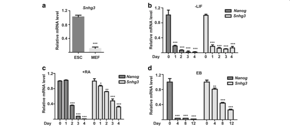
To explore the potential functions of Snhg3 in mESCs, we first compared the expression levels between mESCs and MEFs. qRT-PCR analysis showed that the expression of Snhg3 was much higher in mESCs than in MEFs (~ 9-fold; Fig. 1a). To further investigate the expression pat-terns of Snhg3 during differentiation, we adopted three different methods to induce mESC differentiation, in-cluding LIF withdrawal, RA addition, and EB formation. Similar to levels of the key pluripotency marker Nanog, the expression of Snhg3 was gradually downregulated after differentiation (Fig. 1b). Thus, the enrichment of Snhg3 in an undifferentiated state suggested its potential regulatory function in mESCs.
Knockdown of Snhg3 impairs mESC self-renewal
To understand the role of Snhg3 in mESCs, we first transfected two efficient and specific targeting Snhg3 siRNA oligos into mESCs to knock down its expression (Fig. 2a). After transfection for 48 h, siSnhg3-treated mESCs quickly lost their mESC morphology, became flat, and displayed weaker AP activity, when compared to those features in control cells (Fig. 2b). To further
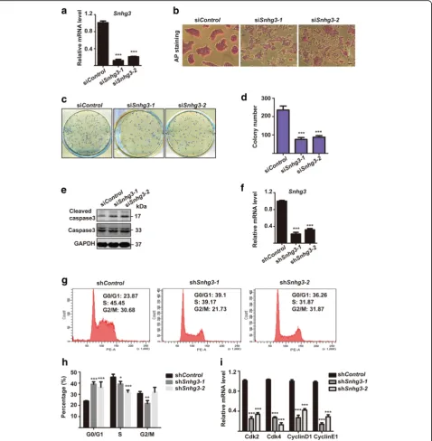
access whether Snhg3 regulates self-renewal in mESCs, we performed secondary colony formation assays by re-plating 800 siControl or siSnhg3-treated mESCs on feeders for 7 days (Fig. 2c). As indicated by AP staining, depletion of Snhg3 significantly impaired the colony for-mation ability of mESCs (Fig. 2d). We hypothesized that this might be a consequence of apoptosis triggered by Snhg3 depletion. To test this hypothesis, we examined the expression of cleaved caspase 3, a marker of apop-tosis, by western blotting. Indeed, cleaved caspase 3 levels were increased upon Snhg3 knockdown, whereas total caspase 3 remained stable (Fig.2e). In addition, cell apoptosis was also analyzed via Annexin V and PI stain-ing. Flow cytometry results showed that Snhg3 knock-down led to more early apoptotic cells, and less viable cells, which suggested that Snhg3 depletion results in apoptosis (Additional file1: Figure S1a-b).
To understand the mechanism associated with sup-pressed self-renewal observed in Snhg3-knockdown mESCs, we first generated stable Snhg3-knockdown mESCs (shSnhg3-1 and shSnhg3-2) using a lentivirus de-livery system; shRNA targeting luciferase served as a nega-tive control (shControl) (Fig.2f ). The cell cycle profile of shSnhg3 mESCs was analyzed by PI staining, followed by flow cytometry (Fig.2g). Notably, shSnhg3mESCs showed a marked increase in the proportion of cells in G0/G1 phase, and a concomitant decreases in S phase popula-tions, as compared to those in the shControlcells, indicat-ing that Snhg3 depletion leads to cell cycle arrest at the G1 phase (Fig. 2h). Consistently, the expression of genes encoding cell cycle-related molecules including Cdk2,
Cdk4, CyclinD1, and CyclinE1 was reduced in shSnhg3
mESCs (Fig.2i). These data indicated that the disruption of mESC self-renewal by Snhg3 depletion was par-tially mediated by cell cycle perturbation and apop-tosis induction.
Snhg3 depletion alters mESC pluripotency
To determine whether Snhg3 also affects the pluripo-tency in mESCs, we performed immunostaining (IF) and western blot to probe for the expression levels of the key pluripotency factors Nanog and Oct4 after knocking Fig. 2Snhg3 depletion impairs mouse embryonic stem cell (mESC) self-renewal.amESCs were transfected with siControlor two different oligos targetingSnhg3for 48 h and subjected to qRT-PCR to access knockdown efficiency.bAlkaline phosphatase (AP) staining of mESCs after siControl
down Snhg3 in mESCs; both markers were dramatically reduced compared to the expression in siControl cells (Fig. 3a, b). qRT-PCR also confirmed that Snhg3 knock-down repressed the expression of several pluripotency markers including Nanog, Oct4, Sox2, Klf4, Tbx3, and
Esrrb(Fig.3c). We then speculated that Snhg3 depletion might lead to mESC differentiation. Therefore, we fur-ther examined the expression patterns of three germ layer markers in these cells by qRT-PCR. As shown in Fig.3d, siSnhg3-treated mESCs were primed for differen-tiation, which was characterized by elevated expression levels of endoderm markers (Gata6, Gata4, Foxa2,
Sox17) and the ectoderm marker Mash1, although
Snhg3 knockdown had only a marginal effect on expres-sion of the ectoderm gene Nestin and mesoderm genes (Goosecoid, Brachyury) (Fig. 3d). Finally, to validate the
differentiation state of Snhg3-knockdown mESCs, we performed an EB formation assay for 12 days to mimic early mouse embryonic development [19]. Importantly, cystic EBs, which contain primitive endoderm-derived cells, appeared early at day 8 after Snhg3 depletion and became larger by day 12 (Fig.3e). In contrast, few cystic EBs appeared in the siControl group even after 12 days (Fig. 3e). These results demonstrated that Snhg3 is re-quired for maintaining the pluripotency of mESCs.
Snhg3 overexpression promotes self-renewal and represses mESC differentiation
Next, we generated stable Snhg3-overexpressing mESCs using a lentivirus delivery system, in which GFP-overexpressing mESCs served as a negative control. After validating the constitutive overexpression ofSnhg3
transcripts (Fig. 4a), we examined the self-renewal of these cells by performing secondary colony formation assays, in which the cells were plated at different dens-ities on feeders for 7 days. Compared to that with GFP-overexpressing mESCs, more AP-stained colonies were observed after plating 400 or 800 Snhg3-overexpressing mESCs (Fig. 4b, c), indicating robust cell proliferation after Snhg3 overexpression. Consistently, increased cell growth after Snhg3 overexpression was also observed via CCK-8 assay (Additional file1: Figure S1c). Additionally, only a slight increase in the expression ofNanogwas ob-served, although the other two pluripotency factors,
namelyOct4 andSox2, were not affected (Fig. 4d). Not-ably, Snhg3 overexpression significantly inhibited ex-pression of markers of the three germ layer, as confirmed by qRT-PCR (Fig. 4e), suggesting that Snhg3 expression is sufficient to perturb differenti-ation. Moreover, after EB formation for 6 days, Snhg3-overexpressing EBs became smaller, dispersed, and irregular when compared to control EBs, indicating that the differentiation process was dysregulated (Fig. 4f ). Thus, these data demonstrated that ectopic Snhg3 expression promotes self-renewal and blocks differentiation in mESCs.
Snhg3 is required for early mouse embryonic development
Currently, the role of Snhg3 in mouse embryo develop-ment was unknown. To assess its potential function, zy-gotes were microinjected with siRNAs against Snhg3 or
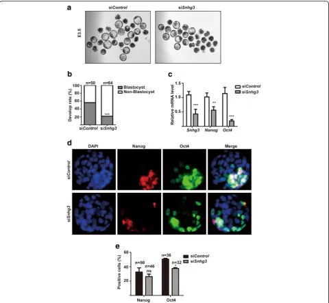
Controloligonucleotides, and the morphology and devel-opment rate were observed and recorded at E3.5. We found that whereas 56% of control embryos developed normally to the blastocyst stage (n= 50), the blastocyst development rate for Snhg3-depleted embryos dropped to 21% (n= 64) (Fig. 5a, b). Moreover, qRT-PCR results indicated that after Snhg3knockdown, the mRNA levels of Nanog and Oct4were decreased to around 50% and 20% of control levels, respectively (Fig. 5c). Similarly, IF staining also showed that the number of Oct4-positive cells was decreased inSnhg3-depleted embryos, whereas the Nanog-positive cell number was only slightly chan-ged (Fig. 5d, e). These data suggested that Snhg3 plays an important role during early embryonic development.
Snhg3 functions with Nanog and Oct4 in mESCs
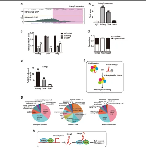
To understand the role of Snhg3 at the molecular level, we first searched for its upstream regulators in mESCs using published ChIP-seq data (GSE73952, GSE53490) [20, 21]. Notably, we found that the active histone marker tri-methylation of histone 3 lysine 4 (H3K4me3) was enriched at the promoter of Snhg3, whereas the re-pressive histone marker tri-methylation of histone 3 ly-sine 27 (H3K27me3) was undetectable, indicating that the Snhg3transcript was activated in mESCs and that it was in part regulated by an epigenetic mechanism (Fig. 6a). Further, increasing evidence suggests that the transcription of lncRNAs is activated by the core pluri-potency transcription factors in ESCs [4,22]. Therefore, we performed ChIP assays to examine the binding of Nanog, Oct4, and Sox2 to theSnhg3 promoter. IgG was used as a negative control (Fig 6b). ChIP-qPCR results confirmed the enrichment of Nanog and Oct4, whereas Sox2 was not found, at the Snhg3 promoter region (Fig. 6b). To further validate the transcriptional activa-tion of Snhg3 by Nanog and Oct4, qRT-PCR analysis was conducted after downregulating Nanog or Oct4
using siRNAs in mESCs. The results showed thatNanog
or Oct4 knockdown significantly decreased the expres-sion ofSnhg3(Fig.6c). Thus, these results suggested that Nanog and Oct4 are upstream regulators of Snhg3 in mESCs.
To determine how Snhg3 modulates its downstream target genes in mESCs, we first performed subcellular fractionation analysis to delineate the localization of Snhg3. qRT-PCR results demonstrated the Snhg3 tran-scripts were distributed similarly in both the cytoplasm and nucleus (Fig.6d), suggesting that Snhg3 might exert its function at both the transcriptional and
post-transcriptional levels. Since we proved that Snhg3 is dir-ectly targeted and regulated by Nanog and Oct4, and that the expression of Nanog and Oct4 are also de-creased after Snhg3 knockdown (Fig. 3b), we inferred that there might be a feedback loop between Snhg3 and these two pluripotency factors in mESCs. To test this, we performed RIP assays using Nanog and Oct4 anti-bodies. Sox2 antibody and IgG were used as controls. qRT-PCR showed that there was a substantial enrich-ment of Snhg3 after the pull-down assays with Nanog and Oct4 antibodies (Fig.6e). In contrast, no significant enrichment of Snhg3 was found with anti-Sox2 (Fig.6e). These data indicated that Snhg3 can bind Nanog and Oct4 in mESCs. Finally, to explore potential interacting partners of Snhg3 in mESCs, we performed RNA pull-down assays followed by mass spectrometry and identi-fied 126 Snhg3-bound proteins (Fig.6f, Additional file3: Table S3). Bioinformatics analysis based on the Gene Ontology (GO) of identified proteins was then per-formed using the PANTHER database (Fig. 6g). Under the “Biological Process” classification, large proportions of genes were involved in metabolic and cellular pro-cesses. For the“Protein Class,”categories of nucleic acid binding and oxidoreductase were significantly enriched, whereas for the “Molecular Function,” catalytic activity and binding were significantly enriched (Fig. 6g). Thus, these data suggested the possible involvement of Snhg3 in metabolic regulation or as a scaffold to recruit pro-teins or ncRNAs in mESCs.
Discussion
Accumulating data have shown that lncRNAs, which were previously considered as transcriptional noise, par-ticipate in multiple biological processes [23]. Although lncRNAs are expressed at a lower level than protein-coding genes, they display more tissue- or cell type-specific expression patterns [24]. Therefore, lncRNAs have been considered essential regulators of ES cell maintenance or differentiation. For example, lncKdm2b
maintains mESC self-renewal via transcriptional activa-tion of the pluripotency factor Zbtb3 [8]. In contrast, lin-cROR functions as a competing endogenous RNA (ceRNA) for miR-145, which targets mRNAs encoding core pluripotency genes, thereby promoting the self-renewal in hESCs [25]. Furthermore, LncPRESS1 aug-ments pluripotency in hESCs by impairing SIRT6-mediated histone H3K56 deacetylation at the promoter regions of pluripotency genes [9]. Thus, these studies in-dicate that lncRNAs can adopt transcriptional, post-transcriptional, and epigenetic regulatory roles to modu-late gene expression in ESCs.
study, in which its depletion impaired mESC morph-ology and downregulated the expression of several pluri-potency factors including Nanog, Oct4, Sox2, Klf4, and Zfp42 [4]. However, how Snhg3 precisely exerts its effect on mESCs was not characterized. Here, we used both loss-of-function and gain-of-function strategies to dem-onstrate that Snhg3 can promote the self-renewal and pluripotency of mESCs, which mainly occurs through associations with the core pluripotency factors Nanog and Oct4 via potential feedback loops (Fig. 6h). In
addition, our data also implied an important role for Snhg3 in early mouse embryonic development.
Mouse Snhg3 is located on chromosome 4 (chr4: 132, 351,934-132,353,633, UCSC Genome Browser/mm10) and is transcribed on the reverse strand. Interestingly, we found that knocking down Snhg3 in mESCs also re-duced the mRNA level of its neighbor gene Rcc1 (data not shown), a guanine-nucleotide exchange factor for Ran GTPase and plays roles in mitosis, nuclear envelope assembly. The role of Rcc1 in ESCs is not characterized Fig. 5Snhg3 is essential for mouse embryonic development.aMorphology of E3.5 mouse embryos after siControlor siSnhg3treatment; × 10 objective.bsiControl- or siSnhg3-injected zygotes were cultured in vitro until E4.5 and the blastocyst/non-blastocyst rate was quantified. siControl,
Fig. 6Snhg3 is regulated by pluripotency factors and its associated protein network.aGenome browser plot of histone methylation related to transcriptional activation (H3K4me3) and repression (H3K27me3) in the gene body region ofSnhg3in mouse embryonic stem cells (mESCs). The gray peaks indicate potential H3K4me3-binding sites in Snhg3. The purple box highlights the proximal promoter regions (−0.5 kb to + 1.5 kb of
before, yet a paper has found it may be a pluripotency marker in embryonal carcinoma cell line [26]. Thus, be-sides interacting with pluripotency factors, Snhg3 may also work with Rcc1 to regulate stemness in ESCs.
Snhg3 was distributed at a similar ratio in both the nu-cleus and cytoplasm. We have studied the functions of nuclear Snhg3; however, the role of cytoplasmic Snhg3 needs to be explored. It is possible that Snhg3 can regu-late the alternative splicing of pre-mRNA transcripts, modulate mRNA transportation, or act as a ceRNA for miRNAs.
LncRNAs also tend to serve as flexible scaffolds and bind multiple chromatin complexes [23]. One previous study using a RIP method showed that Snhg3 can interact with several chromatin-remodeling proteins including PRC2, JARID1B, RING1B, and SUV39H1, which can be characterized as chromatin “writers” (PRC2, SUV39H1), an“eraser”(JARID1B), and a“reader”(RING1B) [4]. How-ever, we failed to identify these proteins in our RNA pull-down and mass spectrometry results. Additionally, there was no significant overlap in affected gene expression pro-files from Snhg3 and its interacting chromatin remodeling proteins [4].
LncRNAs commonly regulate gene expression through their interactions with RNA-binding proteins (RBPs) [27]. Indeed, we found that Snhg3 binds several RBPs based on RNA pull-down and mass spectrometry results, including Hnrnpu, Hnrnpa2b1, Hnrnpa1, Hnrnpr, Hnrnph1, Nucleolin, and Pcbp2. Interestingly, some of these RBPs were reported to be associated with ESC maintenance. For example, Hnrnpu can activate Oct4 expression and is required for proper ESC proliferation [28, 29]. Further, knockdown of Hnrnpa2b1 was found to disrupt ESC proliferation via cell cycle arrest at the G0/G1 phase [30]. Moreover, Nucleolin can form a com-plex with lncRNAs or proteins in ESCs, which in turn maintains self-renewal and represses apoptosis [5, 31,
32]. Based on these data, we speculated that some RBPs such as Hnrnpu, Hnrnpa2b1, and Nucleolin could form complexes with Snhg3 to regulate specific classes of genes in mESCs. Further investigations are needed to confirm these interactions and potential functions.
Finally, Snhg3 may also play roles in induced pluripo-tent stem cell (iPSC) production and regulation. First de-veloped in 2006, iPSCs are pluripotent stem cells reprogrammed from adult somatic cells by induced ex-pression of transcription factors like Oct4/Sox2/Nanog/ Lin28 or Oct4/Sox2/c-Myc/Klf4 [33, 34]. Obviating the ethical concern about the use of ESCs, iPSCs become at-tractive sources for organ regeneration and therapeutic application. Given that Snhg3 could form a feedback loop with Nanog and Oct4 in mESCs, it is possible that Snhg3 has a potential function in iPSCs. Further studies are required to validate the function of Snhg3 in iPSCs.
Conclusions
In this study, we demonstrated the function of Snhg3 in mESC self-renewal, pluripotency, and early mouse em-bryonic development. Snhg3 was found to interact with core pluripotency factors and form a possible feedback loop to exert its function in mESCs. Our findings clarify the importance of long non-coding RNA in balancing the stemness and differentiation and indicate that it is essential for mouse early embryogenesis.
Additional files
Additional file 1:Figure S1.Snhg3 affects apoptosis and proliferation in mESCs. a-b mESCs were transfected with siControl or siSnhg3 for 48 h, followed by Annexin V and PI staining. Representative results of flow cytometry (a) and the statistical analysis (b) showed that Snhg3 depletion resulted in less viable cells (Q3, Annexin V(−) and PI(−)) and more early apoptotic cells (Q4, Annexin V(+) and PI(−)).c The CCK-8 assay was used to evaluate the proliferation of Control or Snhg3 overexpressing mESCs for different time points. Data are presented as mean ± SD;n= 3, two-way ANOVA. **p< 0.01, ***p< 0.001 for all panels. (TIF 16312 kb)
Additional file 2:siRNAs and primer sequences. (DOCX 18 kb)
Additional file 3:Identified Snhg3 interacting proteins. (XLSX 26 kb)
Abbreviations
AP:Alkaline phosphatase; ceRNA: Competing endogenous RNA; ChIP: Chromatin immunoprecipitation; EB: Embryoid body; H3K27me3: Histone marker tri-methylation of histone 3 lysine 27; H3K4me4: Histone marker tri-methylation of histone 3 lysine 4; IF: Immunofluorescence staining; lncRNA: Long non-coding RNA; MEF: Mouse embryonic fibroblast; mESCs: Mouse embryonic stem cells; PI: Propidium iodide; qRT-PCR: Quantitative reverse transcription polymerase chain reaction; RBP: RNA-binding protein; RIP: RNA immunoprecipitation; Snhg3: Small nucleolar RNA host gene 3; WB: Western blotting
Acknowledgements
Not applicable.
Authors’contributions
WSL, ZSY, and JJH conceived and designed the project. WSL, JPY, FTS, RH, and SSY performed the experiments. JNZ analyzed the ChIP-seq data. WSL wrote the manuscript. JJH and WSL contributed to the final approval of the manuscript. All authors read and approved the final manuscript.
Funding
This work is supported by the National Key R&D Program of China (2017YFA0102801 and 2017YFC1001901), the National Nature Science Foundation of China (31671540 and 31601179), the Natural Science Foundation of Guangdong Province (2015A020212005 and 2016A030310209), the Guangzhou Science and Technology Project (201803010020), and the Fundamental Research Funds of State Key Laboratory of Ophthalmology (30306020240020307).
Availability of data and materials
All data generated and/or analyzed in this study are included in this published article.
Ethics approval and consent to participate
All experiments involving animals were performed in accordance with the guidelines approved by the Institutional Animal Care and Use Committee of Sun Yat-Sen University, People’s Republic of China.
Consent for publication
Competing interests
The authors declare that they have no competing interests.
Author details
1The State Key Laboratory of Ophthalmology, Zhongshan Ophthalmic
Center, Sun Yat-sen University, Guangzhou 510060, China.2MOE Key
Laboratory of Gene Function and Regulation, Guangzhou Key Laboratory of Healthy Aging Research and State Key Laboratory of Biocontrol, SYSU-BCM Joint Research Center, School of Life Sciences, Sun Yat-sen University, Guangzhou 510275, China.3Department of Dermatology and Skin Science,
University of British Columbia, Vancouver, BC V5Z 4E8, Canada.4Verna and
Marrs Mclean Department of Biochemistry and Molecular Biology, Baylor College of Medicine, One Baylor Plaza, Houston, TX 77030, USA.
Received: 3 January 2019 Revised: 8 May 2019 Accepted: 20 May 2019
References
1. Young RA. Control of the embryonic stem cell state. Cell. 2011;144(6):940–54. 2. Morey L, Santanach A, Di Croce L. Pluripotency and epigenetic factors in
mouse embryonic stem cell fate regulation. Mol Cell Biol. 2015;35(16):2716–28. 3. Dinger ME, Amaral PP, Mercer TR, Pang KC, Bruce SJ, Gardiner BB, et al.
Long noncoding RNAs in mouse embryonic stem cell pluripotency and differentiation. Genome Res. 2008;18(9):1433–45.
4. Guttman M, Donaghey J, Carey BW, Garber M, Grenier JK, Munson G, et al. lincRNAs act in the circuitry controlling pluripotency and differentiation. Nature. 2011;477(7364):295–300.
5. Lin N, Chang KY, Li Z, Gates K, Rana ZA, Dang J, et al. An evolutionarily conserved long noncoding RNA TUNA controls pluripotency and neural lineage commitment. Mol Cell. 2014;53(6):1005–19.
6. Derrien T, Johnson R, Bussotti G, Tanzer A, Djebali S, Tilgner H, et al. The GENCODE v7 catalog of human long noncoding RNAs: analysis of their gene structure, evolution, and expression. Genome Res. 2012;22(9):1775–89. 7. Guttman M, Amit I, Garber M, French C, Lin MF, Feldser D, et al. Chromatin
signature reveals over a thousand highly conserved large non-coding RNAs in mammals. Nature. 2009;458(7235):223–7.
8. Ye B, Liu B, Yang L, Zhu X, Zhang D, Wu W, et al. LncKdm2b controls self-renewal of embryonic stem cells via activating expression of transcription factor Zbtb3. EMBO J. 2018;37(8).https://doi.org/10.15252/embj.201797174. 9. Jain AK, Xi Y, McCarthy R, Allton K, Akdemir KC, Patel LR, et al. LncPRESS1 is a p53-regulated LncRNA that safeguards pluripotency by disrupting SIRT6-mediated de-acetylation of histone H3K56. Mol Cell. 2016;64(5):967–81. 10. Xu C, Zhang Y, Wang Q, Xu Z, Jiang J, Gao Y, et al. Long non-coding RNA
GAS5 controls human embryonic stem cell self-renewal by maintaining NODAL signalling. Nat Commun. 2016;7:13287.
11. Zhang T, Cao C, Wu D, Liu L. SNHG3 correlates with malignant status and poor prognosis in hepatocellular carcinoma. Tumour Biol. 2016;37(2):2379–85. 12. Huang W, Tian Y, Dong S, Cha Y, Li J, Guo X, et al. The long non-coding RNA
SNHG3 functions as a competing endogenous RNA to promote malignant development of colorectal cancer. Oncol Rep. 2017;38(3):1402–10. 13. Hong L, Chen W, Wu D, Wang Y. Upregulation of SNHG3 expression
associated with poor prognosis and enhances malignant progression of ovarian cancer. Cancer Biomark. 2018;22(3):367–74.
14. Fei F, He Y, He S, He Z, Wang Y, Wu G, et al. LncRNA SNHG3 enhances the malignant progress of glioma through silencing KLF2 and p21. Biosci Rep. 2018;38(5).https://doi.org/10.1042/BSR20180420.
15. Liu L, Ni J, He X. Upregulation of the long noncoding RNA SNHG3 promotes lung adenocarcinoma proliferation. Dis Markers. 2018;2018: 5736716.
16. Lu W, Fang L, Ouyang B, Zhang X, Zhan S, Feng X, et al. Actl6a protects embryonic stem cells from differentiating into primitive endoderm. Stem Cells. 2015;33(6):1782–93.
17. Lee DF, Su J, Sevilla A, Gingold J, Schaniel C, Lemischka IR. Combining competition assays with genetic complementation strategies to dissect mouse embryonic stem cell self-renewal and pluripotency. Nat Protoc. 2012; 7(4):729–48.
18. Zhan S, Zhang X, Cao S, Huang J. Benzo(a)pyrene disrupts mouse preimplantation embryo development. Fertil Steril. 2015;103(3):815–25. 19. Keller GM. In vitro differentiation of embryonic stem cells. Curr Opin Cell
Biol. 1995;7(6):862–9.
20. Liu X, Wang C, Liu W, Li J, Li C, Kou X, et al. Distinct features of H3K4me3 and H3K27me3 chromatin domains in pre-implantation embryos. Nature. 2016;537(7621):558–62.
21. Clouaire T, Webb S, Bird A. Cfp1 is required for gene expression-dependent H3K4 trimethylation and H3K9 acetylation in embryonic stem cells. Genome Biol. 2014;15(9):451.
22. Tu J, Tian G, Cheung HH, Wei W, Lee TL. Gas5 is an essential lncRNA regulator for self-renewal and pluripotency of mouse embryonic stem cells and induced pluripotent stem cells. Stem Cell Res Ther. 2018;9(1):71. 23. Hu W, Alvarez-Dominguez JR, Lodish HF. Regulation of mammalian cell
differentiation by long non-coding RNAs. EMBO Rep. 2012;13(11):971–83. 24. Ghosal S, Das S, Chakrabarti J. Long noncoding RNAs: new players in the molecular mechanism for maintenance and differentiation of pluripotent stem cells. Stem Cells Dev. 2013;22(16):2240–53.
25. Wang Y, Xu Z, Jiang J, Xu C, Kang J, Xiao L, et al. Endogenous miRNA sponge lincRNA-RoR regulates Oct4, Nanog, and Sox2 in human embryonic stem cell self-renewal. Dev Cell. 2013;25(1):69–80.
26. Hoff AM, Alagaratnam S, Zhao S, Bruun J, Andrews PW, Lothe RA, et al. Identification of novel fusion genes in testicular germ cell tumors. Cancer Res. 2016;76(1):108–16.
27. Sun X, Haider Ali MSS, Moran M. The role of interactions of long non-coding RNAs and heterogeneous nuclear ribonucleoproteins in regulating cellular functions. Biochem J. 2017;474(17):2925–35.
28. Vizlin-Hodzic D, Johansson H, Ryme J, Simonsson T, Simonsson S. SAF-A has a role in transcriptional regulation of Oct4 in ES cells through promoter binding. Cell Reprogram. 2011;13(1):13–27.
29. Hasegawa Y, Brockdorff N, Kawano S, Tsutui K, Tsutui K, Nakagawa S. The matrix protein hnRNP U is required for chromosomal localization of Xist RNA. Dev Cell. 2010;19(3):469–76.
30. Choi HS, Lee HM, Jang YJ, Kim CH, Ryu CJ. Heterogeneous nuclear ribonucleoprotein A2/B1 regulates the self-renewal and pluripotency of human embryonic stem cells via the control of the G1/S transition. Stem Cells. 2013;31(12):2647–58.
31. Li H, Wang B, Yang A, Lu R, Wang W, Zhou Y, et al. Ly-1 antibody reactive clone is an important nucleolar protein for control of self-renewal and differentiation in embryonic stem cells. Stem Cells. 2009;27(6):1244–54. 32. Yang A, Shi G, Zhou C, Lu R, Li H, Sun L, et al. Nucleolin maintains
embryonic stem cell self-renewal by suppression of p53 protein-dependent pathway. J Biol Chem. 2011;286(50):43370–82.
33. Takahashi K, Tanabe K, Ohnuki M, Narita M, Ichisaka T, Tomoda K, et al. Induction of pluripotent stem cells from adult human fibroblasts by defined factors. Cell. 2007;131(5):861–72.
34. Yu J, Vodyanik MA, Smuga-Otto K, Antosiewicz-Bourget J, Frane JL, Tian S, et al. Induced pluripotent stem cell lines derived from human somatic cells. Science. 2007;318(5858):1917–20.
Publisher’s Note