R E S E A R C H
Open Access
Variable sensitivity to
complement-dependent cytotoxicity in murine
models of neuromyelitis optica
Yiting Liu
1, Danielle E. Harlow
2, Katherine S. Given
2, Gregory P. Owens
1, Wendy B. Macklin
2,4and Jeffrey L. Bennett
1,3,4*Abstract
Background:Studies of neuromyelitis optica (NMO), an autoimmune disease of the central nervous system (CNS),
have demonstrated that autoantibodies against the water channel aquaporin-4 (AQP4) induce astrocyte damage through complement-dependent cytotoxicity (CDC). In developing experimental models of NMO using cells, tissues or animals from mice, co-administration of AQP4-IgG and normal human serum, which serves as the source of human complement (HC), is required. The sensitivity of mouse CNS cells to HC and CDC in these models is not known.
Methods: We used HC and recombinant monoclonal antibodies (rAbs) against AQP4 to investigate CDC on
mouse neurons, astrocytes, differentiated oligodendrocytes (OLs), and oligodendrocyte progenitors (OPCs) in the context of purified monocultures, neuroglial mixed cultures, and organotypic cerebellar slices.
Results: We found that murine neurons, OLs, and OPCs were sensitive to HC in monocultures. In mixed murine neuroglial cultures, HC-mediated toxicity to neurons and OLs was reduced; however, astrocyte damage induced by an AQP-specific rAb #53 and HC increased neuronal and oligodendroglial loss. OPCs were resistant to HC toxicity in neuroglial mixed cultures. In mouse cerebellar slices, damage to neurons and OLs following rAb #53-mediated CDC was further reduced, but in contrast to neuroglial mixed cultures, astrocyte damage sensitized OPCs to complement damage. Finally, we established that some injury to neurons, OLs, and OPCs in cell and slice cultures resulted from the activation of HC by anti-tissue antibodies to mouse cells.
Conclusions:Murine neurons and oligodendroglia demonstrate variable sensitivity to activated complement
based on their differentiation and culture conditions. In organotypic cultures, the protection of neurons, OLs, and OPCs against CDC is eliminated by targeted astrocyte destruction. The activation of human complement proteins on mouse CNS cells necessitates caution when interpreting the results of mouse experimental models of NMO using HC.
Keywords: NMO, Complement, Cytotoxicity, Mouse model, Neuroglial networks
* Correspondence:[email protected] 1
Department of Neurology, University of Colorado, School of Medicine, 12700 E. 19th Ave, Aurora, CO, USA
3Department of Ophthalmology, University of Colorado, School of Medicine, 12700 E. 19th Ave, Aurora, CO, USA
Full list of author information is available at the end of the article
Background
Neuromyelitis optica (NMO) is a severe autoimmune disease of the central nervous system (CNS) associated with predilection for the spinal cord and optic nerve. Histopathology and animal models demonstrate that complement-dependent cytotoxicity (CDC) plays a key role in the initiation of the NMO pathology [1].
The complement system consists of over 30 compo-nents that are normally found inactive in serum and is an essential immune regulator of host defense to infection, cell integrity, and tissue homeostasis. The complement cascade can be activated by antigen-antibody complexes (classic pathway) or the antibody-independent lectin (lec-tin pathway). Additionally, the alternative pathway serves to amplify the classic and lectin pathways. All three path-ways converge to trigger activation of terminal comple-ment components resulting in cell lysis [2]. In the CNS, the complement cascade regulates synaptic refinement and neuronal survival during development [3, 4] and plays a role in maintaining brain homeostasis in adulthood [5]. There is mounting evidence that complement synthesis and activation are increased in neurodegenerative and neuroinflammatory environments [6].
NMO lesions show perivascular deposition of im-munoglobulin and activated complement [7, 8], and acti-vated complement proteins are eleacti-vated in the serum and cerebrospinal fluid (CSF) of NMO patients [9, 10]. Serum autoantibodies (NMO-IgG) against the water channel aquaporin-4 (AQP4) are found in most NMO patients [11]. In the CSF of NMO-IgG positive patients, there exists a dynamic and enriched population of ex-panded plasmablast clones producing AQP4-specific antibodies [12]. Both serum NMO-IgG and CSF-derived AQP4-specific monoclonal recombinant antibodies re-capitulate NMO pathology when microinjected into the CNS, added to ex vivo spinal cord slices in the presence of human complement proteins [13], introduced into ro-dent models of CNS inflammation [12, 14], or injected into mice pretreated with Freund’s complete adjuvant [15]. Astrocytes are selectively targeted in NMO, as evidenced by the extensive loss of immunoreactivity for the astrocytic proteins AQP4 and glial fibrillary acidic protein (GFAP) [7]. Moreover, inhibition of the classic complement pathway blocks neuropathology in both in vitro and in vivo models of NMO [16].
In developing experimental mouse models of NMO in vivo, ex vivo, or in vitro, co-administration of AQP4-IgG and normal human serum as the source of human com-plement (HC) is required to produce NMO pathological lesions [17]. The weak activity of intrinsic mouse com-plement [18] and the presence of comcom-plement inhibi-tor(s) in the mouse serum [19] may combine to prevent rigorous complement activation in the absence of added HC in NMO models. Additional effects of HC on other
CNS cells have not been characterized in NMO animal models.
In this study, we investigated the toxic effects of HC on murine neurons, astrocytes, differentiated oligodendro-cytes (OLs), and oligodendrocyte progenitors (OPCs) in the presence or absence of AQP4-IgG in several in vitro and ex vivo CNS culture systems (monocultures, neuro-glial mixed cultures, and organotypic cerebellar slice cul-tures). Our results reveal that neurons, OLs, and OPCs derived from mice display differential sensitivity to HC de-pending on the complexity of the neuroglial environment, and that AQP4-IgG-targeted astrocyte destruction results in secondary oligodendrocyte and neuronal loss.
Methods
Recombinant antibodies
The recombinant monoclonal human AQP4 anti-body #53 (rAb #53) was constructed from a clonally ex-panded plasmablast recovered from the cerebrospinal fluid (CSF) of a seropositive NMO patient. The control human antibody was generated from a CSF plasmablast sorted from a chronic meningitis patient. Recombinant antibodies were produced using a dual vector transient transfection system and purified with protein A-sepharose (Sigma-Aldrich, St. Louis, MO) as previously described [12].
Biochemical assays and reagents
Lactate dehydrogenase (LDH) cytotoxicity assays were performed on cell culture supernatants using the LDH cytotoxicity detection kit (TakaRa Bio, Shiga, Japan). Except for IncuCyte live imaging (see below), propidium iodide (PI) (Sigma) was used at 5μg/ml in the culture medium to label dead cells in cell and organotypic slice cultures. Nor-mal, C1q-depleted, C3-depleted, C4-depleted, C5-depleted, and C9-depleted human serum (Complement Technology, Tyler, TX) were used as the source of human complement. Pooled and individual normal human (Sigma and Innova-tive Research), rat (Complement Technology), and mouse (Complement Technology and Innovative Research) sera were used as sources of complement for comparative com-plement experiments. Serum proteins were obtained by passing the serum through Amicon Ultra 3KD centrifugal filters (Millipore, Billerica, MA). Protein A/G plus agarose (Thermo Fisher Scientific, Waltham, MA) was incubated with human serum overnight at 4 °C, then centrifuged and discarded to deplete IgGs in the serum. Heat-inactivated serum was obtained by incubating normal serum at 58 °C for 30 min.
Primary cell cultures
described from mice (CD-1 or PLP-eGFP [20]) or rats (Sprague Dawley) (Harlan, Indianapolis, IN) with some modifications [21–24]. Mixed glial cultures were pre-pared from P0-1 dissociated cortices and plated in poly-D-lysine-coated 24-well plates at a density of one brain per plate. Plates were used for experiments when astro-cyte layer was confluent and oligodendroastro-cyte cell bodies were apparent (day 5–8 in culture). Purified oligo-dendrocyte monocultures were prepared by shaking flasks of mixed glial cultures overnight at 200 rpm to de-tach the OPCs. OPCs were plated on poly-D-lysine/lam-inin coated dishes and maintained in media contapoly-D-lysine/lam-ining 10 ng/ml PDGF (platelet-derived growth factor)/FGF (fibroblast growth factor) (Peprotech, Rocky Hill, NJ) for 48–72 h prior to experiments. To differentiate OPCs into OLs, PDFG/FGF was withdrawn after 48 h and media supplemented with 40 ng/ml T3 (Sigma). OLs were used for experiments 24–72 h after exposure to T3. For neuroglial mixed cultures, P0-1 cortices were dissected, dissociated in 0.25% trypsin, and cells were plated on poly-D-lysine/Laminin coated coverslips (BD Biosciences, Franklin Lakes, NJ) in Neurobasal medium containing B27 supplement, 0.05 mM L-glutamine, penicillin-streptomycin (all from Life Technologies) and 5% heat-inactivated FBS (Sigma). Neuronal monocul-tures were prepared from E15-17 mouse cortex and cultured in Neurobasal medium in the presence of B27 supplement, 0.05 mM L-glutamine and penicillin-streptomycin. On day 2–5 in culture, 2 μM ara-C (Sigma) was added to inhibit the growth of glial cells. Human cortical neuron cultures (iCell Neurons) were cultured in iCell proprietary maintenance medium pur-chased from Cellular Dynamics (Madison, WI). Cell cultures were maintained at 37 °C in 5 or 8.5% CO2 atmospheric conditions.
Cerebellar slice culture
Sagittal slices (300μm) were prepared from the cerebella of C57Bl6 or PLP-eGFP mice at P10 and cultured on MilliCell 0.4μm membrane inserts (Millipore) for 10–14 days in media containing 25% inactivated horse serum [25]. Prior to treatment, slices were switched to a serum-free Neurobasal medium supplemented with B27, 2 mM GlutaMAX, and 28 mM D-glucose (Sigma).
Immunostaining
Differential labeling of cell surface and internalized proteins in cell culture
Cell cultures were washed with fresh culture medium, then incubated with 20μg/ml recombinant antibody at 4 or 37 °C for 1 h. Cells were then washed and fixed in 4% paraformaldehyde (Thermo Scientific) in phosphate-buffered saline (PBS) and rinsed and blocked with 3% normal goat serum (NGS) and 2% bovine serum albumin
(BSA) in PBS without Triton X-100 (all from Sigma). Cells were then probed with Alexa-fluor 488 goat anti-human IgG secondary antibodies (1:500). Subsequently, cells were blocked overnight with unlabeled goat anti-human IgG at 0.25 mg/ml at room temperature and then washed, postfixed, and permeabilized with blocking buffer containing 0.1% Triton X-100. Alexa-fluor 594 goat anti-human IgG (1:500) was used to probe for intra-cellular recombinant antibody. After extensive washing, slides were mounted with ProLong Gold anti-fade reagent with DAPI (Life Technologies).
Immunostaining of fixed cells and brain sclices
Cell cultures or brain slices were fixed in 4% paraformal-dehyde in PBS, rinsed, permeabilized in 0.1% (for cells) or 3% (for slices) Triton X-100 in PBS, and then blocked with 5% normal goat serum (NGS) or normal donkey serum (NDS) in PBS containing 0.1% (for cells) or 0.3% (for slices) Triton X-100 for 1 h. Samples were then in-cubated with primary antibodies in blocking buffer, washed, and then probed with secondary antibodies (Alexa Fluor secondary antibodies, all from Life Tech-nologies). After washing, the coverslips were mounted with ProLong Gold anti-fade reagent (Life Technolo-gies). The brain slices were mounted with Fluoromount-G (Southern Biotech, Birmingham, AL), placed under the glass covers, and sealed. Primary antibodies used in the immunostaining were mouse anti-NeuN (Millipore), mouse anti-β-tubulin (Sigma), rabbit anti-GFAP (DAKO, Carpinteria, CA), mouse anti-Olig1(Millipore), mouse anti-O4 IgM (R&D Systems, Minneapolis, MN ), rabbit anti-LAMP1(Abcam, Cambridge, UK), rabbit anti-Calbindin (Millipore), mouse anti-C3d (a gift from Dr. Joshua Thurman, University of Colorado), rabbit anti-MAC complex (Abcam), and rabbit anti-NG2 and rabbit anti-Olig2 (both are gifts from Dr. Charles Stiles, Harvard University).
Microscopy
Fluorescence images were acquired by Zeiss fluorescence microscope with Axiovision software (Zeiss, Jena, Germany). Confocal images were acquired by Leica SP5 laser scanning microscope (Leica Microsystems GmbH, Wetzlar, Germany) and Zeiss LSM 780 microscope or Olympus FV-1000 confocal microscope (Olympus, Center Valley, PA).
IncuCyte live cell imaging
microscopy using the following filter sets: 460/524 nm (green fluorescence, to detect enhanced green fluorescent protein, eGFP) and 585/635 nm (red fluorescence, to de-tect the DRAQ7 dead cell nuclei; Cell Signaling Technol-ogy, Danvers, MA). Image processing and cell counting were performed using IncuCyte software. Oligodendrocyte lineage cells in the neuroglial mixed cultures were labeled with eGFP in PLP-eGFP mice [20]. In cell culture, OLs and OPCs were defined by their distinct morphologies. In the neuroglia mixed cultures, the majority of oligodendrocytes were bipolar and tripolar OPCs, while a minority of the cells displayed a highly complex network of processes and O4 antigen consistent with differentiating OLs [26]. Neu-rons and astrocytes were identified by their distinct morph-ologies. DRAQ7+ cells were classified as dead neurons or astrocytes based on the different signal intensity and area. Dead oligodendrocytes were identified as DRAQ7+ cells with reduced eGFP signal. The identification of different cell types in mixed cultures was confirmed by immuno-staining with NeuN (neurons), AQP4 and GFAP (astro-cytes), Olig1 (a general oligodendrocyte lineage marker), and O4 (differentiating OLs) (Additional file 1: Figure S1). To assess the total number of the cells in the cultures, cells were stained with Cell Trace Calcein red-orange (Life Technologies) and then scanned and counted using IncuCyte software.
Quantification and statistical analysis
Cell counts in IncuCyte images were quantified by Incu-Cyte software. In each well of the 24-well plate, signals were measured in nine scanned positions and sum-mated. PLP-eGFP positive cell bodies in the brain slices were imaged using a Zeiss fluorescence microscope with 20× objective. Images were quantified with ImageJ (Na-tional Institutes of Health open source). For each slice, 2– 3 images were taken, quantified, and averaged. Slices from three independent experiments were analyzed. Statistical analyses were performed by unpaired Student’st test for single comparisons or by two-way ANOVA for grouped comparisons using GraphPad Prism software. Data are expressed as means ± SD of independent experiments (n≥3). Significance is reported forp< 0.05.
Results
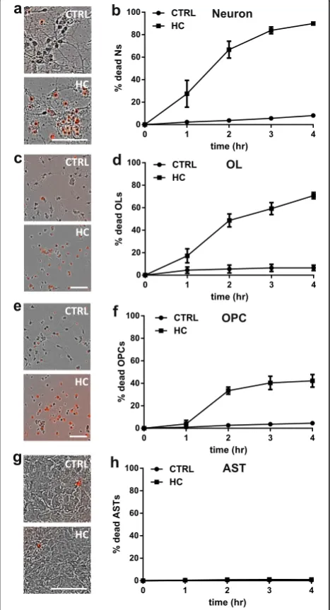
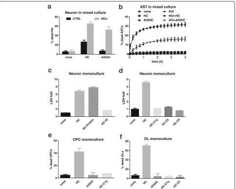
Human serum as the source of human complement is cytotoxic to mouse neurons and oligodendrocytes, but not astrocytes in monocultures
To investigate the effect of HC on murine CNS cells, we assessed the sensitivity of mouse neurons, oligodendro-cytes (OLs), oligodendrocyte progenitor cells (OPCs), and astrocytes to human serum in monocultures. Mono-cultures were exposed to either PBS as a control or 2– 5% (vol/vol) HC. Cell death was measured by the addition of DRAQ7 to the media to label dead/dying
cells, followed by IncuCyte live cell imaging. After 4 h of incubation with 2% HC, 90.0 ± 1.9% of the mouse neur-onal population was DRAQ7+ (Fig. 1a–b). OLs and OPCs were also sensitive to HC exposure with 70.8 ± 2.8% and 42.2 ± 5.6% cell death, respectively, after 4 h incubation (Fig. 1c–f and Additional file 2: Movie 1 and Additional file 3: Movie 2). In contrast, HC did
not induce astrocyte cell death as determined by LDH release assay (Fig. 1g) or IncuCyte live cell im-aging (data not shown). The degree of cell death in these murine CNS monocultures in the presence of HC is summarized in Table 1.
Reduced sensitivity of mouse neurons and OLs to HC in neuroglial mixed cultures
In the CNS, the interaction of neurons and glia may alter their responses to environmental stressors. We in-vestigated how mouse neurons and glial cells responded to HC in mixed cell cultures prepared from PLP-eGFP [20] mouse pups in which both OPCs and differentiated OLs were labeled with eGFP. Cell type specific marker staining showed that the neuroglial mixed cultures con-sisted of 70% astrocytes (GFAP and AQP4), 10–15% oligodendroglial cells (Olig1), and 10–15% neurons (βIII-Tubulin). Most oligodendroglial cells were OPCs, but occasionally maturing OLs (O4+) were noted (Additional file 1: Figure S1).
Cell death in mixed cultures was monitored by Incu-Cyte live imaging. In the mixed cultures, HC was not toxic to astrocytes (Fig. 2a, arrow heads). Neurons remained sensitive to HC in the mixed cultures (Fig. 2a, arrows); however, the magnitude of loss was significantly reduced. The addition of 5% HC caused 31.4 ± 4.0% neuronal death in the mixed culture after 4 h incubation (Fig. 2b and Additional file 4: Movie 3); whereas, HC resulted in greater than 90% neuronal death in the monoculture (Fig. 1b). Neuronal death was confirmed by co-labeling with the dead cell dye propidium iodide (PI) and neuronal marker NeuN (Fig. 2c).
In the neuroglial mixed cultures, the majority of eGFP+ oligodendrocyte lineage cells were bipolar and tripolar progenitors, while a minority of the cells
displayed a highly complex network of processes con-sistent with differentiated OLs [26]. OLs died rapidly in the presence of HC alone with 56.0 ± 1.4% of OLs lost by 4 h (Fig. 2d). OL cell death was reduced, how-ever, in mixed cultures when compared to monocul-tures (70.8 ± 2.8%, Fig. 1e). In contrast to their behavior in monoculture (Fig. 1d–e), OPCs were not sensitive to HC in the neuroglial mixed cultures, even after an extended incubation time of 3 days (Fig. 2e and Additional file 4: Movie 3). As observed in the monocul-ture, astrocytes remained insensitive to HC in the neuro-glial mixed culture (Fig. 2a and Additional file 4: Movie 3). The percent of CNS cell death in mixed cultures is sum-marized in Table 1.
Targeted astrocyte damage increases HC-mediated neuron and OL death in mixed cultures
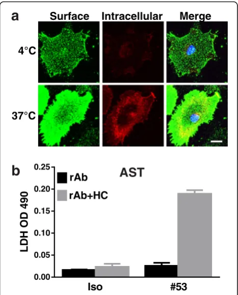
We hypothesized that astrocytes in mixed cultures pro-vided protection against HC to adjacent oligodendro-cytes and neurons. To test this, we used a recombinant monoclonal antibody specific for AQP4 (rAb #53) [12] to target astrocytes via complement-mediated lysis (CDC) [13]. Live staining of rAb #53 of astrocytes at 4 or 37 °C displayed a robust membrane staining and dim cytoplasmic staining, indicating only a small portion of AQP4 being internalized from the cell surface (Fig. 3a). In combination with HC, rAb #53 induced significant astrocyte death, resulting in a marked increase of LDH in the culture medium (Fig. 3b).
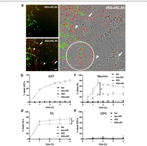
We monitored the kinetics of neuron, oligodendrocyte, and astrocyte death in the mixed cultures following tar-geted rAb #53-mediated astrocyte damage with IncuCyte live cell imaging. No cell death was noted with rAb #53 or isotype control (Iso) without the presence of HC. Treatment with rAb #53+ 5% HC induced rapid cell death, notably among astrocytes (Fig. 4a–b). Dead neu-rons were identified as small round DRAQ7+ particles with high fluorescence intensity (Fig. 4a, arrows), whereas dead astrocytes were distinguished as larger but less intense DRAQ7+ cells (Fig. 4a, arrow head). Dead astrocytes floated into the culture supernatant, resulting in areas of astrocyte depletion (Fig. 4a, circled area). After the peak of astrocyte death (4 h), viable astrocytes repopulated these areas (Additional file 5: Movie 4). Neuronal death increased in the presence of rAb #53-mediated astrocyte destruction, with approximately 70% of neurons dying within 2 h. While some neuronal death may result from their sensitivity to HC alone, only 30% neuronal death was observed with Iso+HC (Fig. 4c) or HC alone (Fig. 2b–c). Astrocyte loss induced by rAb #53 plus HC also increased the death of OLs (Fig. 4d). In contrast, rAb #53-mediated astrocyte destruction caused no loss of OPCs (Fig. 4e). The percent cell death is summarized in Table 1.
Table 1Summary of cell death in monoculture, neuroglial
mixed culture and brain slices
Cell death Monoculturea Mixed cultureb
Brain slicesc
Mouse AST HC No death No death No death
#53+HC 33.9 ± 1.5% 32.0 ± 1.7% Damage from 8 h Mouse N HC 93.4 ± 4.0% 31.4 ± 4.0% 17.4 ± 0.7%
#53+HC 97.7 ± 2.1% 72.3 ± 11.1% 52.3 ± 2.7% Mouse OL HC 70.8 ± 2.8% 56.0 ± 1.4% 26.4 ± 5.7% #53+HC 61.3 ± 7.8% 73.2 ± 5.4% 43.7 ± 3.9% Mouse OPC HC 42.2 ± 5.6% No death No death
#53+HC 36.9 ± 5.6% No death 46.9 ± 16.9% Abbreviations:ASTastrocyte,Nneuron,OLmature oligodendrocyte,OPC oligodendrocyte precursor cell,HChuman complement,#53+HCrAb #53 plus human complement.
a
Percentage of cell death at 4 h
b
Percentage death at 4 h except neuron (N) death with #53+HC which was maximal at 1.5 h
c
The sensitivity of neurons, OL, and astrocytes to HC was not dependent on the source of human sera. Both pooled and single donor samples of HC from different vendors were tested for cytotoxic activity on mixed neu-roglial cultures and produced identical cytotoxic effects
(Additional file 1: Figure S2 and Table S1). Consistent with previous reports [18, 19], we did not detect any cytotoxic effects of mouse serum on neurons or OLs in mixed culture in the presence or absence of AQP4-specific rAb #53 (Additional file 1: Figure S2 and Table S1).
CTRL, 0h
2 h 4 h
0 20 40 60 80 100
%d
e
ad
N
s
CTRL 2%HC 5%HC
b
NeuronCTRL HC
0 20 40 60 80 100
%d
e
a
d
Ns
Neuron
CTRL, 4h
d
c
HC, 4h CTRL, 4h
CTRL HC
0 20 40 60 80 100
%d
e
a
d
O
P
C
s
HC, 4h
OPC
CTRL HC
0 20 40 60 80 100
%d
e
a
dO
L
s
OL
HC, 0h HC, 4h
e
CTRL, 0h CTRL, 4hHC, 0h
a
HC, 0h
HC, 4h
Fig. 2Mixed cultures of neurons, oligodendrocytes, and astrocytes demonstrate reduced complement cytotoxicity. Neuroglial mixed cultures prepared from PLP-eGFP mouse pups were treated with 5% HC for 4 h in the presence of DRAQ7 and imaged using IncuCyte.aPhase contrast images of IncuCyte cultures treated with human complement (HC) at 0 and 4 h.Insert: Fluorescence image showing oligodendrocytes (PLP-eGFP;
Sensitivity to HC is further modified and attenuated in mouse organotypic cerebellar slices
In order to determine the sensitivity of CNS cells to HC in a complex tissue network, cell death was examined in mouse organotypic cerebellar slices. Slices treated with HC alone had no effect on astrocyte morphology and tissue structure (Fig. 5a). A low level of neuronal death was detected at 48 h following HC treatment: 17.4 ± 0.7% of calbindin-positive neurons were PI-positive (Fig. 5b–c). Neuronal death, however, was not limited to Purkinje cells, as coincident NeuN and PI staining showed a general loss of neurons throughout the slice, including the granule cell layer (Additional file 1: Figure S3).
In cerebellar slice cultures, all PLP-eGFP positive cells differentiated into mature oligodendrocytes and co-localized with CC1 (data not shown), a mature oligo-dendrocyte marker [27]; OPCs no longer expressed eGFP [20]. HC caused a significant loss of PLP-eGFP+ cell bodies from 24 h treatment. At 48 h, the number of eGFP+ oligodendrocyte cell bodies was reduced by 25% with some PLP-eGFP+ OLs displaying disorganized
processes (Fig. 5d–e). OPCs were visualized with NG2 and Olig2 co-staining and did not express PLP-eGFP (Fig. 5f, arrows). No loss of OPCs was observed in cere-bellar slices treated with HC alone for 48 h (Fig. 5f–g).
To further investigate the role of astrocyte injury on cell-specific survival within organotypic cultures, we treated cerebellar slices with NMO rAb #53+HC. In rAb #53+HC-treated slices, astrocytes became swollen, and the network of astrocytic processes was disrupted within 8 h. At 48 h, massive destruction of the astrocyte net-work was apparent (Fig. 6c–d). In contrast, no signifi-cant morphological changes in astrocytes were detected using an isotype control rAb+HC or rAb #53 alone (Fig. 6a–b). At 48 h, neuronal death in the slices treated with rAb #53+HC increased about threefold compared to HC alone: 52.3 ± 2.7% calbindin-positive neurons were PI-positive following rAb #53+HC (Fig. 7a–b), as com-pared with 17.4 ± 0.7% with HC alone (Fig. 5b–c). With increasing astrocyte destruction, the loss of OLs rapidly escalated: approximately 30% of OLs were lost at 12 h, whereas, at 48 h, about 50% of OLs were lost (Fig. 7c–d). The loss of OLs at 48 h was increased approximately 1.6-fold compared to HC alone (Fig. 5d–e). Surprisingly, ap-proximately 50% of NG2+ Olig2+ OPCs were lost in slices by 24 h after treatment with rAb #53+HC (Fig. 7e–f, ar-rows), despite their lack of sensitivity to HC in neuroglial mixed cultures following astrocyte loss (Fig. 4e and Additional file 5: Movie 4) and their resistance to HC alone in organotypic cultures. The results are summarized in Table 1.
Sensitivity of murine cells to HC is due to cross-reactive IgGs and requires complement activation
Because of the intrinsically weak activity of mouse com-plement [18] and the presence of comcom-plement inhibi-tor(s) in the mouse serum [19], complement sourced from human serum is required to trigger complement activation in NMO models. Our results demonstrate that murine neurons and oligodendroglia are sensitive to HC in monocultures and mixed cell cultures. Human neu-rons, however, did not demonstrate a similar sensitivity to HC (Additional file 1: Figure S4). Similarly, rat OPCs and OLs showed no sensitivity to rat serum (Additional file 1: Figure S4). These data suggest that human serum may contain cross-reactive antibodies that are mediating murine neuron and oligodendrocyte cell death. Using heat inactivation, which eliminated complement protein activity but maintained human antibody function, we ob-served binding of HC-derived human IgG to murine neurons, OLs, OPCs, and astrocytes (Additional file 1: Figure S5). Furthermore, IgG-depleted HC was not toxic to mouse neurons, OPCs or OLs in the monocultures or in mixed cell culture, and retained its ability to induce CDC on cultured astrocytes in the presence of rAb #53
0.00 0.05 0.10 0.15 0.20 0.25
L
D
HO
D4
9
0
Iso #53
rAb
rAb+HC
Surface Intracellular Merge
4°C
a
b
AST
37°C
(Fig. 8a–b and e–f ). To determine if the complement pathway is required for HC-mediated cell death of mur-ine cells, we heat inactivated or depleted HC of com-plement components (C1q, C3, C5, or C9) and applied it to murine monocultures. Inactivation of the comple-ment pathway prevented HC-mediated cell death of murine cells (Fig. 8c–f ). Taken together, these results suggest cross-reactive antibodies in human sera bind
to the surface of mouse cells and may activate the complement cascade.
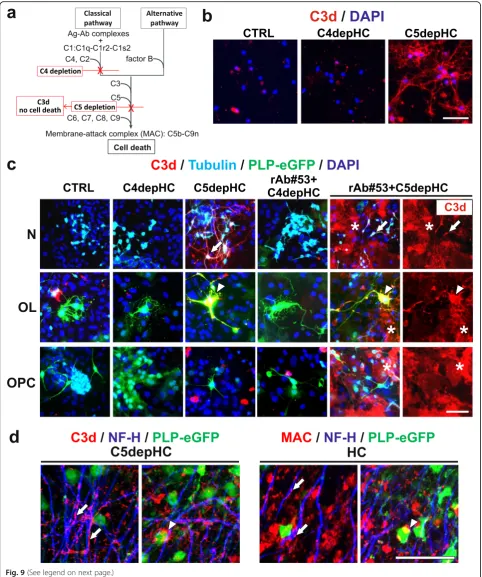
Complete activation of the complement cascade leads to the formation of the terminal membrane attack com-plex (MAC) and cell lysis. To determine which cells are targeted for MAC formation, we used selective depletion of proteins along the classical and common pathways to prevent cell death and localize the deposition of
0 1 2 3 4
0 10 20 30 40
time (h)
%
de
ad
AS
T
s
Iso Iso+HC #53 #53+HC
0 1 2 3 4
0 20 40 60 80 100
time (h)
%d
e
a
dN
s
#53 #53+HC Iso Iso+HC
**
c
b
AST Neuron0 1 2 3 4
0 20 40 60 80 100
time (h)
%d
e
a
d
O
P
Cs
Iso Iso+HC #53 #53+HC
e
OPC
d
OL
0 1 2 3 4
0 20 40 60 80 100
time (h)
%d
e
a
dO
L
s
Iso
#53 Iso+HC
#53+HC **
a
#53+HC,0h#53+HC,4h
#53+HC,4h
activated complement components. Using C5-depleted HC, we arrested complement activation following C3 proteolytic cleavage resulting in C3d deposition on complement-targeted cells (Fig. 9a). In neuronal monocul-tures treated with C5-depleted HC, C3d was detected on the cell surface. When neuronal monocultures were treated with C4-depleted human complement (Fig. 9b),
there was no C3d deposition, indicating that activation of the classical pathway was essential for neuronal injury with HC alone. In neuroglial mixed cultures treated with C5-depleted HC, C3d was detected on the surface of neu-rons and OLs (arrows and arrow heads, respectively), but not OPCs or surrounding astrocytes (Fig. 9c). Since HC-derived IgG bound to murine neurons, OLs, OPCs, and
CTRL HC
a
CTRL HC
0 20 40 60
%
PI+/C
alb
in
d
in
***
Neuron
OPC
d
CTRL HCe
OL
8 h 12 h 24 h 48 h 0
50 100 150
eG
F
P
+
c
el
l
b
od
ie
s
p
e
r
fi
e
ld
CTRL HC
* **
24 h 48 h
0 10 20 30 40
NG
2+
O
li
g
2+
ce
ll
s
pe
r
fi
el
d
CTRL HC
g
f
PLP-eGFP/NG2/Olig2CTRL HC
c
b
CTRL HCPI
Calbindin/PI
astrocytes (Additional file 1: Figure S5), the resistance of OPCs and astrocytes to CDC in mixed culture was either due to the local expression of complement inhibitors or the inability of surface-bound human IgGs to reach a suf-ficient threshold to activate the complement cascade. When mixed cultures were treated with NMO rAb #53 and C5-depleted HC, C3d deposits were found on the face of neurons (arrows), OLs (arrow heads), and sur-rounding astrocytes (stars), but not on OPCs (Fig. 9c). In organotypic cerebellar slices treated with C5-depleted or complete HC, deposits of C3d and MAC were detected along fibers containing neuro-filaments (Fig. 9d, arrows) and surrounding oligodendrocyte cell bodies (Fig. 9d, ar-rowheads). No detectable C3d or MAC deposits were noted in slices exposed to medium alone (data not shown). These results demonstrate that complement in human serum is activated by human anti-murine cross-reactive antibodies in primary cell monocultures, mixed cultures, and cerebellar slice cultures.
Discussion
These studies demonstrated that activated human comple-ment HC is injurious to mouse neurons and OLs in a var-iety of culture conditions. We found that complement activation is necessary for cell injury in primary cell and slice cultures and occurs through the binding and activation of the classical pathway by anti-murine immunoglobulin in
human sera. The susceptibility of neurons and glia to HC in the culture system lessens as the CNS cells coalesce into more complex networks. It may be that the different cul-ture conditions (i.e., mono- or mixed cell culcul-tures or tis-sues) alter the expression profiles of individual cell types resulting in loss of antigen expression, masking of target epitopes, and/or acquisition of complement inhibition. Each of these mechanisms may play a role in cell injury in models of NMO lesions and need to be examined relative to pathology in human tissue.
In primary cell monocultures, the addition of human serum alone is sufficient to cause rapid and substantial oligodendrocyte and neuronal cell death. In the neuro-glial mixed cultures, the effects of activated complement are likely reduced through protective oligodendroglial and neuronal interactions with astrocytes. As a conse-quence, astrocyte injury and loss driven by AQP4-specific rAb and complement-mediated cytotoxicity rapidly in-crease the level of neuronal and oligodendroglial damage. Interestingly, OPCs in the neuroglial cultures are resistant to complement attack following astrocyte damage. In cerebellar tissue slices, cell-cell interactions are more complex, and complement-mediated cell death of neurons and OLs are further reduced. While astrocyte destruction similarly increases oligodendroglial and neuronal cell death, the susceptibility of these populations to comple-ment cytotoxicity independent of astrocyte loss is much less than that observed in cell cultures. OPCs are the not-able exception, becoming sensitized in the presence of astrocyte damage in organotypic slice culture (Table 1). Our results suggest that the increasing complexity of the neuroglia environment, particularly in slice cultures, pro-tects neurons and oligodendrocytes from complement-induced cytotoxicity, and astrocytes play a central role in modulating complement injury. In the CNS of NMO patients, additional factors other than CDC, such as exci-totoxicity, inflammatory cytokines, antibody-dependent cell-mediated cytotoxicity, anaphylaxotoxins, and dis-rupted astrocyte physiology may play critical roles in lesion formation (reviewed in [28]). Whether exposure of tissue epitopes allows CDC to contribute to oligodendro-cyte and neuronal injury in human NMO lesions remains to be determined. Conversely, the use of complement from multiple species will be necessary to validate the relevance of oligodendroglial and neuronal damage ob-served across in vitro, ex vivo, and in vivo NMO models.
Astrocytes are a major producer of complement in the healthy and diseased CNS [29]. While astrocytes are not sensitive to HC alone, astrocyte destruction occurs readily through the production of terminal complement complexes (MAC complex) with AQP4-specific antibodies. Indeed, the current and previous studies demonstrate that inactiva-tion of the classical or alternative pathway affects AQP4-IgG mediated astrocyte cytotoxicity [16, 30]. Antibodies
a
b
c
d
against factor B [31], a key regulator of alternative pathway, or HC depleted of factor B, decrease astrocyte damage [16]. Neurons in monoculture were very sensitive to acti-vated complement, in agreement with the previous observations that neurons, unlike most peripheral cell
types, express complement receptors and regulators, but do not express the most known complement inhibitors [32, 33]. Neuronal damage in our in vitro and ex vivo models occurred as the direct result of activated comple-ment. This may, in part, explain why persistent neurologic
24 h 48 h 0
10 20 30 40
NG
2+
O
li
g
2
+
ce
ll
s
pe
r
fi
e
ld
Iso Iso+HC
#53 #53+HC
** ***
OPC
d
#53, 48 h #53+HC, 12 h #53+HC, 48 h
OL
8 h 12 h 24 h 48 h 0
50 100 150
eG
FP+
c
e
ll
b
odie
s
per
fi
e
ld
Iso Iso+HC
#53 #53+HC
**
ns*
c
PLP-eGFP
/
NG2
/
Olig2
e
f
#53+HC
#53
#53 #53+HC 0
20 40 60
%
PI+/Calb
id
in
Neuron
b
a
#53
#53+HC
PI
Calbindin
/PI
disability is strongly linked with acute relapses, as acti-vated complement is prominent feature of acute NMO lesions [1, 34, 35].
Consistent with previous reports [36], our results dem-onstrated that oligodendrocytes were vulnerable to acti-vated complement. Both in cell culture and brain slices, OLs (O4+) were susceptible to the addition of HC inde-pendent of the presence of astrocytes. Oligodendrocytes, unlike astrocytes, lack the complement regulatory protein CD59, which inhibits the final formation of membrane attack complex [37–39]. Complement resistance in rat oligodendrocytes can be restored by the incorporation of purified rat CD59 into the cell membrane. Furthermore,
neutralization of rat CD59 on complement-resistant astro-cytes rendered them susceptible to CDC [37]. When stud-ied in mixed cultures or slice cultures with astrocytes and neurons, OL susceptibility to complement was partially de-creased (Table 1). One explanation is that the presence of astrocytes or neurons may reduce the amount of the acti-vated complement available for OL binding, mask OL membrane epitopes, or alter OL membrane epitope expres-sion. Alternatively, astrocytes or neurons may secrete, ex-change, or modulate complement regulatory protein expression on the oligodendrocyte surface. It has been reported that in a mixed culture system of rat oligodendro-cytes and dorsal root ganglion neurons, OLs are remarkably
less sensitive to complement. Incubation of OLs with neuron-conditioned medium afforded a similar protection against complement-mediated lysis [40].
Our results demonstrate that OPCs remain resistant to activated complement independent of the astrocyte damage in mixed cultures. Unexpectedly, in organotypic cerebellar slice cultures, astrocyte destruction enhanced OPC loss. Although the neuroglia mixed cultures are prepared from the cortex and the organotypic slices from cerebellum, the differential sensitivity of OPCs to astrocyte damage does not appear to result from distinc-tions between cortical and cerebellar glia. In cell cultures prepared from cerebellum, there are no significant differ-ences in astrocyte death or OPC survival in the presence of NMO rAb #53 and HC (unpublished data). Thus, it appears that astrocyte damage in the complex neuroglia network of cerebellar slices sensitizes OPCs to injury. In addition, activated microglia, inflammatory mediators, and excitotoxic molecules may combine to produce a toxic milieu for OPCs in brain slices. Alternatively, the state of differentiation of OPCs in that environment may result in novel susceptibility to neuroinflammatory injury.
Following astrocyte damage, loss of oligodendrocytes, both mature cells and progenitors, is observed earlier than neuronal death in cerebellar slices. This is consist-ent with reports that the loss of OLs and OPCs are early pathologic features after astrocyte depletion in human and experimental NMO lesions [41]. Vulnerability of oli-godendrocytes is found in many neurological conditions. In patients with relapsing-remitting multiple sclerosis (RRMS), oligodendrocytes in affected tissue show apop-totic features and myelin sheaths stain with activated complement [42]. In a rat model of hypoxia-ischemic in-jury and in a mouse model of amyotrophic lateral scler-osis (ALS), extensive degeneration of differentiated oligodendrocytes (O4+) is found before the loss of OPCs [43, 44]. Although new oligodendrocytes are formed from NG2+ OPCs in the spinal cord of ALS mice, they fail to mature [44]. These observations in combination with our results suggest a selective vulnerability of ma-ture oligodendrocytes to multiple forms of injury. With
extreme conditions, such as massive astrocyte destruc-tion in NMO, OPCs may also be affected.
Taken together, our findings demonstrate that in mouse monoculture, activated human complement is targeted to neurons, OLs and OPCs through the action of anti-murine immunoglobulins. In mixed cul-tures, surrounding astrocytes ameliorate CDC; tar-geted destruction of astrocytes with AQP4-specific recombinant monoclonal antibodies restores neuronal and oligodendroglia susceptibility to HC. Increasing complexity of neuroglial networks in murine cerebel-lar slices further reduces the effects of activated hu-man complement alone and alters the secondary effects of astrocyte injury on OPC survival. Experi-mental models of NMO require complement activa-tion to initiate CNS lesions. Initial models involved administration of AQP4-IgG to rats with pre-existing neuroinflammation produced by experimental auto-immune encephalomyelitis [12, 14, 45]. Although some pathology resulted from the influx of myelin-reactive T cells, lesions demonstrated hallmark fea-tures of NMO pathology including perivascular astro-cyte destruction and complement deposition. Subsequent passive-transfer models of NMO in mice required intracranial injection of NMO-IgG and hu-man complement to recapitulate key pathological findings in human lesions [30, 46, 47]. The use of hu-man complement was required due to the presence of inhibitors in mouse complement preparations [18, 19]. The current studies indicate that one should use caution in interpreting the cause of secondary injury to CNS cells following AQP4-IgG mediated CDC when using human serum in murine models.
Conclusions
Our study demonstrates that mouse neurons, OLs, and OPCs display variable sensitivity to activated HC based on culture conditions and tissue complexity. In cell and slice cultures, the protection of neurons, OLs, and OPCs against HC is eliminated by targeted astrocyte destruc-tion. Because of the targeted activation of HC on mouse (See figure on previous page.)
CNS cells, caution is needed in interpreting the relation-ship between damage in experimental mouse models of NMO and human lesions.
Additional files
Additional file 1: Figure S1.Immunostaining with cell type specific markers to identify different cell types in the neuroglial co-cultures.Figure S2. Cytotoxic effects of human and mouse complement on neuroglial mixed cultures.Figure S3.Confocal images of PI-stained cells double stained with neuronal marker NeuN in slices treated with 10% HC or rAbs (NMO#53) plus HC for 48 h in slices.Figure S4.Human neurons and rat OPCs and OLs were not sensitive to human serum or rat serum.Figure S5.Binding of HC-derived human IgG to the surface of murine neurons, OLs, OPCs and astrocytes.Table S1.Cytotoxic effects of multiple sources of human (HC) and mouse complement (MC) in neuroglial mixed cultures. (PDF 648 kb)
Additional file 2: Movie 1.Pure OL with HC. IncuCyte live cell imaging in purified differentiated OL mono-culture treated with 5% HC. Cell cultures were scanned for 24 hr. Images were captured at 15 min intervals with high definition phase contrast and red fluorescence (to detect the dead cell nuclei dye DRAQ7). (MOVIE 5345.28 kb)
Additional file 3: Movie 2.Pure OPC with HC. IncuCyte live cell imaging in purified OPC mono-culture treated with 5% HC. Cell cultures were scanned for 24 hr. Images were captured at 15 min intervals with high definition phase contrast and red fluorescence (to detect the dead cell nuclei dye DRAQ7). (MOVIE 4218.88 kb)
Additional file 4: Movie 3.Co-culture with HC. IncuCyte live cell imaging in neuro-glial co-cultures prepared from PLP-eGFP mouse pups and treated with 5% HC. Cell cultures were scanned for 66 hr. Images were captured at 15 min intervals with high definition phase contrast and green (to detect eGFP + OPC and differentiated OL) and red fluorescence (to detect the dead cell nuclei dye DRAQ7). (MOVIE 18534.4 kb)
Additional file 5: Movie 4.Co-culture with NMO53 + HC. IncuCyte live cell imaging in neuro-glial co-cultures prepared from PLP-eGFP mouse pups and treated with NMO rab#53 plus 5% HC. Cell cultures were scanned for 66 hr. Images were captured at 15 min intervals with high definition phase contrast and green (to detect eGFP + OPC and differentiated OL) and red fluorescence (to detect the dead cell nuclei dye DRAQ7). Around the area marked with "A": dead astrocytes floated into the culture supernatant, resulting in focal areas of astrocyte-depletion (also shown in Fig. 4a circled area). After the peak of astrocyte death, astrocytes repopulated the depleted areas. (MOVIE 34099.2 kb)
Abbreviations
AQP4:Aquaporin-4; AQP4-IgG: Aquaporin-4 autoantibody; AST: Astrocyte; BSA: Bovine serum albumin; C4-C5depHC: C4 or C5 depleted human serum; CDC: Complement-dependent cytotoxicity; CNS: Central nervous system; CSF: Cerebrospinal fluid; CTRL: Control; eGFP: Enhanced green fluorescent protein; FBS: Fetal bovine serum; FGF: Fibroblast growth factor; GFAP: Glial fibrillary acidic protein; HC: Human complement; IgG: Immunoglobulins; LDH: Lactate dehydrogenase; MAC: Membrane attack complex; NDS: Normal donkey serum; NF-H: Neurofilament heavy chain; NGS: Normal goat serum; NMO: Neuromyelitis optica; OL: Oligodendrocytes; OPC: Oligodendrocyte progenitors; PBS: Phosphate-buffered saline; PDGF: Platelet-derived growth factor; PI: Propidium iodide; PLP: Proteolipid protein; rAb: Recombinant antibody
Acknowledgements
We are grateful to the shared resource in Protein Production/Mab/Tissue Culture Core (supported by the Cancer Center Support Grant (CCSG) P30CA046934), University of Colorado Cancer Center for assistance with IncuCyte live imaging. And we are indebted to Hannah Schumann and Andre Navarro for the preparation of isotype control, NMO anti-AQP4 #53 rAbs, and anti-C3d antibody.
Funding
This work was supported by the Collaborative Research Grant from the National Multiple Sclerosis Society (W.B.M.), NEI EY022936 (J.L.B.), UM1AI110498 (J.L.B.), NINDS NS072141 (G.P.O.), NS25304 (W.B.M.), and the Guthy-Jackson Charitable Foundation (J.L.B.).
Availability of data and materials The relevant raw data are freely available.
Authors’contributions
YL designed and coordinated the study, performed the experiments, analyzed the data, and drafted the manuscript and revision. DH and KS performed the slice culture preparation and immunohistochemistry, and primary OPC and OL culture preparation. GO participated in the review the data and manuscript. WM and JB designed the study and reviewed the data and the manuscript and revision. All authors read and approved the manuscript.
Competing interests
The authors declare that they have no competing interests.
Consent for publication Not applicable.
Ethics approval and consent to participate
The care and euthanization of animals were in accordance with the University of Colorado IUCAC policy for animal use, which is in agreement with the NIH Guide for the Care and Use of Laboratory Animals.
Author details 1
Department of Neurology, University of Colorado, School of Medicine, 12700 E. 19th Ave, Aurora, CO, USA.2Department of Cell & Developmental Biology, University of Colorado, School of Medicine, 12700 E. 19th Ave, Aurora, CO, USA.3Department of Ophthalmology, University of Colorado, School of Medicine, 12700 E. 19th Ave, Aurora, CO, USA.4Program in Neuroscience, University of Colorado, School of Medicine, 12700 E. 19th Ave, Aurora, CO, USA.
Received: 16 August 2016 Accepted: 21 November 2016
References
1. Papadopoulos MC, Bennett JL, Verkman AS. Treatment of neuromyelitis optica: state-of-the-art and emerging therapies. Nat Rev Neurol. 2014;10:493–506. 2. Ricklin D, Lambris JD. Complement in immune and inflammatory disorders:
therapeutic interventions. J Immunol. 2013;190:3839–47.
3. Benoit ME, Tenner AJ. Complement protein C1q-mediated neuroprotection is correlated with regulation of neuronal gene and microRNA expression. J Neurosci. 2011;31:3459–69.
4. Peterson SL, Nguyen HX, Mendez OA, Anderson AJ. Complement protein C1q modulates neurite outgrowth in vitro and spinal cord axon regeneration in vivo. J Neurosci. 2015;35:4332–49.
5. Stephan AH, Barres BA, Stevens B. The complement system: an unexpected role in synaptic pruning during development and disease. Annu Rev Neurosci. 2012;35:369–89.
6. Orsini F, De Blasio D, Zangari R, Zanier ER, De Simoni MG. Versatility of the complement system in neuroinflammation, neurodegeneration and brain homeostasis. Front Cell Neurosci. 2014;8:380.
7. Roemer SF, Parisi JE, Lennon VA, Benarroch EE, Lassmann H, Bruck W, Mandler RN, Weinshenker BG, Pittock SJ, Wingerchuk DM, Lucchinetti CF. Pattern-specific loss of aquaporin-4 immunoreactivity distinguishes neuromyelitis optica from multiple sclerosis. Brain. 2007;130:1194–205. 8. Bruck W, Popescu B, Lucchinetti CF, Markovic-Plese S, Gold R, Thal DR, Metz
I. Neuromyelitis optica lesions may inform multiple sclerosis heterogeneity debate. Ann Neurol. 2012;72:385–94.
9. Kuroda H, Fujihara K, Takano R, Takai Y, Takahashi T, Misu T, Nakashima I, Sato S, Itoyama Y, Aoki M. Increase of complement fragment C5a in cerebrospinal fluid during exacerbation of neuromyelitis optica. J Neuroimmunol. 2013;254:178–82.
10. Nytrova P, Potlukova E, Kemlink D, Woodhall M, Horakova D, Waters P, Havrdova E, Zivorova D, Vincent A, Trendelenburg M. Complement activation in patients with neuromyelitis optica. J Neuroimmunol. 2014;274:185–91.
11. Lennon VA, Kryzer TJ, Pittock SJ, Verkman AS, Hinson SR. IgG marker of optic-spinal multiple sclerosis binds to the aquaporin-4 water channel. J Exp Med. 2005;202:473–7.
13. Zhang H, Bennett JL, Verkman AS. Ex vivo spinal cord slice model of neuromyelitis optica reveals novel immunopathogenic mechanisms. Ann Neurol. 2011;70:943–54.
14. Bradl M, Misu T, Takahashi T, Watanabe M, Mader S, Reindl M, Adzemovic M, Bauer J, Berger T, Fujihara K, et al. Neuromyelitis optica: pathogenicity of patient immunoglobulin in vivo. Ann Neurol. 2009;66:630–43.
15. Kinoshita M, Nakatsuji Y, Kimura T, Moriya M, Takata K, Okuno T, Kumanogoh A, Kajiyama K, Yoshikawa H, Sakoda S. Anti-aquaporin-4 antibody induces astrocytic cytotoxicity in the absence of CNS antigen-specific T cells. Biochem Biophys Res Commun. 2010;394:205–10.
16. Phuan PW, Zhang H, Asavapanumas N, Leviten M, Rosenthal A, Tradtrantip L, Verkman AS. C1q-targeted monoclonal antibody prevents complement-dependent cytotoxicity and neuropathology in in vitro and mouse models of neuromyelitis optica. Acta Neuropathol. 2013;125:829–40.
17. Li M, Yan Y. Experimental models of neuromyelitis optica: current status, challenges and future directions. Neurosci Bull. 2015;31:735–44.
18. Bergman I, Basse PH, Barmada MA, Griffin JA, Cheung NK. Comparison of in vitro antibody-targeted cytotoxicity using mouse, rat and human effectors. Cancer Immunol Immunother. 2000;49:259–66.
19. Ratelade J, Verkman AS. Inhibitor(s) of the classical complement pathway in mouse serum limit the utility of mice as experimental models of neuromyelitis optica. Mol Immunol. 2014;62:104–13.
20. Mallon BS, Shick HE, Kidd GJ, Macklin WB. Proteolipid promoter activity distinguishes two populations of NG2-positive cells throughout neonatal cortical development. J Neurosci. 2002;22:876–85.
21. Liu Y, Vidensky S, Ruggiero AM, Maier S, Sitte HH, Rothstein JD. Reticulon RTN2B regulates trafficking and function of neuronal glutamate transporter EAAC1. J Biol Chem. 2008;283:6561–71.
22. McCarthy KD, de Vellis J. Preparation of separate astroglial and oligodendroglial cell cultures from rat cerebral tissue. J Cell Biol. 1980;85:890–902.
23. Harlow DE, Saul KE, Komuro H, Macklin WB. Myelin proteolipid protein complexes with alphav integrin and AMPA receptors in vivo and regulates AMPA-dependent oligodendrocyte progenitor cell migration through the modulation of cell-surface GluR2 expression. J Neurosci. 2015;35:12018–32. 24. Gudz TI, Schneider TE, Haas TA, Macklin WB. Myelin proteolipid protein
forms a complex with integrins and may participate in integrin receptor signaling in oligodendrocytes. J Neurosci. 2002;22:7398–407.
25. Sheridan GK, Dev KK. S1P1 receptor subtype inhibits demyelination and regulates chemokine release in cerebellar slice cultures. Glia. 2012;60:382–92.
26. Osterhout DJ, Wolven A, Wolf RM, Resh MD, Chao MV. Morphological differentiation of oligodendrocytes requires activation of Fyn tyrosine kinase. J Cell Biol. 1999;145:1209–18.
27. Emery B, Agalliu D, Cahoy JD, Watkins TA, Dugas JC, Mulinyawe SB, Ibrahim A, Ligon KL, Rowitch DH, Barres BA. Myelin gene regulatory factor is a critical transcriptional regulator required for CNS myelination. Cell. 2009;138:172–85. 28. Kowarik MC, Soltys J, Bennett JL. The treatment of neuromyelitis optica.
J Neuroophthalmol. 2014;34:70–82.
29. Veerhuis R, Nielsen HM, Tenner AJ. Complement in the brain. Mol Immunol. 2011;48:1592–603.
30. Saadoun S, Waters P, Bell BA, Vincent A, Verkman AS, Papadopoulos MC. Intra-cerebral injection of neuromyelitis optica immunoglobulin G and human complement produces neuromyelitis optica lesions in mice. Brain. 2010;133:349–61.
31. Thurman JM, Kraus DM, Girardi G, Hourcade D, Kang HJ, Royer PA, Mitchell LM, Giclas PC, Salmon J, Gilkeson G, Holers VM. A novel inhibitor of the alternative complement pathway prevents antiphospholipid antibody-induced pregnancy loss in mice. Mol Immunol. 2005;42:87–97.
32. Cahoy JD, Emery B, Kaushal A, Foo LC, Zamanian JL, Christopherson KS, Xing Y, Lubischer JL, Krieg PA, Krupenko SA, et al. A transcriptome database for astrocytes, neurons, and oligodendrocytes: a new resource for understanding brain development and function. J Neurosci. 2008;28:264–78.
33. O’Barr SA, Caguioa J, Gruol D, Perkins G, Ember JA, Hugli T, Cooper NR. Neuronal expression of a functional receptor for the C5a complement activation fragment. J Immunol. 2001;166:4154–62.
34. Lucchinetti CF, Bruck W, Lassmann H. Evidence for pathogenic heterogeneity in multiple sclerosis. Ann Neurol. 2004;56:308.
35. Misu T, Hoftberger R, Fujihara K, Wimmer I, Takai Y, Nishiyama S, Nakashima I, Konno H, Bradl M, Garzuly F, et al. Presence of six different lesion types suggests diverse mechanisms of tissue injury in neuromyelitis optica. Acta Neuropathol. 2013;125:815–27.
36. Wren DR, Noble M. Oligodendrocytes and oligodendrocyte/type-2 astrocyte progenitor cells of adult rats are specifically susceptible to the lytic effects of complement in absence of antibody. Proc Natl Acad Sci U S A. 1989;86:9025–9.
37. Wing MG, Zajicek J, Seilly DJ, Compston DA, Lachmann PJ. Oligodendrocytes lack glycolipid anchored proteins which protect them against complement lysis. Restoration of resistance to lysis by incorporation of CD59. Immunology. 1992;76:140–5.
38. Piddlesden SJ, Morgan BP. Killing of rat glial cells by complement: deficiency of the rat analogue of CD59 is the cause of oligodendrocyte susceptibility to lysis. J Neuroimmunol. 1993;48:169–75.
39. Scolding NJ, Morgan BP, Compston DA. The expression of complement regulatory proteins by adult human oligodendrocytes. J Neuroimmunol. 1998;84:69–75.
40. Agoropoulou C, Piddlesden SJ, Lachmann PJ, Wing MG. Neuronal protection of oligodendrocytes from antibody-independent complement lysis. Neuroreport. 1998;9:927–32.
41. Wrzos C, Winkler A, Metz I, Kayser DM, Thal DR, Wegner C, Bruck W, Nessler S, Bennett JL, Stadelmann C. Early loss of oligodendrocytes in human and experimental neuromyelitis optica lesions. Acta Neuropathol. 2014;127:523–38. 42. Barnett MH, Prineas JW. Relapsing and remitting multiple sclerosis: pathology
of the newly forming lesion. Ann Neurol. 2004;55:458–68.
43. Back SA, Han BH, Luo NL, Chricton CA, Xanthoudakis S, Tam J, Arvin KL, Holtzman DM. Selective vulnerability of late oligodendrocyte progenitors to hypoxia-ischemia. J Neurosci. 2002;22:455–63.
44. Kang SH, Li Y, Fukaya M, Lorenzini I, Cleveland DW, Ostrow LW, Rothstein JD, Bergles DE. Degeneration and impaired regeneration of gray matter oligodendrocytes in amyotrophic lateral sclerosis. Nat Neurosci. 2013;16:571–9. 45. Kinoshita M, Nakatsuji Y, Kimura T, Moriya M, Takata K, Okuno T, Kumanogoh A, Kajiyama K, Yoshikawa H, Sakoda S. Neuromyelitis optica: passive transfer to rats by human immunoglobulin. Biochem Biophys Res Commun. 2009;386:623–7. 46. Asavapanumas N, Ratelade J, Papadopoulos MC, Bennett JL, Levin MH, Verkman AS. Experimental mouse model of optic neuritis with inflammatory demyelination produced by passive transfer of neuromyelitis optica-immunoglobulin G. J Neuroinflammation. 2014;11:16.
47. Zhang H, Verkman AS. Longitudinally extensive NMO spinal cord pathology produced by passive transfer of NMO-IgG in mice lacking complement inhibitor CD59. J Autoimmun. 2014;53:67–77.
• We accept pre-submission inquiries
• Our selector tool helps you to find the most relevant journal
• We provide round the clock customer support
• Convenient online submission
• Thorough peer review
• Inclusion in PubMed and all major indexing services
• Maximum visibility for your research
Submit your manuscript at www.biomedcentral.com/submit