Medical Journal of the
Islamic Republic of Iran Volume 17 Number 3
Fall 1382 November 2003
� _____________
O
__n
_
g
_
in
_
a
_
l
_
A
__n
_
k
_
l
_
e
s
_
______________J
EFFECT OF CLINICAL IN FORMATION ON BRAIN
CT SCAN IN T ERPRETATION: A BLIN DED
DOUBLE CROSSOVER ST UDY
MOTAHAREH ZHIANPOUR, M.D.,
ANDMOHSEN JANGHORBANI, Ph.D.
From the Departments of Radiology and Epidemiology, Isfahan University of Medical Sciences and Heath Services, Isfahan, Iran.
ABSTRACT
Errors and variations in interpretation can happen in clinical imaging. Few
studies have examined the biased effect of clinical information on reporting of
brain CT scans. In a blinded double crossover design, we studied whether three
radiologists were biased by clinical information when making CT scan diagnosis
of the brain. Three consultant radiologists in three rounds with at least a one
month interval assessed
100
consecutive cases of brain CT scan. In the first round,
clinical information was not available and
100
films without clinical information
were given to radiologists. In the second round, the same
100
films were given
and true clinical information was available. In the third round, the same
100
films
were given and false clinical information was allocated. In
180
cases
(60%)
the
evaluation resulted in the same diagnosis on all three occasions
(95%
confidence
interval (CI):
54.5,65.5),
whereas
120 (400/0; 95%
CI:
34.5,45.5)
sets were evalu
ated differently.
48
cases
(16%; 95%
CI:
11.9, 20.1)
had discordant evaluation
with true and
33 (11 %; 950/0
CI:
7.5,14.5)
with false clinical information. Discor
dance without and with true and false clinical information was
39 (13%; 95%
CI:
9.2, 16.8).
Correct clinical information improves the brain CT report, while the
report became less accurate after false clinical information was allocated. These
results indicate that radiologists are biased by clinical information when report
ing brain CT scans.
MJIRI, Vol. 17, No.3, 173-177, 2003.
Keywords: Clinical information, radiology, CT scan, bias, Iran.
INTRODUCTION
The value of a CT scan in the diagnosis and
manage-Address for correspondence: Prof. M. Janghorbani, School of Public Health, Isfahan University of Medical Sciences, Isfahan, Iran. Tel: (+98) 311-2334893, Fax: (+98) 311-6682509, e.mail:[email protected]
ment of patients with brain disease is widely recognized, and the CT scan diagnosis of it is considered very reli able. However, as in other diagnostic tests, errors and variations in interpretation should be expected to occur.
Bias can influence the evaluation of every diagnostic test.1-3 Bias among radiologists is especially an area in which no attention has been given in Iran. Although it has been argued for
3
decades that the usefulness of anyimaging procedure can only be measured objectively when radiologists are blinded to all clinical informa tion,4,5 only a few reports have investigated this influ ence on subjective diagnosis,6.8 Previous reports on the influence of clinical information in plain radiographic reporting have shown inconsistent results,9.11
The radiologist who interprets the brain CT scan of ten has access to the radiography requisition form, which contains clinical information concerning signs and symp toms as reported by the treating clinician, While some radiologists prefer, at least initially, to inspect films with out knowledge of the clinical information, others believe that such information is essential for adequate interpre tation, It is reasonable to hypothesize that the reporting may be biased, whether consciously or unconsciously, by this information, To our knowledge, however, only two studies from the UK and USA have examined the existence, direction, and magnitude of this bias and evaluated its potential impact on the diagnosis of by CT scan 12,13 and concluded that clinical information affects the CT report. To our knowledge, there are no studies describing the possible effect of clinical information on brain CT scan reporting in developing countries,
In a blinded double crossover design we studied whether three radiologists were biased by clinical infor mation when making brain CT scan diagnoses, The pur pose was not to evaluate the accuracy of the diagnoses,
MATERIAL AND METHODS
The study comprises
100
consecutive brain CT scans, The cases were identified prospectively from those re ferring to the CT scan Department of Shahid Bahonar Hospital, affiliated to Kerman University of Medical Sci ences and Health Services, Iran, which covered a period from December3, 1999
to October27,2000,
The
100
films were assessed three times with inter vals of at least one month, by three consultant radiolo gists who were blinded to their own and the other ob servers' previous assessments, In the first round, clini cal information was not available, One-hundred brain CT scan films without clinical information were given, In the second round, the same100
films were given to radiologists and true clinical information was available, In the third round, the same100
films were given, and false clinical information was allocated as in the first and second rounds, Each reader recorded any abnormal findings and their interpretation of these findings in each round. Any changes in interpretation without and with false and true clinical information were noted, The true clinical information was taken from the medical records of the patients and interview with patients. One of the investigators (MZ) made the false clinical information. Any scans requiring review of the images at the time ofinvestigation were excluded.
The diagnosis of
100
cases were as follows:7
infarctions
(6
CVA,1
post-meningitis), 4 infections(3
abscesses, I encephalitis),17
tumors(2
metastases,5
meningiomas,
1
meduloblastoma,1
ependymoma,3
glio blastoma multiforme,1 5th
nerve neuroma,2
astrocyto mas,1
craniopharyngioma),2
encephaloceles(1
fron tal,1
occipital), I basilar artery aneurysm,7
normal,7
post-craniotomy complications (including: intracerebral hemorrhage, subdural hemorrhage, pneumocephalus, bone defect, edema, brain herniation through the bone defect, infection), 4 complicating shunt tube (such as: compressed ventricles, intracerebral hematoma, intra ventricular hemorrhage, poor functioning shunt tube),
1
massive basal ganglia calcification,
1
pseudotumor cerebri,1
senile brain atrophy,1
m assive hydroceph alus and47
head injuries (including: contusion, hemor rhagic contusion, intracerebral hematoma, subdural he matoma, subdural hygroma, epidural hematoma, sub arachnoid hemorrhage, fracture (linear, depressed), soft tissue hematoma, deep axonal injury, generalized brain edema, midline shift, hydrocephalus, sinus fracture, pneumocephalus)The three radiologists were told that the study was about inter- and intra-observer variation, They did not know that some of the clinical information was mislead ing or the 100 films were repeated. The radiologists were asked to read the films as routine. They did not keep notes from previous rounds. Each reader recorded any abnormal findings and their interpretation of these find ings without clinical information. The images were then reviewed with true and false knowledge of the clinical information on the request form. Any further abnormal findings or changes in interpretation were noted by one of us (M.Z.) who did not participate in the interpreta tion of CT scans.
For the assessment of bias, the material was regarded as
100
different sets of brain CT scan films. Each of these sets were examined three times by each radiolo gist, without and with a true and false clinical history attached on each occasion, summing to a total of300
sets of evaluation.Evaluations resulting in different diagnoses could be long to one of two categories: (a) those in which there was concordance at all occasions between radiological diagnoses; and (b) those in which there was discordance at one occasion between the radiological diagnosis and the diagnosis which might be inferred from the other occasions, Patients in whom the report was changed af ter knowledge of true or false clinical information were followed up to determine whether the amended report was more or less accurate.
The reproducibility of the three observers' diagnoses was assessed separately for the
100
films. Two of theM.
Zhianpour and M.
Janghorbani
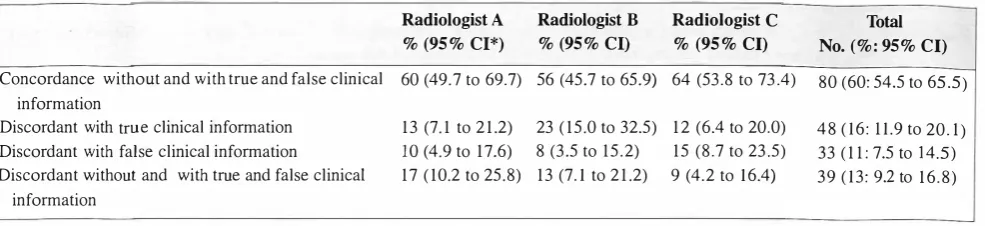
Table J. Distribution of 100 sets of brain CT scan in which different diagnoses were made in three rounds by three radiologists.
Radiologist A
% (95%
CI*)Radiologist B
% (95%
CI)Radiologist C
% (95%
CI)Total No.
(%: 95%
CI)Concordance without and with true and false clinical 60 (49.7 to 69.7) 56 (45.7 to 65.9) 64 (53.8 to 73.4) 80 (60: 54.5 to 65.5) information
Discordant with true clinical information Discordant with false clinical information
Discordant without and with true and false clinical
13 (7.1 to 21.2) 23 (15.0 to 32.5) 12 (6.4 to 20.0) 10 (4.9 to 17.6) 8 (3.5 to 15.2) 15 (8.7 to 23.5) 17 (10.2 to 25.8) 13 (7.1 to 21.2) 9 (4.2 to 16.4)
48 (16: 11.9 to 20.1) 33 (11: 7.5 to 14.5) 39 (13: 9.2 to 16.8) information
*CI indicates confidence interval
Table II. Changes made by the reader after knowledge of true clinical information in 100 sets of brain CT scans.
Radiologist No. of CTs reported correctly without clinical information
No. reported correctly
%
C hange(95 %
confidence interval) with true clinical informationA B C Total
*p<O.OOI
60 56 64 180
consultant radiologists had
11
years of radiological ex perience and the third had9
years. All of the radiolo gists were faculty members of Kerman University of Medical Sciences and trained to consultant level in cross sectional imaging.Statistical analysis
The biasing effect of the clinical information on the diagnosis of brain CT scan was evaluated by the sign test (two-sided). 95% confidence interval (CI), based on the normal approximation to the binominal distribution was calculated by confidence interval analysis software. 14
RESULTS
Seventy-one of the
100 patients imaged were male
and 29 were female. Each scan was triple reported giv ing a total of300
CT reports. One-hundred and eighty(60%; 95%
CI:54.5, 65.5)
evaluations resulted in the same diagnosis on all three occasions, whereas 120 (40%;95% CI: 34.5,45.5) sets were evaluated differently. These
120 were split into 48 (16%; 95% CI:
11.9,20.1) discor
dant evaluations with true and33 (11 %; 95%
CI:7.5,14.5)
with false clinical information. Discordance without and with true and false clinical information was39 (13%: 95% CI:
9.2, 16.8) (Table I). Tables II and III
show the percentage of reports changed for each reader after knowing true and false clinical information.Twenty-90 92 85 267
30 (18.7 to 41.3)* 36 (24.9 to 47.1)* 21 (9.3 to 32.7)* 29 (22.4 to 35.6)*
nine percent
(95%
CI:22.4, 35.6)
of brain CT reports were changed to more accurate diagnoses after true clini cal information was known and 24% (95% CI:16.2,31.8)
were changed to a less accurate diagnosis after false clini cal information was allocated. This was a statistically significant departure from the expected equal split(p<0.00l).
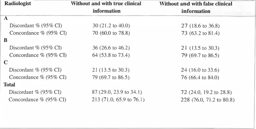
Thus, when the radiologists made different evaluation of a given radiological material, they were prone to do this in accordance with the clinical informa tion. The overall inter-observer agreement for radiologi cal diagnosis of brain CT scans did not change when true or false clinical information was added (Table IV). The difference was not statistically significant.DISCUSSION
In this study 40% of interpretations were changed by knowledge of the true or false clinical information. Twenty-nine percent of brain CT reports were more ac curate after true clinical information was known and 24% were less accurate after false clinical information was allocated. It is a well-known problem that clinicians may be biased by clinical information in the scientific evalu ation of a diagnostic test and this is generally consid ered undesirable. One out of two doctors was si,gnifi cantly biased by his knowledge of the radiological diag nosis of deformity of the bulbous when making an endo scopic diagnosis. IS Neurologists were asked to judge a
Table III. Changes made by reader after knowledge of false clinical information in 100 sets of brain CT scans.
Radiologist No. of CTs reported correctly No. reported correctly
%
Change(95%
confidence interval) without clinical information with false clinical informationA B C Total
60 56 64 180
33 35 40 108
27 (13.7 to 40.3)* 21 (7.5 to 34.5)* 24 (10.6 to 37.4)* 24 (16.2 to 31.8)*
Table IV. Comparison of 100 sets of brain CT scans in which different diagnoses were made in round 1, 2 and 3 by three radiologists.
Radiologist Without and with true clinical Without and with false clinical
information information A
Discordant % (95% CI) 30 (21.2 to 40.0) 27 (18.6 to 36.8)
Concordance % (95% CI) 70 (60.0 to 78.8) 73 (63.2 to 81.4)
B
Discordant % (95% CI) 36 (26.6 to 46.2) 21 (13.5 to 30.3)
Concordance % (95% CI) 64 (53.8 to 73.4) 79 (69.7 to 86.5) C
Discordant % (95% CI) 21 (13.5 to 30.3) 24 (16.0 to 33.6) Concordance % (95% CI) 79 (69.7 to 86.5) 76 (66.4 to 84.0) Total
Discordant % (95% CI) 87 (29.0, 23.9 to 34.
I)
72 (24.0, 19.2 to 28.8)Concordance % (95% Cl) 213 (71.0, 65.9 to 76.1) 228 (76.0, 71.2 to 80.8)
*C[ indicates confidence interval
number of plantar responses on films preceded by ficti tious abstract of history and examination. I Two films,
showing equivocal toe movements were presented twice with opposing information as to the probability of a plan tar reflex (Babinski sign). Interpretation of these identi cal pictures differed significantly conforming to the in formation given. In another study, bias from previous knowledge of the duration of amenorrhea was demon strated in gynecologists' estimation of the size of the uterus in pregnant women.2 The interpretation of histo pathologic findings among pathologists was biased by clinical information given. 3 The review of numerous stud ies regarding the effect of clinical information on plain radiographic reporting showed inconsistent results. Sev eral of these studies showed a positive effect7·9,16.17 while others showed no effect. II Knowledge of a previous di
agnosis influenced all the observers in a study evaluat ing plain roentgenograms of hands.16 In another study, radiologists' diagnosis was significantly influenced by the context of interpretation, even when spectrum and verification bias are avoided.17 Doubilet and Herman9
found that appropriate clinical information increases the rate of positive reading in chest radiograph. Berbaum et al. 8 found clinical information improved detection and interpretation of abnormalities in a series of pediatric chest and abdomen radiographs. However, Good et aLII in a larger series found clinical information did not af fect the accuracy of chest radiography reporting and Babcook et a1.7 found that while appropriate informa tion improved the true positive detection, inappropriate information increased false positive detection,
Clinical information plays an extremely important role in the analysis and interpretation of CT scans. Clini cal information must be accurate to improve radiologi cal reports. While clinical information has an inconsis tent effect on plain radiograph reporting, it was much less marked than that of clinical information on CT re porting even in the studies where it was shown to have a beneficial influence. We found only two other studies that assessed the effect of clinical information on CT reporting. In one of these studies Eldevik et a1. 13 consid ered the effect of clinical bias on the interpretation of
M.
Zhianpour and
M.
Janghorbani
myelography and spinal CT. In the other study Leslie et al. 12 reported the effect of clinical information on the in terpretation of CT reports. In both of these studies clini cal information was found to bias the reports.
We do not believe that our findings can be explained by a practice experience impact in the present study, as the radiologists who made different diagnoses in one set of x-ray films had about I I years of radiology practice and they were relatively as good as each other.
It would not be well founded to recommend that x ray diagnoses be made without clinical information; rather, we recommend that x-ray diagnosis be made first without clinical information and then with it. Some knowledge of the clinical problem is necessary to choose the region to be examined.
In conclusion, we demonstrated that radiologists were biased by clinical information when reporting brain CT scans and the biasing effect of clinical information de clined when accurate clinical information was added to the requisition form and as Leslie et al.12 stated "it is the responsibility of the referring physician to ensure that the radiologist is gi ven accurate and legible information".
ACKNOWLEDGEMENT
This work was supported by a grant from the Kerman University of Medical Sciences. The authors are grate ful to Dr. R. Foladi, Dr. B. Fahimi and Dr. S. Naroiee for their assistance in reading x-ray films and Prof. Ali Sadeghi-Hassanabadi for his comments.
REFERENCES
I.Yan Grijn J, Bonke B: Interpretation of plantar reflexes: bi asing effect of other signs and symptoms. J Neurol Neurosurg P sychiatry 40: 787-789, 1977.
2. Gjorup T, Saurbrey N, Hermann N: Clinical estimation of the duration of pregnancy in legal abortion-are doctors biased by their knowledge of the duration of amenorrhoea? Meth Inform Med 23: 96-98, 1984.
3. Skov B G, Braendstrup 0, Hirsch FR, Lauritzen AF, Nielsen HW, Skov T: Are pathologists biased by clini cal information? A blinded crossover study of the histo pathological diagnosis of mesothelial tumors versus pul monary adenocarcinoma. Lung Cancer I I : 365-372, 1994.
4. Schreiber MH: The clinical history as a factor in roent genogram interpretation. JAMA 185:137-139, 1963. 5. Chalmers TC: PET Scans and technology assessment.
JAM A 260: 2713-2715, 1988.
6. McNeil BJ, Hanley JA, Funkenstein HH, Wallman J: Paired receiver operating characteristic curves and the effect of history on radiographic interpretation. Radiol ogy 149: 75-77, 1983.
7. Babcook CJ, Norman GR, Cobl entz CL: Effect o f clinical history on the interpretation o f chest radio graphs in childhoo d bronchit i s . I nvest Radiol 2 8: 214-217, 1993.
8. Berbaum KS, el Khoury GY, Franken EAJ, Kathol M, Montgomery WJ, Hesson W: Impact of clinical history on fracture detection with radiography. Radiology. 168: 507-511, 1988.
9. Doubilet P, Harman po. Interpretation of radiographs: ef fect of clinical history: AJR 137: 1055-8, 1981.
10. Berbaum KS, Franken EA Jr, Dorfman DD, Lueben KR: Influence of clinical history on perception of abnormali ties in pediatrics radiographs. Acad Radiol 1: 217-23,
1994.
I I. Good BC, Cooperstein LA, DeMarino GB, Miketic LM, Gennari RC, Rockette HE, et al: Does knowledge of the clinical history affect the accuracy of chest radiographs interpretation? AJR 154: 709-I 2, 1990.
12. Leslie S, Jones AJ, Goddard PR: The influence of clini cal information on the reporting of CT by radiologists. Br J Radiol 73: 1052-I 055, 2000.
13. Eldevik OP, Dugstad G, Orrison WW, Haughton YM: The effect of clinical bias on the interpretation of myel ography and spinal computed tomography. Radiology 145: 85-89, 1982.
14. Gardner MJ, Altman DG: Statistics with confidence. Lon don: British Medical Association, 1989.
15. Gjoup T, Agner E, Jensen AM, Molmann KM: The en doscopic diagnosis of duodenal ulcer disease. A randomised clinical trial of bias and inter-observer varia tion. Scand J Gastroentrol 21: 261-267, 1986.
16. Bland JH, Soule AB, van Buskirk FW, Brown E, Clayton RY: A study of inter- and intra-observer error in reading plain roentgenograms of the hands. Am J Rad L05: 853-859, 1969.
17. Egglin TKP, Feinstein AR: Context bias. A problem in diagnostic radiology. JAMA 276: 1752-1755, 1996.