P R I M A R Y R E S E A R C H
Open Access
Dysfunction of orbitofrontal and dorsolateral
prefrontal cortices in children and adolescents
with high-functioning pervasive developmental
disorders
Tetsuji Sawa
1, Masaki Kodaira
2, Arata Oiji
1*, Daimei Sasayama
3, Yoshitaka Iwadare
4, Hirokage Ushijima
4,
Masahide Usami
4, Kyota Watanabe
4and Kazuhiko Saito
2Abstract
Background:Several lines of evidence suggest that dysfunction of the dorsolateral prefrontal cortex (DLPFC) and orbitofrontal cortex (OFC) contributes to the pathophysiology of pervasive developmental disorders (PDD). The purpose of this study was to investigate neuropsychological dysfunctions in both the DLPFC and OFC of children and adolescents with high-functioning PDD.
Methods:The Iowa gambling task (IGT), which reflects OFC function, and the Wisconsin Card Sorting Test (WCST), which reflects DLPFC function, were assigned to 19 children and early adolescents with high-functioning PDD and 19 healthy controls matched for gender, age, and intelligence.
Results:Compared to healthy controls, patients with high-functioning PDD displayed poorer performance on the IGT and the WCST.
Conclusions:These results indicate that both the DLPFC and OFC could be impaired in children and early adolescents with high-functioning PDD.
Keywords:Pervasive developmental disorders, Childhood, Adolescence, Iowa gambling task, Wisconsin card sorting test, Orbitofrontal cortex, Dorsolateral prefrontal cortex
Background
Pervasive developmental disorders (PDDs) are a group of mental disorders that are characterized by multiple devel-opmental delays in several basic functions such as cogni-tion, socializacogni-tion, and communication. PDDs include autistic disorder, Asperger's disorder, Rett's disorder, childhood disintegrative disorder, and pervasive devel-opment disorder not otherwise specified (PDD-NOS) by the Diagnostic and Statistical Manual of Mental Disor-ders (DSM-IV-TR). In the DSM-5 classification, the diagnosis of autistic disorder spectrum disorder encom-passes the previous DSM-IV-TR diagnoses of autistic
disorder, Asperger's disorder, childhood disintegrative dis-order, and PDD-NOS. Autistic disorder is characterized by severe and pervasive impairment of several developmental functions, such as reciprocal social interaction skills, ver-bal and nonverver-bal communication, and the presence of stereotyped behavior, interests, and activities. Asperger's syndrome is similar to autistic disorder in that it is charac-terized by social and verbal impairments but differs in that it shows relatively normal language and cognitive development.
The etiology of PDD is hitherto unknown, but brain dys-functions have been implicated in children and adults with PDD. Many studies have investigated whether functions of various brain regions are altered in PDD. However, none indicates a unified view on the specific regions that are af-fected in PDD. Considering that the core symptoms of PDD include impaired communication and social skills * Correspondence:[email protected]
1Department of Developmental Psychiatry, Kitasato University Graduate
School of Medical Science, 1-15-1 Kitasato, Minami-Ku, Sagamihara 252-0374, Kanagawa, Japan
Full list of author information is available at the end of the article
and that the frontal lobes are known to participate in these functions, it is likely that frontal lobe dysfunctions play a major role in PDD.
Several neuropsychological studies have revealed im-paired executive functions in PDD patients. Executive functions are a set of mental processes that controls goal-directed behavior, which includes planning, working memory, attention, problem solving, verbal reasoning, in-hibition, mental flexibility, task switching, and initiation and monitoring of actions. The Wisconsin Card Sorting Test (WCST) is among the most frequently administered neuropsychological tests that assess executive functions. Positron emission tomography (PET) studies have indi-cated that WCST performance is associated with activity in the dorsolateral prefrontal cortex (DLPFC) [1]. PDD pa-tients, particularly those with autistic disorder, tend to perform worse on the WCST than healthy subjects by committing a significantly higher number of perseverative errors and achieving fewer categories [2-4].
Recently, altered function of the orbitofrontal cortex (OFC) has been implicated in the neuropsychological pathophysiology of PDD, in the context that PDD pa-tients exhibit impaired social behavior, social inter-action, and attention. Bachevalier [5] hypothesized that abnormalities in the orbitofrontal-amygdala circuit may be a fundamental underlying mechanism in PDD. The OFC, especially its right lateral subdivision, appears to play an important role in social cognition [6,7] and in the pathophysiology of autistic disorder [5,8,9]. Per-formance on the Iowa gambling task (IGT), which evalu-ates real-life decision-making abilities under ambiguous conditions, is particularly sensitive to changes in OFC function [10,11]. Nevertheless, only a few studies have used the IGT to examine individuals with PDD.
In the present study, we hypothesized that children and adolescents with high-functioning PDD have lower DLPFC and OFC functions than healthy individuals. To test this hypothesis, we assigned the WCST and IGT to children and adolescents with high-functioning PDD and compared their performance on these tests with that of gender-, age-, and intelligence-matched healthy subjects.
Methods
Subjects
Study participants included 19 Japanese patients with aut-istic disorder or Asperger's disorder (aged 10–15 years) who were admitted as either inpatients or outpatients at Kohnodai Hospital, National Center for Global Health and Medicine. In order to include only patients with overt clin-ical features of PDD, those diagnosed as PDD-NOS were not included in the study. Two trained child and adoles-cent psychiatrists diagnosed their condition according to the DSM-IV-TR criteria [12]. Patients with a current or past diagnosis of mood disorders, schizophrenia,
substance-related disorders, attention-deficit and disrup-tive behavior disorders, or mental retardation were ex-cluded from this study. Eleven patients (ten males and one female) were diagnosed with autistic disorder, and eight patients (seven males and one female) were diagnosed with Asperger's syndrome. Normal healthy controls were recruited from two local public schools by word of mouth, and a total of 47 students volunteered to participate. A child psychiatrist conducted a 30-min clinical interview with each potential control subject to rule out any preva-lent or past history of psychiatric disorders. Subjects with prevalent or past history of psychiatric diagnosis, those who underwent treatment for psychiatric or psychological disorders, or those who had a history of absenteeism from school were excluded from the control group. Two of the 47 volunteers were excluded because they exhibited obses-sive symptoms: one because of tic disorder, and three be-cause of PDD. After matching these subjects with the patients for gender, age, handedness, and intelligence, 19 volunteers were enrolled as control subjects.
Assessment
each block of 20 selections. Four clinical psychologists in-dividually administered the Wechsler Intelligence Scale for Children-Third Edition and the Keio version of the WCST [17]. The WCST was used to determine executive function or abstract reasoning that involves working memory [1] and to examine DLPFC function [18]. In the WCST, we evaluated the number of categories achieved (CA), total errors (TE), and perseverative errors (PE) as reported by Nelson [19]. Categories achieved indicated the ability to change abstract categories, and PEs represent perseveration of preceding errors or the inability to inhibit preceding incorrect responses.
Statistical analyses
Data were analyzed using the Statistical Package for the So-cial Sciences (SPSS; IBM, Armonk, NY, USA) version 16 for Windows (Microsoft, Redmond, WA, USA). We com-pared continuous variables (i.e., age and IQ) using ttests and ordinal variables using the Mann-Whitney test. The results were considered statistically significant atp< 0.05.
Ethical considerations
This study was designed in accordance with the Declar-ation of Helsinki and approved by the Ethics Committee of the National Center for Global Health and Medicine. Written informed consent was obtained from all partici-pants and their parents after they had received a descrip-tion of the study.
Results
Demographic and clinical characteristics of the study par-ticipants are summarized in Table 1. PDD patients had not been taking medications at the time of the study. The following comorbidities were observed in the patients: stereotypic movement disorder (three patients), tic disor-ders (two patients), trichotillomania (one patient), selective
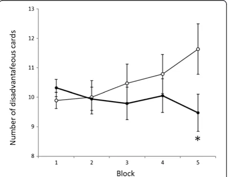
mutism (one patient), and specific phobias (one patient). In the WCST, PDD patients achieved significantly fewer categories (CA) and committed significantly more TEs and PEs than the controls (Table 2). During the last selec-tion of the IGT (selecselec-tions 81–100), the number of disad-vantageous card selected was significantly higher in PDD patients than in controls (Figure 1). During the other se-lections in IGT, we observed no significant differences be-tween PDD patients and the controls.
Discussion
In the present study, PDD patients committed significantly more perseverative errors and achieved fewer categories in the WCST than the controls. Our results are consistent with those of previous studies in which WCST was assigned to individuals with high-functioning autistic dis-order in late adolescence and early adulthood [3,4]. Pa-tients with DLPFC damage also show more perseverative errors in the WCST than controls [20], and DLPFC activa-tion measured by PET [21] and funcactiva-tional magnetic res-onance (fMRI) [22] is increased in subjects performing the WCST. Proton magnetic resonance spectroscopy has re-vealed similar dysfunction in the left DLPFC and anterior cingulate cortex in children with autistic disorder [23]. Courchesne et al. [24] reported that the mean number and size of DLPFC and medial prefrontal cortex neurons are higher than those of control subjects. Thus, the results of the WCST in the present study are consistent with the findings of previous studies employing anatomical or neu-roimaging techniques.
In the present study, PDD patients showed a signifi-cantly stronger tendency toward selecting disadvantageous cards in the IGT than the controls, even during the late examination period. To our knowledge, only one other study has investigated the results of IGT assigned to chil-dren and adolescents with PDD, which showed no
Table 1 Subject characteristics
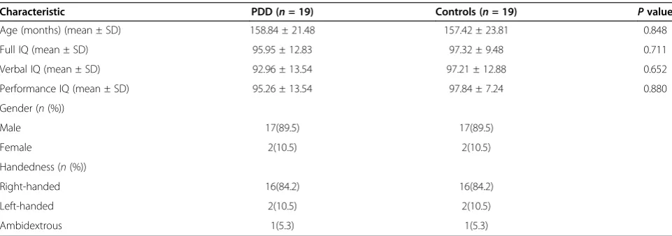
Characteristic PDD (n= 19) Controls (n= 19) Pvalue
Age (months) (mean ± SD) 158.84 ± 21.48 157.42 ± 23.81 0.848
Full IQ (mean ± SD) 95.95 ± 12.83 97.32 ± 9.48 0.711
Verbal IQ (mean ± SD) 92.96 ± 13.54 97.21 ± 12.88 0.652
Performance IQ (mean ± SD) 95.26 ± 13.54 97.84 ± 7.24 0.880
Gender (n(%))
Male 17(89.5) 17(89.5)
Female 2(10.5) 2(10.5)
Handedness (n(%))
Right-handed 16(84.2) 16(84.2)
Left-handed 2(10.5) 2(10.5)
Ambidextrous 1(5.3) 1(5.3)
significant difference between patients with Asperger's syn-drome and controls [25]. This study showed that the per-centage for selecting disadvantageous cards in the final selection in the control group was 31.6%, whereas that of the Asperger's syndrome group was 43.6%. We suspect that the nonsignificant results could be because of a smaller sample size of patients or the low severity of PDD that they exhibited. Future studies with larger sample sizes are needed to elucidate this relationship. Other studies have reported that OFC-injured patients also show a ten-dency to select disadvantage cards continuously in the later selections [10,11].
Using morphometric MRI, Hardan et al. [26] observed that the total volume (i.e., gray plus white matter) in the right lateral OFC was decreased in children and adoles-cents with autistic disorder but was increased in adults with autistic disorder. Moreover, a recent fMRI study of healthy adults reported that the processing theory of mind tasks, a function believed to be impaired in individuals with autistic disorder, was associated with increased activation
of the right lateral OFC [7]. The results of the IGT in this study seem to fit with the findings of these previous neuro-imaging studies. Thus, considering the results of this study and those of previous studies, PDD patients may have some structural and functional abnormalities in the OFC. Further-more, the results of this study indicate that both DLPFC and OFC functions are impaired in high-functioning PDD patients in childhood and early adolescence.
The limitations of this study are as follows: (1) the sam-ple size was small, and the PDD subjects were recruited from only one facility; (2) because of a narrow age range and a smaller number of female patients, we could not examine any effects of sex/age differences; (3) only neuro-psychological tests were employed in this study. These neuropsychological tests cannot verify frontal lobe dys-functions directly.
Conclusions
We assigned the WCST and IGT to high-functioning PDD patients in childhood and early adolescence with controls matched for gender, age, handedness, and intelligence. We identified that both DLPFC and OFC functions are im-paired in PDD patients. This study is the first to examine the impairment of both DLPFC and OFC functions in chil-dren and adolescents with high-functioning PDD. The findings of this study will contribute toward the under-standing of brain dysfunction and clinical features of PDD.
To achieve this end, joint studies involving multiple fa-cilities with larger sample sizes and wider age ranges need to be conducted. Moreover, studies evaluating the relationships between frontal lobe functions, and mind tasks and social skills also need to be conducted.
Competing interests
The authors declare that they have no competing interests.
Authors’contributions
TS wrote the manuscript. MK and AO designed the study. MK, YI, HU, MU, and KW recruited and screened the study participants. MK and YI diagnosed the study participants. MK administered the IGT. TS undertook the statistical analysis. AO and KS supervised the data analysis and manuscript writing. DS gave critical comments on the manuscript. All authors have contributed to and approved the final manuscript.
Acknowledgements
This study was funded by a research grant for Nervous and Mental Disorders (20B-6) of the Japanese Ministry of Health, Labour, and Welfare. We would like to express our deep gratitude to Professor Katsutoshi Tanaka, Professor Hirokuni Tagaya, and Professor Yumi Iwamitsu for giving us good opinions and advice. Moreover, we feel deeply grateful for the children, caregivers, and staff members who participated in this study.
Author details 1
Department of Developmental Psychiatry, Kitasato University Graduate School of Medical Science, 1-15-1 Kitasato, Minami-Ku, Sagamihara 252-0374, Kanagawa, Japan.2Department of Child and Adolescent Mental Health, Aiiku Hospital, 5-6-8 Minamiazabu, Minato-Ku 106-8580, Tokyo, Japan.3Department of Neuropsychiatry, Shinshu University School of Medicine, 3-1-1 Asahi, Matsumoto 390-8621, Nagano, Japan.4Department of Child and Adolescent Psychiatry, National Center for Global Health and Medicine, Kohnodai Hospital, 1-7-1 Kohnodai, Ichikawa 272-0836, Chiba, Japan.
Table 2 Subject performance on the Wisconsin card scoring test
PDD (n= 19) Controls (n= 19) P
value
Mean ± SD Mean ± SD
CA 2.53 ± 1.96 4.21 ± 1.44 0.004
TE 23.58 ± 10.47 15.79 ± 8.19 0.015
PE 7.89 ± 4.52 3.58 ± 4.75 0.025
Subject performances on the Wisconsin Card Scoring Test are shown.CA categories achieved,TEtotal errors,PEperseverative errors.
Received: 22 August 2013 Accepted: 17 September 2013 Published: 8 October 2013
References
1. Berman KF, Ostrem JL, Randolph C, Gold J, Goldberg TE, Coppola R, Carson RE, Herscovitch P, Weinberger DR:Physiological activation of a cortical network during performance of the Wisconsin card sorting test: a positron emission tomography study.Neuropsychologia1995,33:1027–1046.
2. Liss M, Fein D, Bullard S, Robins D:Brief report: cognitive estimation in individuals with pervasive developmental disorders.J Autism Dev Disord 2000,30:613–618.
3. Rumsey JM:Conceptual problem-solving in highly verbal, nonretarded autistic men.J Autism Dev Disord1985,15:23–36.
4. Rumsey JM, Hamburger SD:Neuropsychological divergence of high-level autism and severe dyslexia.J Autism Dev Disord1990,20:155–168. 5. Bachevalier J, Loveland KA:The orbitofrontal-amygdala circuit and
self-regulation of social-emotional behavior in autism.Neurosci Biobehav Rev 2006,30:97–117.
6. O’Doherty J, Kringelbach ML, Rolls ET, Hornak J, Andrews C:Abstract reward and punishment representations in the human orbitofrontal cortex.Nat Neurosci2001,4:95–102.
7. Völlm BA, Taylor ANW, Richardson P, Corcoran R, Stirling J, McKie S, Deakin JFW, Elliott R:Neuronal correlates of theory of mind and empathy: a functional magnetic resonance imaging study in a nonverbal task. Neuroimage2006,29:90–98.
8. Dawson G, Munson J, Estes A, Osterling J, McPartland J, Toth K, Carver L, Abbott R:Neurocognitive function and joint attention ability in young children with autism spectrum disorder versus developmental delay. Child Dev2002,73:345–358.
9. Salmond CH, De Haan M, Friston KJ, Gadian DG, Vargha-Khadem F: Investigating individual differences in brain abnormalities in autism. Philos Trans R Soc London Ser B Biol Sci2003,358:405–413.
10. Bechara A, Damasio H, Tranel D, Anderson SW:Dissociation of working memory from decision making within the human prefrontal cortex. J Neurosci1998,18:428–437.
11. Grant S, Contoreggi C, London ED:Drug abusers show impaired performance in a laboratory test of decision making. Neuropsychologia2000,38:1180–1187.
12. American Psychological Association:Diagnostic and Statistical Manual of Mental Disorders.4th edition. Washington DC: American Psychological Association; 2000.
13. Chapman LJ, Chapman JP:The measurement of handedness. Brain Cognition1987,6:175–183.
14. Bechara A, Damasio AR, Damasio H, Anderson SW:Insensitivity to future consequences following damage to human prefrontal cortex. Cognition1984,50:7–15.
15. Fukui H, Murai T, Fukuyama H, Hayashi T, Hanakawa T:Functional activity related to risk anticipation during performance of the Iowa gambling task.Neuroimage2005,24:253–259.
16. Kodaira M, Iwadare Y, Ushijima H, Oiji A, Kato M, Sugiyama N, Sasayama D, Usami M, Watanabe K, Saito K:Poor performance on the Iowa gambling task in children with obsessive-compulsive disorder.Ann Gen Psychiatry 2012,11:25.
17. Igarashi K, Oguni H, Osawa M, Awaya Y, Kato M, Mimura M, Kashima H: Wisconsin card sorting test in children with temporal lobe epilepsy. Brain Dev2002,24:174–178.
18. Nelson HE:A modified card sorting test sensitive to frontal lobe defects. Cortex1976,12:313–324.
19. Lezak MD:Neuropsychological Assessment.Oxford: Oxford University Press; 1995. 20. Milner B:Effects of different brain lesions on card sorting: the role of the
frontal lobes.Arch Neurol1963,9:90–100.
21. Cabeza R, Nyberg L:Imaging cognition II: an empirical review of 275 PET and fMRI studies.J Cogn Neurosci2000,12:1–47.
22. Konishi S, Nakajima K, Uchida I, Kameyama M, Nakahara K, Sekihara K, Miyashita Y:Transient activation of inferior prefrontal cortex during cognitive set shifting.Nat Neurosci1998,1:80–84.
23. Fujii E, Mori K, Miyazaki M, Hashimoto T, Harada M, Kagami S:Function of the frontal lobe in autistic individuals: a proton magnetic resonance spectroscopic study.JMI2010,57:35–44.
24. Courchesne E, Mouton PR, Calhoun ME, Semendeferi K, Ahrens-Barbeau C, Hallet MJ, Barnes CC, Pierce K:Neuron number and size in prefrontal cortex of children with autism.JAMA2011,306:2001–2010. 25. Johnson SA, Yechiam E, Murphy RR, Queller S, Stout JC:Motivational
processes and autonomic responsivity in Asperger's syndrome: evidence from the Iowa gambling task.JINS2006,12:668–676.
26. Hardan AY, Girgis RR, Lacerda ALT, Yorbik O, Kilpatrick M, Keshavan MS, Minshew NJ:Magnetic resonance imaging study of the orbitofrontal cortex in autism.J Child Neurol2006,21:866–871.
doi:10.1186/1744-859X-12-31
Cite this article as:Sawaet al.:Dysfunction of orbitofrontal and dorsolateral prefrontal cortices in children and adolescents with high-functioning pervasive developmental disorders.Annals of General Psychiatry201312:31.
Submit your next manuscript to BioMed Central and take full advantage of:
• Convenient online submission
• Thorough peer review
• No space constraints or color figure charges
• Immediate publication on acceptance
• Inclusion in PubMed, CAS, Scopus and Google Scholar
• Research which is freely available for redistribution