R E S E A R C H
Open Access
Product inhibition of cellulases studied with
14
C-labeled cellulose substrates
Hele Teugjas and Priit Väljamäe
*Abstract
Background:As a green alternative for the production of transportation fuels, the enzymatic hydrolysis of lignocellulose and subsequent fermentation to ethanol are being intensively researched. To be economically feasible, the hydrolysis of lignocellulose must be conducted at a high concentration of solids, which results in high concentrations of hydrolysis end-products, cellobiose and glucose, making the relief of product inhibition of cellulases a major challenge in the process. However, little quantitative information on the product inhibition of individual cellulases acting on cellulose substrates is available because it is experimentally difficult to assess the hydrolysis of the heterogeneous polymeric substrate in the high background of added products.
Results:The cellobiose and glucose inhibition of thermostable cellulases fromAcremonium thermophilum,
Thermoascus aurantiacus, andChaetomium thermophilumacting on uniformly14C-labeled bacterial cellulose and its derivatives,14C-bacterial microcrystalline cellulose and14C-amorphous cellulose, was studied. Cellulases from
Trichoderma reeseiwere used for comparison. The enzymes most sensitive to cellobiose inhibition were glycoside hydrolase (GH) family 7 cellobiohydrolases (CBHs), followed by family 6 CBHs and endoglucanases (EGs). The strength of glucose inhibition followed the same order. The product inhibition of all enzymes was relieved at higher temperatures. The inhibition strength measured for GH7 CBHs with low molecular-weight model substrates did not correlate with that measured with14C-cellulose substrates.
Conclusions:GH7 CBHs are the primary targets for product inhibition of the synergistic hydrolysis of cellulose. The inhibition must be studied on cellulose substrates instead of on low molecular-weight model substrates when selecting enzymes for lignocellulose hydrolysis. The advantages of using higher temperatures are an increase in the catalytic efficiency of enzymes and the relief of product inhibition.
Keywords:Cellulase, Cellulose, Cellobiose, Glucose, Inhibition,Acremonium thermophilum,Thermoascus aurantiacus,
Chaetomium thermophilum,Trichoderma reesei
Background
Cellulose is the most abundant biopolymer on Earth and has great potential as a renewable energy source. In nature, cellulose is degraded mainly by fungi and bacteria, which secrete cellulolytic enzymes [1]. These enzymes include cellulases, hemicellulases, and enzymes involved in lignin breakdown. Cellulases are divided into cellobiohydrolases (CBHs), endoglucanases (EGs) and β-glucosidases (BGs). CBHs are processive enzymes that liberate consecutive cellobiose units from cellulose chain ends, whereas EGs non-processively attack cellulose chains at random po-sitions. β-Glucosidases hydrolyze cellobiose to glucose,
thus relieving the product inhibition of CBHs [2]. One of the most efficient and best-characterized cellulolytic systems is that of the soft rot fungusTricoderma reesei
(Tr). The major component of theTrcellulolytic system is the glycoside hydrolase (GH) family 7 [3,4] CBH,
TrCel7A (formerly CBH I).Tralso secretes a less abun-dant CBH,TrCel6A (CBH II), and a number of EGs, in-cluding TrCel7B,TrCel5A and TrCel12A (EG I, EG II and EG III, respectively).
Cellulases are used in many biotechnological applica-tions, such as fiber modification in the paper and textile industries, but they also have great potential in the emerging industry of ethanol production from lignocel-lulose. To decrease the water consumption and reduce the costs of equipment and distillation, the hydrolysis of
* Correspondence:priit.valjamae@ut.ee
Institute of Molecular and Cell Biology, University of Tartu, Riia 23b–202, Tartu 51010, Estonia
lignocellulose must be conducted at a high concentra-tion of solids. This approach inevitably results in high concentrations of the hydrolysis end-products cellobiose and glucose, and it has been proposed that the end-product inhibition of cellulases is rate limiting for ligno-cellulose hydrolysis in high-solid conditions [5]. Thus, relieving the product inhibition is a major challenge in the process, as well as in enzyme engineering [6]. The end-product inhibition can be relieved in a simultaneous saccharification and fermentation process, where the fermenting organism is added in parallel with hydrolytic enzymes, but one drawback is the need for different condi-tions for optimal hydrolysis and fermentation. The optimal temperature for yeast fermentation is approximately 35°C, whereas temperatures near 50°C are optimal for the performance of cellulases. A process concept using high temperature liquefaction with thermostable enzymes pre-ceding simultaneous saccharification and fermentation has been developed [7], and this has triggered the search for novel thermostable enzymes [8,9].
Despite intensive efforts, little quantitative information about the end-product inhibition of cellulases is available. Many of the studies can be classified as“semi-quantitative”. Most often, the rates of cellulose hydrolysis measured in the presence and absence of β-glucosidase are compared [10-13]. In some studies, the experimental setup enabling the continuous elimination of end-products has been used [6]. The numerical values of inhibition constants have been obtained by the fitting of hydrolysis data to the complex equations derived for the full time-course [14-20]. The val-idity of these figures depends on the valval-idity of the model [21]. Another problem lies in the possible interplay be-tween parameters in trials, where values of multiple param-eters are approximated by a single fit. The inhibition types reported include competitive, non-competitive, uncompeti-tive and mixed inhibition, whereas the values of inhibition constants vary over several orders of magnitude. One rea-son for the variation of reported inhibition types and the values of inhibition constants is that complex cellulase mixtures are often used instead of purified cellulase com-ponents in experiments. Different cellulase comcom-ponents may be inhibited to different extents and by different mechanisms, which clearly complicates the interpretation of the data. For literature reviews of earlier and more re-cent studies, see [22] and [23], respectively.
An inherent problem in measuring the strength of product inhibition is associated with difficulties in meas-uring the initial rates of product formation in the high background of the product added as an inhibitor. Three approaches can be used to overcome this: (i) measure-ment of the initial rates of substrate consumption in-stead of product formation [24]; (ii) measurement of the hydrolysis rate with a method that does not rely on measuring the concentration of the substrate or product;
thermophilum (At), Thermoascus aurantiacus (Ta), and
Chaetomium thermophilum(Ct).Cellulases from these or-ganisms have great potential in biotechnological applica-tions [34-39]. Well-characterized cellulases from Tr were used for comparison.
Results and discussion
Measuring the strength of inhibition
The best parameter for describing the inhibitory strength of an inhibitor is Ki, the equilibrium dissociation con-stant of an enzyme-inhibitor complex. Ki is a funda-mental parameter of enzyme kinetics that is directly related to the thermodynamic stability of the enzyme-inhibitor complex. The conventional approach for the measurement of Ki involves the measurement of kcat and KM values for the substrate at different concentra-tions of an inhibitor. The plotting of kcat and KM or their combination as a function of inhibitor concentra-tion allows the determinaconcentra-tion of both the type of inhib-ition and the Ki value. However, this approach is not applicable to cellulases acting on cellulose. The com-plex, multiple-mode binding of cellulases to the solid substrate obeys the so-called double-saturation charac-ter [1].KMvalues measured for cellulose depend on the enzyme concentration, and therefore, KM has not its usual meaning. Because of the non-productive binding and strong time dependency, the measurement of the
kcatvalue is also not straightforward [40-42].
A simplified approach for assessing the inhibitory strength is to measure the IC50, the inhibitor concen-tration that halves the rate of the enzyme-catalyzed reaction. The IC50 is measured at one substrate con-centration by varying the concon-centration of the inhibi-tor. Data are plotted as vi/v0 versus [I], where vi and v0 are the rates measured in the presence and ab-sence of inhibitor, respectively, and [I] is the concen-tration of inhibitor. To find the IC50, the data are first fitted to hyperbolae in the following form:
vi v0¼
S ½ þC1
S
½ þC1 1þC½ I2
ð1Þ
In the fitting of the data, the substrate concentration ([S]) is fixed to the value used in the experiments. The above value of [S] and the values of empirical constants
C1andC2found by the fitting are further used to calcu-late theIC50value using Equation 2:
IC50¼C2 1þ
S ½ C1
ð2Þ
The IC50is an empirical parameter and its value may depend on the concentration of the substrate (relative to its KM value for the enzyme) used in the measurement
of the IC50. If and how the IC50 value depends on [S]/KM depends on the type of inhibition. In the case of competitive inhibition, the relationship among IC50, Ki and [S]/KMis given as follows:
IC50¼Ki 1þ S ½ KM
ð3Þ
Thus, if the inhibition is competitive and the [S] used in the measurement of the IC50 is well below its KM value, the resulting IC50 value is close to the true Ki value. However, if [S] is near saturating for the enzyme, the inhibition appears to be weak, as the resultingIC50is much higher than Ki. The situation is opposite in the case of un-competitive inhibition, as in this case we have the following:
IC50¼Ki 1þ KM
S ½
ð4Þ
In the case of mixed inhibition, the interplay among
IC50, Ki (there are two different Kis now) and [S]/KM is more complicated, and whether the inhibition appears to be stronger at low or high [S]/KM ratio depends on which type of inhibition (competitive or un-competitive) is dominating. However, in the case of pure non-competitive inhibition, IC50 =Ki, so IC50 represents the value of the true Kiat any substrate concentration used for its measurement.
GH family 7 cellobiohydrolases
GH7 CBHs are major components of efficient fungal cel-lulase systems. They are processive enzymes that are responsible for the degradation of crystalline cellulose [43]. Because of their central role in cellulose degrad-ation, the inhibition of GH7 CBHs is of utmost import-ance. Here, we undertook a study of the inhibition of GH7 CBHs acting on14C-BC. Thermostable GH7 CBHs
AtCel7,TaCel7A, andCtCel7A [44], along withTrCel7A, were characterized in terms of cellobiose and glucose inhibition.Tmvalues of 75°C, 69°C, 75°C and 65°C have been reported for TaCel7A, AtCel7A, CtCel7A and
GH7 CBHs were thus provided with the EG, TrCel5A (10% on a molar basis).
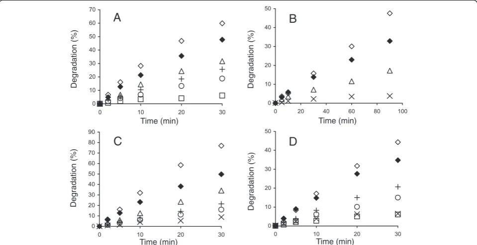
Figure 1 shows the time courses for the synergistic hydrolysis of 14C-BC by CBHs (supplemented with
TrCel5A and β-glucosidase, N188BG) at different tem-peratures. With all CBHs, the time courses of 14C-BC degradation measured at lower temperatures (25°C – 35°C) were nearly linear, whereas the time courses mea-sured at higher temperatures gradually deviated from linearity. With TaCel7A as an exception, the degree of conversion after 30 min of hydrolysis measured at 60°C was less than that measured at 50°C (Figure 1). How-ever, such a decrease in the degree of conversion with increasing temperature was not observed after 5 min of hydrolysis. Similar observations have also been made for the hydrolysis of pre-treated lignocellulose [36], suggesting that this phenomenon is not 14C-BC spe-cific. The simplest explanation would be the thermal inactivation of enzymes that progresses with time. We tested the possible thermal inactivation of enzymes in an experiment where the hydrolysis began at 55°C, and after 30 min, the temperature was decreased to 40°C.
TrCel7A was used as the CBH because of its lowestTm value among the CBHs studied. Figure 2 demonstrates that despite a 15°C drop in temperature, the rate of cel-lulose hydrolysis actually increased. This finding rules
out the irreversible inactivation of enzymes as the pri-mary cause of the non-linearity in time curves observed at higher temperatures. However, the contribution of the reversible denaturation of enzymes cannot be ruled out. The hydrolysis of cellulose by CBH is a multi-step process including binding to cellulose, the capture of the cellulose chain-end, processive degradation, and dis-sociation [32,50]. Therefore, another possibility is that some kinetic property of CBHs is negatively affected by temperature. Whatever the underlying mechanisms, the change in the linearity of time curves depending on temperature may also result in a change in the apparent inhibition strength with hydrolysis time.
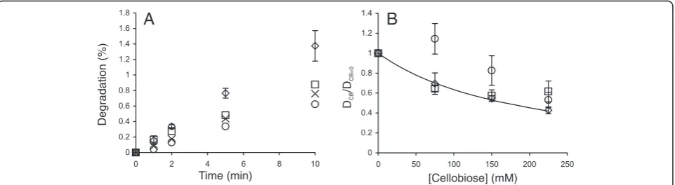
To study the cellobiose inhibition of GH7 CBHs, the synergistic hydrolysis of14C-BC in the presence of added cellobiose was followed (Figure 3, Additional file 1: Figures S1 and S2). Because the cellobiose inhibition of the EGTrCel5A is much weaker than that of GH7 CBHs [28,31], the inhibition of synergistic hydrolysis apparently reflects the inhibition of CBH. The strength of cellobiose inhibition was analyzed using plots of (DCB/DCB=0) versus [cellobiose], where DCB and DCB=0 represent the degree of conversion of14C-BC in the presence and ab-sence of cellobiose, respectively (Figure 4, Additional file 1: Figure S3). In the case of experiments without added cellobiose, the reactions were provided withN188BG to
A
B
C
D
Time (min) Time (min)
Time (min) Time (min)
Degradation (%)
Degradation (%)
Degradation (%)
Degradation (%)
Figure 1Synergistic hydrolysis of14C-BC by GH7 CBHs at different temperatures.14C-BC (0.25 mg ml-1) was incubated with 0.25μM CBH,
supplemented with 0.025μM EG (TrCel5A) and 0.06μMN188BG, at 25°C (□), 35°C (Δ), 40°C (◊), 50°C (×), and 60°C (○). CBH was(A)TrCel7A,
prevent the inhibition of the CBH by the cellobiose re-leased during hydrolysis. Experiments with no added cellobiose and without BG were also conducted. Com-parison of the results obtained with and withoutN188BG (both without added cellobiose) demonstrates that the
inhibition of CBHs by the cellobiose released during hydrolysis was significant (Figure 3, Additional file 1: Figures S1 and S2). Therefore, the concentration of the cellobiose released during hydrolysis was added to the concentration of externally supplied cellobiose in generating the plots in Figure 4 and Additional file 1: Figure S3. For the calculation ofIC50values, the data were first fitted to hyperbolae:
DCB DCB¼0¼
14C BC
þC1
1−H
ð Þ
14C BC
þC1 1þ½ CBC2
þH ð5Þ
where [CB] is the concentration of cellobiose; [14CBC] is the concentration of14C-BC used in the experiment; and
C1, C2 and H are empirical constants. Equation 5 differs from Equation 1 by the presence of constantH.Hwas in-cluded to improve the fit and is a constant that accounts for the background radioactivity (the degree of conversion that is independent of CBH). The degree of conversion resulting from the activity of the EG was measured in a separate experiment (Additional file 1: Figure S4) and was subtracted from the degree of conversion resulting from the synergistic hydrolysis. Thus, in the case of complete in-hibition,Hshould have a value of zero. Non-zeroHvalues are indicative of partial inhibition. However, because theH
values remained between 0 and 0.2 and were even negative in some cases, they may also be a result of experimental
Degradation (%)
Degradation (%)
Degradation (%)
Degradation (%)
Time (min)
Time (min) Time (min)
Time (min)
A
B
C
D
Figure 3Inhibition of GH7 CBHs by cellobiose.14C-BC (0.25 mg ml-1) was incubated with a mixture of 0.25μM CBH and 0.025μM EG
(TrCel5A) at 35°C. The concentration of cellobiose added was 0 mM + 0.06μMN188BG (◊), 0 mM (♦), 0.5 mM (Δ), 1.0 mM (+), 2.0 mM (○), 5.0 mM (×) or 10 mM (□). CBH was(A)TrCel7A,(B)TaCel7A,(C)AtCel7A, and(D)CtCel7A.
Time (min)
Degradation (%)
uncertainty. Provided with the values ofC1,C2andH, the value ofIC50was calculated as follows:
IC50¼
14C BC
þC1 C1
C2ð1−2HÞ
ð6Þ
Using the time course data measured in the presence and absence of cellobiose, theIC50values were first found separately for each time point (Figure 4A) [32]. Table 1 lists the averageIC50values over all time points. In some cases, a systematic drift of IC50 values with hydrolysis time was observed, which may indicate that different rate-limiting factors with different IC50values may con-trol the hydrolysis rate in different time or conversion frames. An apparent decrease in inhibition strength with increasing hydrolysis time was also observed for the cel-lobiose inhibition of EGTrCel7B [28]. A more systematic analysis of the time dependency ofIC50values remained outside the scope of the present study. The enzyme most sensitive to cellobiose inhibition appeared to beTaCel7A, followed by AtCel7A, TrCel7A and CtCel7A (Table 1). However, the differences between enzymes were not very
prominent, especially considering error limits. With all enzymes, the strength of cellobiose inhibition decreased significantly with increasing temperature.
The cellobiose inhibition of GH7 CBHs is most often studied on low-Mw model substrates. However, it has been shown that the inhibition of CBHs acting on low-Mw substrates appears to be much stronger than that on cellulose substrates [31,33]. TheKivalues for cellobi-ose inhibition of GH7 CBHs measured on low-Mw sub-strates are in the micromolar range [44,51,52], whereas those measured on cellulose are in the low- to high-millimolar range [28,31,32]. An interesting exception is Cel7A from Trichoderma harzianum, which shows a 7.2 mM Ki value for the cellobiose inhibition of the hydrolysis of chloro-nitrophenyl lactoside [53]. Unfor-tunately, the inhibition of this enzyme on cellulose has not been studied. We also studied the cellobiose inhib-ition of GH7 CBHs acting on MUL. The initial rates of MUL hydrolysis measured in the presence and ab-sence of added cellobiose were first analyzed according to Equation 1, and the IC50 values were found using Equation 2. As cellobiose was shown to be a competitive inhibitor for these CBHs acting on MUL [44] and the con-centration of MUL used in the experiments (5μM) was far below itsKMvalue (approximately 300μM [44]), the mea-sured IC50 value represents the true Ki (see Equation 3). The resulting Ki values are listed in Table 2. Van´t Hoff analysis of the temperature dependency of theIC50andKi values ofTrCel7A resulted in standard enthalpy changes of 63.6 ± 2.6 kJ mol-1 (for IC50 values on 14C-BC, Table 1) and approximately 63 kJ mol-1 (for Ki values on MUL, Table 2). The inhibition of MUL hydrolysis is attributable to the binding of cellobiose to the product sites (+1/+2) of TrCel7A [52]. Similar standard enthalpy changes thus suggest that the cellobiose inhibition of the synergistic
[Cellobiose] (mM) [Cellobiose] (mM)
D/
D
CB
CB=0
D/
D
CB
CB=0
A
B
Figure 4Analysis of the inhibition of GH7 CBHs by cellobiose. (A)Data for the hydrolysis of14C-BC by the mixture ofTrCel7A andTrCel5A at
35°C (Figure 3A) were rearranged in the coordinates (DCB/DCB=0) versus [cellobiose], where DCBand DCB=0represent the degree of conversion of 14C-BC in the presence and absence of cellobiose, respectively. The ratio of (D
CB/DCB=0) was found after different times of hydrolysis, which were
2 min (◊), 5 min (□), 10 min (Δ), 20 min (○), and 30 min (×).(B)Data for the hydrolysis of14C-BC by the mixture of CBH andTrCel5A at 35°C
(Figure 3) in the coordinates (DCB/DCB=0) versus [cellobiose]. (DCB/DCB=0) values for all hydrolysis time points are shown. CBH wasTrCel7A (□), TaCel7A, (◊),AtCel7A (Δ), andCtCel7A (×). Solid lines are from the non-linear regression according to Equation 5.
Table 1 Inhibition of GH7 CBHs by cellobiose and glucose studied with14C-BC substrate
IC50for cellobiose (mM) IC50for glucose (mM)
25°C 35°C 50°C 35°C
TrCel7A 0.38 ± 0.03a 0.68 ± 0.24b 2.61 ± 0.10 420 ± 230c
AtCel7A 0.19 ± 0.10c 0.44 ± 0.10 2.12 ± 1.40b 420 ± 180c
CtCel7A 0.41 ± 0.06 1.08 ± 0.22 2.48 ± 0.91b 360 ± 170c
TaCel7A 0.58 ± 0.35 0.93 ± 0.10
a
Data from [32]. b
IC50increased with hydrolysis time. c
hydrolysis of 14C-BC is also attributable to the binding of cellobiose to sites +1/+2. Nonetheless, for all CBHs, the Ki values found for the cellobiose inhibition of MUL hydrolysis (Table 2) were smaller than the corresponding
IC50 values for the inhibition of 14C-BC hydrolysis (Table 1). The reason for this difference may lie in the different modes of action used by CBHs with low-Mw model substrates and cellulose and therefore the different types of inhibition [32]. Another possible explanation is that the cellobiose inhibition of CBHs on cellulose is competitive and that the concentration of cellulose chain ends used in the measurement of theIC50value is higher than the corresponding KM value. In this case, the observed IC50 is expected to be higher than the Ki (see Equation 3), and the inhibition of cellulose hy-drolysis appears to be weak. This scenario has been proposed to explain the differences in the inhibitory strength of xylo-oligosaccharides toward CBHs acting on MUL and cellulose [33]. The binding of xylo-oligosaccharides with DP 8–10 is expected to mimic the binding of the cellulose chain to the active site of
TrCel7A, resulting in competitive inhibition. In contrast, despite the strong binding of cellobiose to the product
sites (+1/+2) of TrCel7A [52,54,55], the cellulose chain can still bind to the substrate sites (from−7 to−1), and this predicts non-competitive inhibition [23,31,32]. The results of our previous studies of the inhibition of
TrCel7A under single-turnover and steady-state condi-tions suggested that cellobiose might be a mixed-type in-hibitor of TrCel7A acting on cellulose. The binding of cellobiose to the product and substrate binding sites was proposed to be responsible for the non-competitive and competitive components of inhibition, respectively [32]. Observations that the binding affinity of TrCel7A and
TrCel6A towards cellulose increased in the presence of cellobiose also suggest an inhibition mode that is not competitive [56,57]. From the practical point of view, it is important to note that for different CBHs, the differences in inhibition strength observed on MUL and cellulose were not of the same magnitude (Figure 5). This result can be exemplified best byTaCel7A, which appeared to be most resistant to cellobiose inhibition on MUL sub-strate (Table 2) but was most sensitive to cellobiose in-hibition on cellulose (Table 1). This finding stresses the importance of the use of“as native as possible”screening systems for selecting cellulases [58].
The glucose inhibition of CBHs with 14C-BC as the substrate was also studied. CBHs were provided with EG TrCel5A (TrCel5A is not significantly inhibited by glucose [28]) and also with BG in the experiments with-out added glucose. The time courses of 14C-BC hy-drolysis in the presence and absence of added glucose are shown in Figure 6. As revealed by the scattering of data points in the plot of (DGlc/DGlc=0) versus [glucose] (Figure 6D), the inhibition by the cellobiose released during hydrolysis was significant. This result was accounted for by adding the term [CB]/IC50(CB) ([CB] is the concentration
14
IC
for CB on
C-BC (mM)
50
K
ifor CB on MUL (mM)
Figure 5Relative strength of cellobiose inhibition of GH7 CBHs depends on the substrate.Kivalues measured for MUL hydrolysis andIC50
values measured for the hydrolysis of14C-BC, both at 35°C and 50°C, were taken from Table 2 and Table 1, respectively. CBH wasTrCel7A (□), TaCel7A, (◊),AtCel7A (Δ), and CtCel7A (×).
Table 2 Inhibition of GH7 CBHs by cellobiose studied with MUL substrate
Kifor cellobiose (mM)
35°C 50°C
TrCel7A 0.040 0.124
AtCel7A 0.095 0.233
CtCel7A 0.110 0.346
of the released cellobiose, and IC50(CB) is the IC50 for cellobiose previously determined) to Equation 5 to create Equation 7:
DGlc DGlc¼0¼
14C BC
þC1
1−H
ð Þ
14C BC
þC1 1þIC½ CB50ð ÞCB þ½GlcC2
þH ð7Þ
DGlcand DGlc=0represent the degree of conversion of 14
C-BC in the presence and absence of added glucose, respectively; [Glc] is the concentration of added glucose; [14CBC] is the14C-BC concentration used in the experi-ment; and C1, C2 and H are empirical constants. The values ofC1,C2andHobtained by the fitting of the data to Equation 7 were used to calculate theIC50for glucose according to Equation 6. The glucose inhibition of GH7 CBHs was more than two orders of magnitude weaker than cellobiose inhibition (Table 1). Although relatively weak, glucose inhibition may become significant in the separate hydrolysis and fermentation of lignocellulose at a high dry matter consistency, where glucose may accu-mulate to well above 50 g/l (0.28 M) [5,23].
GH family 6 cellobiohydrolases
GH6 CBHs are the second most abundant components of fungal cellulase systems. They are inverting CBHs that preferentially attack cellulose chains from non-reducing ends. To date, there are no good chromo-or fluchromo-orogenic model substrates fchromo-or GH6 CBHs [59]. Because of the different chain-end preferences, inhibition studies on reducing-end-labeled cellulose substrates are also not applicable [31]. Therefore, little is known about the strength of the product inhibition of GH6 CBHs. From the reported binding constants measured using fluorophore competition experiments [60,61] and ana-lysis of the progress curves of cellotriose hydroana-lysis [51,62], Kivalues in a sub- to low-millimolar range can be calculated for the interaction ofTrCel6A with cellobi-ose and gluccellobi-ose.
Here, we characterized the cellobiose and glucose inhibition of TrCel6A and its thermophilic counter-part, CtCel6A [9,63]. First, the cellobiose inhibition of the synergistic hydrolysis of 14C-BC by TrCel6A and
TrCel5A was studied (Figure 7A). AsTrCel6A was less sensitive to cellobiose inhibition than TrCel7A, the con-tribution of the cellobiose released during hydrolysis was
Time (min) Time (min)
Time (min) [Glucose] (M)
Degradation (%)
Degradation (%)
Degradation (%)
D/
D
Glc
Glc=0
A
C
D
B
Figure 6Inhibition of GH7 CBHs by glucose.14C-BC (0.25 mg ml-1) was incubated with a mixture of 0.25μM CBH and 0.025μM EG (TrCel5A) at 35°C. The concentration of added glucose was as follows: 0 M + 0.06μMN188BG (◊), 0 M (♦), 0.05 M (Δ), 0.125 M (+), 0.25 M (○), 0.5 M (×) or 1.0 M (□). CBH was as follows:(A)TrCel7A,(B)AtCel7A, and(C)CtCel7A.(D)Hydrolysis data in the coordinates (DGlc/DGlc=0) versus [glucose],
where DGlcand DGlc=0represent the degree of conversion of 14
not significant, and an average (DCB/DCB=0) over all time points was used in plotting (DCB/DCB=0) versus [cellobi-ose] (Figure 7D). No significant systematic variation of DCB/DCB=0 depending on hydrolysis time was observed. As in the case of GH7 CBHs, theIC50value was found using Equations 5 and 6. Because theIC50value for syn-ergistic hydrolysis (Table 3) was of the same order as the apparentKivalue reported for TrCel5A [31], we further tested the inhibition of individual TrCel6A. BC is not a good substrate for TrCel6A, but its acid-treated deriva-tive, BMCC, is readily degraded by the enzyme. There-fore, we prepared14C-BMCC by the heterogeneous acid hydrolysis of14C-BC. The time courses of14C-BMCC hy-drolysis byTrCel6A andCtCel6A are shown in Figures 7B
and 7C. Without supplied cellobiose, both enzymes had similar activity with the 14C-BMCC substrate, but
CtCel6A was somewhat more resistant to cellobiose in-hibition (Figure 7D, Table 3). TheIC50value forTrCel6A by itself was similar to that found for the synergistic hydrolysis. This result suggests that the inhibition of
TrCel6A was responsible for the cellobiose inhibition of the synergistic hydrolysis of 14C-BC. The glucose inhib-ition ofTrCel6A andCtCel6A with14C-BMCC as a sub-strate was also studied (Figure 8). Because the inhibition by cellobiose released during hydrolysis was not signifi-cant, a simpler equation, Equation 5 (the terms referring to cellobiose were replaced with corresponding terms for glucose), was used instead of Equation 7 to analyze the glucose inhibition of GH6 CBHs. Glucose appeared to be an approximately 10 times weaker inhibitor of TrCel6A and CtCel6A than cellobiose (Table 3), but comparison with corresponding figures for GH7 CBHs (Table 1) re-veals that glucose is a relatively stronger inhibitor of GH6 than GH7 CBHs. The same result was also observed in a recent calorimetry study of the inhibition ofTrcellulases acting on amorphous cellulose [28]. However, the IC50 values found by Murphy et al. [28] for the cellobiose in-hibition of TrCel7A and TrCel6A were approximately one order of magnitude higher than ours. Whether the
Time (min) Time (h)
Time (h)
Degradation (%) Degradation (%)
Degradation (%)
D/
D
CB
CB=0
[Cellobiose] (mM)
A
C
D
B
Figure 7Inhibition of GH6 CBHs by cellobiose. (A)14C-BC (0.25 mg ml-1) was incubated with a mixture of 0.25μMTrCel6A and 0.025μM EG
(TrCel5A) at 25°C.(BandC)14C-BMCC (0.25 mg ml-1) was incubated with 0.25μMTrCel6A (panel B) or with 0.25μMCtCel6A(panelC)at 50°C.
The concentration of added cellobiose was 0 mM + 0.06μMN188BG (◊), 1.0 mM (*), 5.0 mM (+), 10 mM (□), 20 mM (Δ), 50 mM (×), or 100 mM (○).
(D)Hydrolysis data (from panels A-C) in the coordinates (DCB/DCB=0) versus [cellobiose], where DCBand DCB=0represent the degree of conversion of 14C-cellulose in the presence and absence of cellobiose, respectively. The average (D
CB/DCB=0) values over hydrolysis time points are plotted. Solid lines
are from the non-linear regression according to Equation 5.TrCel6A +TrCel5A on14C-BC (□),TrCel6A on14C-BMCC (◊), andCtCel6A on14C-BMCC (×).
Table 3 Inhibition of GH6 CBHs by cellobiose and glucose studied with14C-BC and14C-BMCC substrates
IC50(mM)
Cellobiose Glucose
TrCel6Aa 16 ± 0.5
TrCel6Ab 20 ± 1.4 240 ± 26
CtCel6Ab 28 ± 4.5 301 ± 30
a
Synergistic hydrolysis of14
C-BC byTrCel6A and EG,TrCel5A, at 25°C. b
differences in the strengths of cellobiose inhibition reflect the differences in substrates or the methods used for the measurement of inhibition is not known. Comparison of the IC50 values measured here with Ki values derived from binding constants measured using low-Mw sub-strates and ligands as competitors [60-62] reveals the same trend as in the case of GH7 CBHs: the binding of cellobiose and glucose appears to be weaker when assessed on polymeric substrates.
Endoglucanases
EGs are a diverse group of enzymes present in all effi-cient cellulase systems. Their best recognized function is their synergism with CBHs. Depending on the condi-tions, the degree of synergistic effect may be more than 10-fold [32,64]. Therefore, the inhibition of the EG com-ponent may result in a drastic decrease in the rate of the synergistic hydrolysis of cellulose. The main soluble
product of the EG-catalyzed cellulose hydrolysis is cellobiose, but some glucose and higher-order oligosac-charides are also produced [65]. Here, we studied the cel-lobiose inhibition of the EGs TrCel7B, TrCel5A and
TrCel12A with 14C-amorphous cellulose substrate. The enzyme concentrations and hydrolysis times were ad-justed so that the linear region of the time course was studied. The time courses for the hydrolysis of 14 C-amorphous cellulose byTrCel7B in the presence and ab-sence of added cellobiose are shown in Figure 9A. For the results with TrCel5A andTrCel12A, see Additional file 1: Figure S5. The “conventional” inhibition pattern was observed only in the case of TrCel7B, with an IC50 value of 168 ± 2 mM. This figure is reasonably well in line with that measured forTrCel7B on amorphous cellu-lose using isothermal titration calorimetry [28]. Calorim-etry measures the amount of glycosidic bonds that are cleaved irrespective of the solubility of the products [41].
Time (h)
Degradation (%)
[Glucose] (M)
D/
D
Glc
Glc=0
A
B
Figure 8Inhibition of GH6 CBHs by glucose. (A)14C-BMCC (0.25 mg ml-1) was incubated with 0.25μMTrCel6A (opened symbols) or with 0.25
μMCtCel6A (filled symbols) 50°C. The concentration of added glucose was as follows: 0 M + 0.06μMN188BG (◊,♦), 0.25 M (□,■), 0.5 M (Δ,▲), or 1.0 M (○,●).(B)Hydrolysis data in coordinates (DGlc/DGlc=0)versus[glucose] where DGlcand DGlc=0represent the degree of conversion of14C-BMCC
in the presence and absence of added glucose, respectively. (DGlc/DGlc=0) values for all hydrolysis time points are shown. Solid lines are from the
non-linear regression according to Equation 5 (the terms referring to cellobiose were replaced with corresponding terms for glucose). CBH was TrCel6A (◊) or CtCel6A (×).
Time (min)
Degradation (%)
D/
D
CB
CB=0
[Cellobiose] (mM)
A
B
Figure 9Inhibition of EGs by cellobiose. (A)14C-amorphous cellulose (0.5 mg ml-1) was incubated with 2.5 nMTrCel7B at 35°C. The concentration of
added cellobiose was 0 mM (◊), 75 mM (□), 150 mM (×), or 225 mM (○).(B)Hydrolysis data from panel A and Additional file 1: Figure S5 in the coordinates (DCB/DCB=0) versus [cellobiose] where DCBand DCB=0represent the degree of conversion of14C-cellulose in the presence and absence of
cellobiose, respectively. Average (DCB/DCB=0) values over hydrolysis time points are plotted. The solid line is from the non-linear regression according to
Thus, the agreement between the IC50 values from calorimetric measurements and those reported here sug-gests that the inhibition of the release of soluble products represents the inhibition of the total activity ofTrCel7B. However, we have previously reported an apparent Ki value of 11 ± 3 mM for TrCel7B with a 3H-reduced amorphous cellulose substrate [31]. Thus, the cellobiose inhibition of TrCel7B on uniformly 14C-labeled amorph-ous cellulose was much weaker. The same was also true for TrCel5A. The inhibition of TrCel5A and TrCel12A was not accountable by Equation 5 (Figure 9B). In the case ofTrCel5A, the initial drop in activity was followed by a slight increase at the highest cellobiose concentra-tion tested. In the case of TrCel12A, there was an ap-parent activation at a lower cellobiose concentration of 75 mM, followed by a decrease in activity with in-creasing cellobiose concentration (Figure 9B). We pre-viously observed the apparent activation of TrCel12A in the cellobiose concentration range of 1 mM–100 mM acting on a 3H-reduced amorphous cellulose substrate [31]. Glucose concentration dependent apparent activation or inhibition of pNPG-ase activity of BGs has also been observed [66-70]. The concentration-dependent apparent activation or inhibition most likely reflects the complex kinetics with competing hydrolytic and transglycosylation reactions [28,31]. Whether the sugar appears to be an inhibitor or an activator may depend on the rate-limiting step, which may also change depending on the sugar concentration and the experiment conditions, e. g., the method used for rate measurement. Although the IC50 values cannot be calculated for TrCel5A and TrCel12A, approximate figures in a few hundred milli-molar range can be estimated by visual inspection of the data in Figure 9B. TheKivalue of 424μM has been reported for the cellobiose inhibition of TrCel5A acting on cellohexaose [27]. Thus, the strong dependence of inhibition strength on the type of substrate used seems to also be true for EGs. Despite some discrepancies in
IC50 values, the inhibition of EGs is far weaker than that of CBHs and is not responsible for the cellobiose inhibition of synergistic hydrolysis.
Conclusions
Our data presented here, together with those from the literature, strongly suggest that the inhibition of cellu-lases must be studied on cellulose substrates instead of on low-Mw model substrates. The enzymes most sensitive to cellobiose inhibition were GH7 CBHs, followed by GH6 CBHs and EGs. The strength of glu-cose inhibition followed the same order. Thus, the GH7 CBHs are primary targets for product inhibition of the synergistic hydrolysis of cellulose. With all en-zymes, the strength of the product inhibition de-creased with increasing temperature.
Methods
Materials
Glucose, MUL, pNPL, Novozyme®188, and BSA were purchased from Sigma-Aldrich. Cellobiose (≥ 99%) was from Fluka. D-[U-14C] glucose with a specific activity of 262 mCi mmol-1 was from Hartmann Analytic GmbH. Scintillation cocktail was from Merck. All chemicals were used as purchased.
14
C-cellulose substrates
14
C-BC was prepared by laboratory fermentation of the
Gluconobacter xylinum strain ATCC 53582 [71] in the presence of [U-14C] glucose carbon source [32]. 14C-BC had a specific activity 450,000 DPM mg-1.14C-BMCC was prepared by the limited acid hydrolysis of 14C-BC, and 14
C-amorphous cellulose was prepared from 14C-BMCC by dissolution and regeneration from phosphoric acid [71]. The total concentration of cellulose was determined by the anthrone sulfuric acid method.
Enzymes
TrCel7A was purified from the culture filtrate ofTrQM 9414 as described previously [72]. Culture filtrates containing AtCel7A, CtCel7A or TaCel7A were kindly provided by Terhi Puranen from Roal Oy (Rajamäki, Finland). CBHs were heterologously expressed in a Tr
strain lacking the genes of four major cellulases [34,44]. The natively carbohydrate-binding module-lessTaCel7A was provided with the carbohydrate binding module of
TrCel7A [34,44]. CBHs were purified on a Q-Sepharose column after buffer exchange on a Toyopearl HW-40 col-umn. For ion-exchange chromatography on Q-Sepharose, the column was equilibrated with 20 mM sodium phos-phate, pH 6.0 (in the case of AtCel7A and TaCel7A) or with 20 mM sodium phosphate, pH 6.5 (in the case of
CtCel7A). CBHs were eluted with a linear gradient of 0– 0.3 M NaCl in equilibration buffer.
TrCel6A was purified from the culture filtrate ofTrQM 9414 as described previously [72,73]. The culture filtrate of
CtCel6A heterologously expressed in Tr originated from Roal OY (Rajamäki, Finland) and was kindly provided by Matti Siika-Aho from VTT (Espoo, Finland).CtCel6A was purified on a DEAE-Sepharose column after buffer ex-change on a Toyopearl HW-40 column. For ion-exex-change chromatography on DEAE-Sepharose, the column was equilibrated with 20 mM sodium phosphate (pH 7.0), and
CtCel6A was eluted with a linear gradient of 0 – 0.5 M NaCl in equilibration buffer.
TrCel7B,TrCel5A, and TrCel12A were purified from the culture filtrate ofTrQM 9414 as described previously [72,74,75]. N188BG was purified from Novozyme®188 as described in [76].
Activity and inhibition of GH7 CBHs
The activity and inhibition of GH7 CBHs were assessed by following the synergistic hydrolysis of 14C-BC. For that,14C-BC (0.25 g l-1) was incubated (without stirring) with a mixture of CBH (0.25 μM),TrCel5A (0.025 μM) andN188BG (0.06μM) in 50 mM sodium acetate buffer pH 5.0 containing BSA (0.1 g l-1). At selected times, 0.2-ml aliquots were withdrawn and added to 20 μl 1 M NaOH to stop the reaction. Residual cellulose was sepa-rated by centrifugation (2 min, 104xg), and radioactivity in the supernatant was quantified using liquid scintilla-tion counting. The degree of cellulose degradascintilla-tion was calculated from the ratio of radioactivity in the super-natant to the total radioactivity in the hydrolysis mix-ture. In the case of inhibition studies, the reactions were supplied with cellobiose and glucose at different concen-trations, andN188BG was omitted.
For the inhibition of enzyme acting on the low-Mw substrate, the initial rates of the hydrolysis of MUL in the presence and absence of added cellobiose were followed. MUL (5μM) was incubated with CBH (10 nM) in 50 mM sodium acetate buffer, pH 5.0, containing BSA (0.1 g l-1). Reactions were stopped by the addition of NH3 (final concentration 0.1 M), and the released 4-methylumbelliferone was quantified by fluorescence using excitation and emission wavelengths of 360 nm and 450 nm, respectively.
Activity and inhibition of GH6 CBHs
GH6 CBHs were assessed by observing the hydrolysis of 14
C-BMCC. 14C-BMCC (0.25 g l-1) was incubated (with shaking at 350 rpm) with CBH (0.25 μM) andN188BG (0.06 μM) in 50 mM sodium acetate buffer, pH 5.0, containing BSA (0.1 g l-1). The remainder of the proced-ure was identical to that described for GH7 CBHs. In the case of inhibition studies, the reactions were sup-plied with cellobiose and glucose at different concentra-tions, andN188BG was omitted.
The cellobiose inhibition of the synergistic hydrolysis of 14C-BC was performed identically to the procedure described for GH7 CBHs, but the CBH component was 0.25μMTrCel6A.
Activity and inhibition of EGs
EGs were assessed on 14C-amorphous cellulose. 14 C-amorphous cellulose (0.5 g l-1) was incubated (with shaking at 700 rpm) with EG in 50 mM sodium acet-ate buffer, pH 5.0, containing BSA (0.1 g l-1) in the presence and absence of added cellobiose. The concen-tration of EG was 2.5 nM, 5.0 nM, and 50 nM for
TrCel7B, TrCel5A, and TrCel12A, respectively. The re-mainder of the procedure was identical to that described for GH7 CBHs.
Additional file
Additional file 1: Figure S1. Inhibition of GH7 CBHs by cellobiose at 50°C. Figure S2. Inhibition of GH7 CBHs by cellobiose at 25°C. Figure S3. Analysis of the inhibition of GH7 CBHs by cellobiose at 25°C and 50°C. Figure S4. Hydrolysis of14C-BC by EG,TrCel5A. Figure S5. Inhibition of
EGs,TrCel5A andTrCel12A, by cellobiose.
Abbreviations
At:Acremonium thermophilum; BG:β-glucosidase; BSA: Bovine serum albumin; CB: Cellobiose;14C-BC:14C-labeled bacterial cellulose;
CBH: Cellobiohydrolase;14C-BMCC:14C-labeled bacterial microcrystalline cellulose;Ct:Chaetomium thermophilum; Di: Degree of conversion in the
presence of inhibitori; D0: Degree of conversion in the absence of inhibitor;
DP: Degree of polymerization; EG: Endoglucanase; GH: Glycoside hydrolase; Glc: Glucose; MUL: 4-methylumbelliferyl-β-lactoside; pNP: Para-nitrophenol; pNPL: Para-nitrophenyl-β-lactoside;Ta:Thermoascus aurantiacus; Tr:Trichoderma reesei.
Competing interests
The authors declare that they have no competing interests.
Authors’contributions
HT and PV designed and performed the experiments. PV wrote the paper. Both authors read and approved the final manuscript.
Acknowledgements
This work was funded by the EU Commission (FP7/2007-2013, grant agreement no. 213139). Dr. Terhi Puranen from Roal Oy (Rajamäki, Finland) and Dr. Matti Siika-Aho from VTT (Espoo, Finland) are acknowledged for crude preparations of TaCel7A,AtCel7A,CtCel7A, andCtCel6A. Among our colleagues from the University of Tartu, we thank Jürgen Jalak and Mihhail Kurašin for their assistance in protein purification and in preparing figures, and we thank Dr. Silja Kuusk for critical reading.
aThis work is dedicated to lecturer Hele Teugjas, who passed away during
the preparation of this paper.
Received: 20 May 2013 Accepted: 11 July 2013 Published: 24 July 2013
References
1. Lynd LR, Weimer PJ, van Zyl WH, Pretorius IS:Microbial cellulose utilization: fundamentals and biotechnology.Microbiol Mol Biol Rev2002,66:506–577. 2. Singhania RR, Patel AK, Sukumaran RK, Larroche C, Pandey A:Role and
significance of beta-glucosidases in the hydrolysis of cellulose for bioethanol production.Bioresour Technol2013,127:500–507. 3. CAZy database.http://www.cazy.org.
4. Cantarel BL, Coutinho PM, Rancurel C, Bernard T, Lombard V, Henrissat B:
The carbohydrate-active enzymes database (CAZy): an expert resource for glycogenomics.Nucleic Acid Res2009,37:D233–238.
5. Kristensen JB, Felby C, Jorgensen H:Yield-determining factors in high-solids enzymatic hydrolysis of lignocellulose.Biotechnol Biofuels2009,2:11. 6. Andric P, Meyer AS, Jensen PA, Dam-johansen K:Reactor design for
minimizing product inhibition during enzymatic lignocelluloses hydrolysis: II. Quantification of inhibition and suitability of membrane reactors.Biotechnol Adv2010,28:407–425.
7. Öhgren K, Vehmaanperä J, Siika-aho M, Galbe M, Viikari L, Zacchi G:High temperature enzymatic prehydrolysis prior to simultaneous saccharification and fermentation of steam pretreated corn stover for ethanol production.Enzyme Microb Technol2007,40:607–613. 8. Viikari L, Alapuranen M, Puranen T, Vehamaanperä J, Siika-aho M:
Thermostable enzymes in lignocellulose hydrolysis.Adv Biochem Engin/ Biotechnol2007,108:121–145.
9. Heinzelman P, Snow CD, Wu I, Nguyen C, Villalobos A, Govindarajan S, Minshull J, Arnold FH:A family of thermostable fungal cellulases created by structure-guided recombination.Proc Natl Acad Sci USA2009,
106:5610–5615.
substrates–evidence for the role of accessory enzymes.Enzyme Microb Technol2005,37:175–184.
11. Berlin A, Maximenko V, Gilkes N, Saddler J:Optimization of enzyme complexes for lignocelluloses hydrolysis.Biotechnol Bioeng2007,97:287–296.
12. O`Dwyer JP, Zhu L, Granda CB, Holtzapple MT:Enzymatic hydrolysis of lime-pretreated corn stover and investigation of the HCH-1 model: inhibition pattern, degree of inhibition, validity of simplified HCH-1 model.Bioresour Technol2007,98:2969–2977.
13. Kumar R, Wyman CE:Effect of enzyme supplementation at moderate cellulase loadings on initial glucose and xylose release from corn stover solids pretreated by leading technologies.Biotechnol Bioeng2009,
102:457–467.
14. Howell JA, Stuck JD:Kinetics of Solka Floc cellulose hydrolysis by
Trichoderma viridecellulase.Biotechnol Bioeng1975,17:873–893. 15. Ryu DDY, Lee SB:Enzymatic hydrolysis of cellulose: determination of
kinetic parameters.Chem Eng Commun1986,45:119–134.
16. Kadam KL, Rydholm EC, McMillan JD:Development and validation of a kinetic model for enzymatic saccharification of lignocellulosic biomass.
Biotechnol Prog2004,20:698–705.
17. Bezerra RMF, Dias AA:Enzymatic kinetic of cellulose hydrolysis. Inhibition by ethanol and cellobiose.Appl Biochem Biotechnol2005,126:49–59. 18. Bezerra RMF, Dias AA, Fraga I, Pereira AN:Cellulose hydrolysis by
cellobiohydrolase Cel7A shows mixed hyperbolic product inhibition.
Appl Biochem Biotechnol2011,165:178–189.
19. Levine SE, Fox JM, Clark DS, Blanch HW:A mechanistic model for rational design of optimal cellulase mixtures.Biotechnol Bioeng2011,
108:2561–2570.
20. Khodaverdi M, Jeihanipour A, Karimi K, Taherzadeh MJ:Kinetic modeling of rapid enzymatic hydrolysis of crystalline cellulose after pretreatment by NMMO.J Ind Microbiol Biotechnol2012,39:429–438.
21. Gusakov AV, Sinitsyn AP:A theoretical analysis of cellulase product inhibition: effect of cellulase binding constant, enzyme/substrate ratio, andβ-glucosidase activity on the inhibition pattern.Biotechnol Bioeng 1992,40:663–671.
22. Holtzapple M, Cognata M, Shu Y, Hendrickson C:Inhibition ofTrichoderma reeseicellulase by sugars and solvents.Biotechnol Bioeng1990,36:275–287. 23. Andric P, Meyer AS, Jensen PA, Dam-johansen K:Reactor design for
minimizing product inhibition during enzymatic lignocelluloses hydrolysis: I. Significance and mechanism of cellobiose and glucose inhibition on cellulolytic enzymes.Biotechnol Adv2010,28:308–324. 24. Du FY, Wolger E, Wallace L, Liu A, Kaper T, Kelemen B:Determination of
product inhibition of CBH1, CBH2, and EG1 using a novel cellulase activity assay.Appl Biochem Biotechnol2010,161:313–317.
25. Maurer SA, Bedbrook CN, Radke CJ:Cellulase adsorption and reactivity on a cellulose surface from flow ellipsometry.Ind Eng Chem Res2012,
51:11389–11400.
26. Suchy M, Linder MB, Tammelin T, Campbell JM, Vuorinen T, Kontturi E:
Quantitative assessment of the enzymatic degradation of amorphous cellulose by using a quartz crystal microbalance with dissipation monitoring.Langmuir2011,27:8819–8828.
27. Karim N, Okada H, Kidokoro S:Calorimetric evaluation of the activity and the mechanism of cellulases for the hydrolysis of cello-oligosaccharides accompanied by the mutarotation reaction of the hydrolyzed products.
Thermochim Acta2005,431:9–20.
28. Murphy L, Bohlin C, Baumann MJ, Olsen SN, Sorensen TH, Anderson L, Borch K, Westh P:Product inhibition of fiveHypocrea jecorinacellulases.
Enzyme Microb Technol2013,52:163–169.
29. Murphy L, Baumann MJ, Borch K, Sweeney M, Westh P:An enzymatic signal amplification system for calorimetric studies of cellobiohydrolases.
Anal Biochem2010,404:140–148.
30. van Tilbeurgh H, Claeyssens M:Detection and differentiation of cellulase components using low molecular mass fluorogenic substrates.FEBS Lett 1985,187:283–288.
31. Gruno M, Väljamäe P, Pettersson G, Johansson G:Inhibition of the
Trichoderma reeseicellulases by cellobiose is strongly dependent on the nature of the substrate.Biotechnol Bioeng2004,86:503–511.
32. Jalak J, Kurašin M, Teugjas H, Väljamäe P:Endo-exo synergism in cellulose hydrolysis revisited.J Biol Chem2012,287:28802–28815.
33. Baumann MJ, Borch K, Westh P:Xylan oligosaccharides and cellobiohydrolase I (TrCel7A) interaction and effect on activity.
Biotechnol Biofuels2011,4:45.
34. Vehamaanperä J, Alapuranen M, Puranen T, Siika-aho M, Kallio J, Hooman S, Voutilainen S, Halonen T, Viikari L:Treatment of cellulosic material and enzymes useful therein.Patent application FI 20051318, WO2007071818. Priority 22.12.2055.
35. Zhang J, Tuomainen P, Siika-aho M, Viikari L:Comparison of the synergistic action of two thermostable xylanases from GH families 10 and 11 with thermostable cellulases in lignocellulose hydrolysis.Bioresour Technol 2011,102:9090–9095.
36. Szijarto N, Horan M, Zhang J, Puranen T, Siika-aho M, Viikari L:Thermostable endoglucanases in liquefaction of hydrothermally pretreated wheat straw.
Biotechnol Biofuels2011,4:2.
37. Maijala P, Mäkinen M, Galkin S, Fagerstedt K, Härkäsalmi T, Viikari L:
Enzymatic modification of flaxseed fibers.J Agric Food Chem2012,
60:10903–10909.
38. McClendon SD, Batth T, Petzold CJ, Adams PD, Simmons BA, Singer SW:
Thermoascus aurantiacusis a promising source of enzymes for biomass deconstruction under thermophilic conditions.Biotechnol Biofuels2012,
5:54.
39. Skovgaard PA, Jorgensen H:Influence of high temperature and ethanol on thermostable lignocellulolytic enzymes.J Ind Microbiol Biotechnol2013,
40:447–456.
40. Jalak J, Väljamäe P:Mechanism of initial rapid rate retardation in cellobiohydrolase catalyzed cellulose hydrolysis.Biotechnol Bioeng2010,
106:871–883.
41. Murphy L, Cruys-Bagger N, Damgaard HD, Baumann MJ, Olsen SN, Borch K, Lassen SF, Sweeney M, Tatsumi H, Westh P:Origin of initial burst in activity forTrichoderma reeseiendoglucanases hydrolyzing insoluble cellulose.J Biol Chem2012,287:1252–1260.
42. Cruys-Bagger N, Elmerdahl J, Praestgaard E, Tatsumi H, Spodsberg N, Borch K, Westh P:Pre-steady state kinetics for the hydrolysis of insoluble cellulose by cellobiohydrolase Cel7A.J Biol Chem2012,
287:18451–18458.
43. Teeri TT:Crystalline cellulose degradation: new insight into the function of cellobiohydrolases.Trends Biotechnol1997,15:160–167.
44. Voutilainen SP, Puranen T, Siika-Aho M, Lappalainen A, Alapuranen M, Kallio J, Hooman S, Viikri L, Vehmaanpera J, Koivula A:Cloning, expression, and characterization of novel thermostable family 7 cellobiohydrolases.
Biotechnol Bioeng2008,101:515–528.
45. Santa-Maria M, Jeoh T:Molecular scale investigations of cellulose microstructure during enzymatic hydrolysis.Biomacromolecules2010,
11:2000–2007.
46. Quirk A, Lipkowski J, Vandenende C, Cockburn D, Clarke AJ, Dutcher JR, Roscoe SG:Direct visualization of the enzymatic digestion of a single fiber of native cellulose in an aqueous environment by atomic force microscopy.Langmuir2010,26:5007–5013.
47. Hidayat BJ, Felby C, Johansen KS, Thygesen LG:Cellulose is not just cellulose: a review of dislocations as reactive sites in enzymatic hydrolysis of cellulose microfibrils.Cellulose2012,19:1481–1493.
48. Samejima M, Sugiyama J, Igarashi K, Eriksson KEL:Enzymatic hydrolysis of bacterial cellulose.Carbohydr Res1998,305:281–288.
49. Väljamäe P, Sild V, Nutt A, Pettersson G, Johansson G:Acid hydrolysis of bacterial cellulose reveals different modes of synergistic action between cellobiohydrolase I and endoglucanase I.Eur J Biochem 1999,266:327–334.
50. Beckham GT, Bomble YJ, Bayer EA, Himmel ME, Crowley MF:Applications of computational science for understanding enzymatic deconstruction of cellulose.Curr Opin Biotechnol2011,22:231–238.
51. Vonhoff S, Piens K, Pipelier M, Braet C, Claeyssens M, Vasella A:Inhibition of cellobiohydrolases fromTrichoderma reesei. Synthesis and evaluation of some glucose-, cellobiose-, and cellotriose-derived hydroximolactams and imidazoles.Helv Chim Acta1999,82:963–980.
52. Von Ossowski I, Ståhlberg J, Koivula A, Piens K, Becker D, Boer H, Harle R, Harris M, Divne C, Mahdi S, Zhao Y, Driguez H, Cleayssens M, Sinnott ML, Teeri TT:Engineering the exo-loop ofTrichoderma reeseicellobiohydrolase, Cel7A. A comparison withPhanerochaete chrysosporiumCel7D.J Mol Biol 2003,333:817–829.
53. Textor LC, Colussi F, Silveira RL, Serpa V, de Mello BL, Muniz JRC, Squina FM, Pereira N, Skaf MS, Polikarpov I:Joint X-ray crystallographic and molecular dynamics study of cellobiohydrolase I fromTrichoderma harzianum: deciphering the structural features of cellobiohydrolase catalytic activity.
54. Mulakala C, Reilly PJ:Hypocrea jecorina(Trichoderma reesei) Cel7A as a molecular machine: A docking study.Proteins Struct Funct Bioinform2005,
60:598–605.
55. Bu L, Beckham GT, Shirts MR, Nimlos MR, Adney WS, Himmel ME, Crowley MF:
Probing carbohydrate product expulsion from a processive cellulase with multiple absolute binding free energy methods.J Biol Chem2011,
286:18161–18169.
56. Herner ML, Melnick MS, Rabinovich ML:Enhancement of the affinity of cellobiohydrolase I and its catalytic domain to cellulose in the presence of the reaction product–cellobiose.Biochem Mosc1999,64:1204–1213. 57. Palonen H, Tenkanen M, Linder M:Dynamic interaction ofTrichoderma
reeseicellobiohydrolases Cel6A and Cel7A and cellulose at equilibrium and during hydrolysis.Appl Environ Microbiol1999,65:5229–5233. 58. Zhang YHP, Himmel ME, Mielenz JR:Outlook for cellulase improvement:
screening and selection strategies.Biotechnol Adv2006,24:452–481. 59. Wu M, Nerinckx W, Piens K, Ishida T, Hansson H, Sandgren M, Ståhlberg J:
Rational design, synthesis, evaluation and enzyme-substrate structures of improved fluorogenic substrates for family 6 glycoside hydrolases.
FEBS J2013,280:184–198.
60. van Tilbeurgh H, Pettersson G, Bhikabhai R, DeBoeck H, Claeyssens M:
Studies of the cellulolytic system ofTrichoderma reeseiQM9414. Reaction specificity and thermodynamics of interactions of small substrates and ligands with the 1,4-β-glucan cellobiohydrolase II.Eur J Biochem1985,148:329–334.
61. van Tilbeurgh H, Loontiens FG, Engelborgs Y, Claeyssens M:Studies of the cellulolytic system ofTrichoderma reeseiQM9414. Binding of small ligands to the 1,4-β-glucan cellobiohydrolase II and influence of glucose to their affinity.Eur J Biochem1989,184:553–559.
62. Teleman A, Koivula A, Reinikainen T, Valkeajärvi A, Teeri TT, Drakenberg T, Teleman O:Progress-curve analysis shows that glucose inhibits the cellotriose hydrolysis catalysed by cellobiohydrolase II fromTrichoderma reesei.Eur J Biochem1995,231:250–258.
63. Thompson AJ, Heu T, Shaghasi T, Benyamino R, Jones A, Friis EP, Wilson KS, Davies GJ:Structure of the catalytic core module of theChaetomium thermophilumfamily GH6 cellobiohydrolase Cel6A.Acta Crystallogr D: Biol Crystallogr2012,68:875–882.
64. Chundawat SPS, Bellesia G, Uppugundla N, Sousa LC, Gao D, Cheh AM, Agarwal U, Bianchetti C, Phillips GN Jr, Langan P, Balan V, Gnanakaran S, Dale BE:Restructuring the crystalline cellulose hydrogen bond network enhances its depolymerization rate.J Am Chem Soc2011,133:11163–11174. 65. Karlsson J, Siika-aho M, Tenkanen M, Tjerneld F:Enzymatic properties of
the low molecular mass endoglucanases Cel12A (EG III) and Cel45A (EG V) ofTrichoderma reesei.J Biotechnol2002,99:63–78.
66. Zanoelo FF, Polizeli MLTM, Terenzi HF, Jorge JA:β-glucosidase activity from the thermophilic fungusScytalidium thermophilumis stimulated by glucose and xylose.FEMS Microbiol Lett2004,240:137–143.
67. Uchiyama T, Miyazaki K, Yaoi K:Characterization of a novelβ-glucosidase from a compost microbial metagenome with strong transglycosylation activity.J Biol Chem2013. 288:18325–18334.
68. Zemin F, Fang W, Liu J, Hong Y, Peng H, Zhang X, Sun B, Xiao Y:Cloning and characterization ofβ-glucosidase from marine microbial metagenome with excellent glucose tolerance.J Microbiol Biotechnol 2010,20:1351–1358.
69. Pei J, Pang Q, Zhao L, Fan S, Shi H:Thermoanaerobacterium
thermosaccharolyticumβ-glucosidase: a glucose-tolerant enzyme with high specific activity for cellobiose.Biotechnol Biofuels2012,5:31. 70. Mai Z, Yang J, Tian X, Li J, Zhang S:Gene cloning and characterization of
a novel salt-tolerant and glucose-enhancedβ-glucosidase from marine
Streptomycete.Appl Biochem Biotechnol2013,169:1512–1522. 71. Velleste R, Teugjas H, Väljamäe P:Reducing end-specific fluorescence
labelled celluloses for cellulase mode of action.Cellulose2010,17:125–138. 72. Bhikhabhai R, Johansson G, Pettersson G:Isolation of cellulolytic enzymes
fromTrichoderma reeseiQM 9414.J Appl Biochem1984,6:336–345. 73. Kipper K, Väljamäe P, Johansson G:Processive action of cellobiohydrolase
Cel7A fromTrichoderma reeseiis revealed as”burst”kinetics on fuorescent polymeric model substrates.Biochem J2005,385:527–535. 74. Saloheimo M, Lehtovaara P, Penttilä M, Teeri TT, Ståhlberg J, Johansson G,
Pettersson G, Claeyssens M, Tomme P, Knowles JKC:EG III a new endoglucanase fromTrichoderma reesei: the characterization of both gene and enzyme.Gene1988,63:11–21.
75. Håkansson U, Fägerstam L, Pettersson G, Andersson L:Purification and characterization of a low molecular weight 1,4-beta-glucan glucanohydrolase from the cellulolytic fungusTrichoderma reesei
QM9414.Biochim Biophys Acta1978,524:385–392.
76. Sipos B, Benkö Z, Reczey K, Viikari L, Siika-aho M:Characterisation of specific activities and hydrolytic properties of cell-wall-degrading enzymes produced byTrichoderma reeseiRut C30 on different carbon sources.Appl Biochem Biotechnol2010,161:347–364.
doi:10.1186/1754-6834-6-104
Cite this article as:Teugjas and Väljamäe:Product inhibition of cellulases studied with14C-labeled cellulose substrates.Biotechnology
for Biofuels20136:104.
Submit your next manuscript to BioMed Central and take full advantage of:
• Convenient online submission
• Thorough peer review
• No space constraints or color figure charges
• Immediate publication on acceptance
• Inclusion in PubMed, CAS, Scopus and Google Scholar
• Research which is freely available for redistribution

![Figure 4 Analysis of the inhibition of GH7 CBHs by cellobiose. (A) Data for the hydrolysis of 14C-BC by the mixture of TrCel7A and TrCel5A at35°C (Figure 3A) were rearranged in the coordinates (DCB/DCB=0) versus [cellobiose], where DCB and DCB=0 represent](https://thumb-us.123doks.com/thumbv2/123dok_us/408650.2038262/6.595.54.291.624.708/analysis-inhibition-cellobiose-hydrolysis-rearranged-coordinates-cellobiose-represent.webp)