R E S E A R C H
Open Access
D409H GBA1 mutation accelerates the
progression of pathology in A53T
α
-synuclein transgenic mouse model
Donghoon Kim
1,2, Heehong Hwang
1, Seulah Choi
1, Sang Ho Kwon
1, Suhyun Lee
1, Jae Hong Park
1,
SangMin Kim
1and Han Seok Ko
1,2,3,4*Abstract
Heterozygous mutations in glucocerebrosidase 1 (GBA1) are a major genetic risk factor for Parkinson’s disease and Dementia with Lewy bodies. Mutations inGBA1leads to GBA1 enzyme deficiency, and GBA1-associated parkinsonism has an earlier age of onset and more progressive parkinsonism. To investigate a potential influence of GBA1 deficiency caused by mutations inGBA1on the disease progression of PD, GBA1 mice carrying D409H knock-in mutation were crossbred with the human A53T (hA53T)α-synuclein transgenic mice. Here, we show that GBA1 enzyme activity plays a significant role in the hA53Tα-synuclein inducedα-synucleinopathy. The expression of D409H GBA1 markedly shortens the lifespan of hA53Tα-synuclein transgenic mice. Moreover, D409H GBA1 expression exacerbates the formation of insoluble aggregates ofα-synuclein, glial activation, neuronal degeneration, and motor abnormalities in the hA53Tα-synuclein transgenic mice. Interestingly, the expression of D409H GBA1 results in the loss of dopaminergic neurons in the substantia nigra pars compacta of hA53T transgenic mice. Taken together, these results indicate that GBA1 deficiency due to D409H mutation affects the disease onset and course in hA53Tα-synuclein transgenic mice. Therefore, strategies aimed to maintain GBA1 enzyme activity could be employed to develop an effective novel therapy for GBA1 linked-PD and relatedα-synucleinopathies.
Keywords:Parkinson’s disease, Gaucher’s disease, Glucocerebrosidase 1, D409H GBA1 mutation,α-synuclein
Introduction
Parkinson’s Disease (PD) is a neurodegenerative disorder that affects approximately 1-2% of the elderly population [33]. Common characteristics of PD include the selective loss of dopaminergic neurons and the formation of Lewy bodies (LBs) and Lewy neurites (LNs) in surviving neu-rons in the substantia nigra pars compacta (SNpc) and locus coeruleus (LC), which eventually result in motor impairment [39]. Many pathologically sequestered pro-tein aggregates are found in LBs in whichα-synuclein is a dominating component [43]. Despite the remaining mystery of its exact function ofα-synuclein, it has been known to foster neurodegeneration in several diseases
such as PD, Dementia with Lewy Bodies (DLBs) and multiple system atrophy (MSA) [13].
Lysosomal glucocerebrosidase 1 (GBA1) enzyme cata-lyzes the breakdown of glycosylceramide into ceramide and glucose [18]. Homozygous mutations inGBA1cause a lysosomal storage disorder, Gaucher disease, whereas heterozygous mutations in GBA1 are implicated in PD and DLB [9, 12, 42]. Mutations inGBA1 lead to GBA1 enzyme deficiency and result in α-synuclein accumula-tion [27, 41]. Clinical pathology of PD, in which GBA1 mutations are present, displayed the presence of a greater number of LBs and LNs [4, 25]. Recent studies have revealed that GBA1 enzyme activity and the steady-state level of wild type GBA1 protein are both reduced in the postmortem of PD patients with and without
GBA1mutations [1,2,11,31,36], indicating the pivotal role of GBA1 on the development of sporadic PD.
To date, the relationship between Gaucher disease and α-synucleinopathies such as PD, DLBs, and MSA has
* Correspondence:[email protected] 1
Neuroregeneration and Stem Cell Programs, Institute for Cell Engineering, The Johns Hopkins University School of Medicine, Baltimore, MD 21205, USA
2Department of Neurology, The Johns Hopkins University School of
Medicine, Baltimore, MD 21205, USA
Full list of author information is available at the end of the article
been unraveled to some extent such that PD and LBDs patients with GBA1 mutations typically show an earlier onset of the diseases and more severe symptoms than control group [32]. In addition, the early-onset PD was identified in the patients with low GBA1 enzyme activity through an imaging study [21]. Several studies attempted to define the effect of GBA1 deficiency on α-synuclein accumulation, turn over and its consequent pathology in vivo [8, 10, 30, 37, 44]. Although the studies have par-tially relationship among GBA1, α-synuclein, and PD, the animal models fail to represent GBA1-associated Parkinsonism, lacking an earlier age of PD onset and dopaminergic neurodegeneration. To investigate the linkage between GBA1 deficiency and PD, we crossbred GBA1 mice harboring D409H knock-in mutation with human A53T α-synuclein transgenic (Tg) mice. These mice have exhibited severe motor impairments and neuropathology accompanying typical alpha-synuclein pathology including serine 129 phosphorylation, the formation of synuclein fibrils and truncated alpha-synuclein, as well as biochemical defects including mito-chondrial defects and endoplasmic reticulum stress, but there are no obvious neuropathological changes in the SNpc region [5,6,19,24]. Using these mice, we assessed the effects of D409H GBA1 mutation on the major phe-notypes such as neurodegeneration, accumulation of α -synuclein aggregates, endoplasmic reticulum (ER) stress, and neuroinflammation as well as shortened lifespan were all observed in the A53Tα-synuclein Tg mice with the disease onset. Notably, the expression of D409H GBA1 mutation in the A53Tα-synuclein Tg mice accel-erated the PD progression.
Materials and methods
Animals
All experimental procedures were followed according to the guidelines of Laboratory Animal Manual of the National Institute of Health Guide to the Care and Use of Animals, which were approved by the Johns Hopkins Medical Institute Animal Care and Use Committee. GBA1 D409H knock-in (KI) mice were kindly provided by Dr. Gregory A. Grabowski [45] and human alpha-Syn (A53T) transgenic mice were purchased at the Jackson Lab (Stock#: 006823). The mice were back-crossed with C57BL/6 mice (Jackson Lab), and human A53T α -synuclein Tg mice with D409H GBA1 knock-in mice were generated for the present study.
Stereological assessment
For stereological assessment [47], mice were perfused with PBS followed by 4% paraformaldehyde. After post-fixed with 4% paraformaldehyde for 12 h, the tissue samples were cryoprotected with 30% sucrose, and processed for immunohistochemistry. 50 μm coronal
sections were cut throughout the brain including sub-stantia nigra and every 4th section was used for ana-lysis. The rabbit polyclonal anti-TH (1:1000; Novus) was incubated in blocking solution. The signals were visualized using DAB kit (Vector Laboratories) followed by incubation with biotinylated secondary antibodies and streptavidin-conjugated horseradish peroxidase (HRP) (Vectastain ABC kit, Vector Laboratories). The stained tissue sections were mounted onto slides and counterstained with thionin for Nissl substance. The total number of TH-, and Nissl-positive neurons in the SNpc was counted using Optical Fractionator probe of Stereo Investigator software (MicroBrightfield).
Immunostaining
α-synuclein pathology in the brainstem and the SNpc re-gion were visualized by staining with anti-pS129antibody (1:1000; Abcam). The semi-quantitative grading of p-α -Syn pathology of the SNpc was quantified as previously described [16] with minor modification. The samples were graded using a 0-3 semi-quantitative density scale.
Microglia and astrocyte were stained with anti-Iba-1 (1:1000; Wako) or anti-GFAP (1:2000; Dako), antibodies followed by incubation with biotin-conjugated anti-rabbit antibody and ABC reagents (Vector Laboratories). Then, sections were developed using SigmaFast DAB Peroxidase Substrate (Sigma-Aldrich, St. Louis, MO, USA). The number of microglia and densities of astro-cyte in the SNpc region were measured using ImageJ software. The GlcCer-positive signals were stained with anti-GlcCer antibody (1:500, Glycobiotech), followed by incubation with CY3-conjugated anti-donkey secondary antibody. The fluorescent images were acquired through a Zeiss confocal microscope (LSM 710, Zeiss Confocal).
Western blotting
4 °C overnight with anti-α-synuclein (1:1000; Sigma S5566), anti-α-synuclein (1:1000; BD Biosciences), or anti-grp78 (1:500; Santa Cruz; sc-1050) antibodies, followed by HRP-conjugated secondary antibody (1:5000; GE Healthcare) for 1 h at RT. Finally, the membranes were re-probed with HRP-conjugated β-actin antibody (1:50,000; Sigma-Aldrich, St. Louis, MO, USA) after the blots were stripped.
GBA1 enzyme (GCase) activity assay
The GCase activity assay has been performed as previ-ously described [3,27]. Mouse ventral midbrain tissues were homogenized in the buffer containing 0.25 M sucrose, 10 mM HEPES (pH 7.4) and 0.1 M EDTA, centrifuged at 6800 × g, 4 °C, for 5 min, and the super-natant was collected. The supersuper-natant was centrifuged at 17,000 × g for 10 min, and the pellet enriched with lysosomes was collected in 50μl of activity assay buffer 0.25% Triton X-100 (Sigma-Aldrich), 0.25% Taurocholic acid (Sigma-Aldrich), 1 mM EDTA, in citrate/phos-phate buffer, pH 5.4. The GCase activity was measured by adding 50 μl of 1% BSA, adding 1 mM 4-Methylumbelliferylβ-glucophyranoside (4-MU; M3633, Sigma-Aldrich) and/or 10 mM conduritol B epoxide (CBE, Sigma-Aldrich). The samples were incubated for 40 min at 37 °C, followed by the addition of 50 μl (equi-volume) of 1 M glycine at pH of 12.5 to terminate the reaction. Sample volume of 100 μL per well was prepared on 96 well plate (Nunc, # 136101). The fluorescence was measured via a Perkin Elmer plate reader (ex = 355 nm, em = 460 nm, 0.1 s). GCase1 ac-tivity was obtained by subtracting the GCase acac-tivity in presence of CBE from the total GCase activity of each sample. 95-97% of GCase activity was reduced by CBE treatment.
Dot-blot assay
Samples were loaded onto the pre-wetted nitrocellulose membrane using Bio-Dot microfiltration apparatus (Bio-rad). After washing each sample with tris-buffered saline, samples were blocked with 5% non-fat dry milk in tris-buffered saline containing 0.1% tween-20. Mem-branes were incubated with anti-α-synuclein filament antibody (1:1000; Abcam) or GlcCer antibody (1:500, Glycobiotech) at 4 °C overnight, followed by HRP-conjugated rabbit secondary antibody (GE Healthcare) for 1 h at RT.
Behavioral test
For the pole test [47], the mice were trained for two consecutive days before the actual test. Each training session consisted of three test trials. Animals were placed on the top of the pole (75 cm of metal rod at diameter of 9 mm) facing the head up direction. The
time taken to turn and total time taken to reach the base of the pole were recorded. The maximum cutoff time to stop was 120 s. For the rotarod test [23], the mice were trained for three consecutive days (four 5-min trials, 5-min apart) to acclimate them to the rotarod apparatus. During the test period, mice were placed on the rotarod with increasing speed, from 4 rpm to 40 rpm in 300 s. The latency to fall off was recorded under blind condi-tion to different groups.
Statistical analysis
Data were presented as mean ± SEM with at least 3 inde-pendent experiments. Representative morphological im-ages were taken out of at least 3 experiments with parallel results. An unpaired two-tailed Student’s test or an ANOVA test followed by Bonferroni post hoc analysis was conducted to assess the statistical significance. As-sessments withp< 0.05 were considered significant.
Results
GBA1 enzyme deficiency caused by GBA1 D409H mutation increases the levels ofα-synuclein
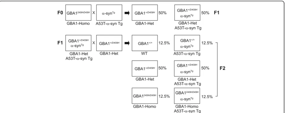
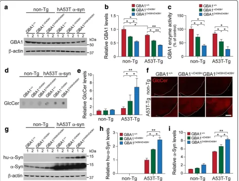
To test our hypothesis that decreased GBA1 enzyme activ-ity due to mutation in GBA1 affects neurodegeneration in the hA53T α-synuclein transgenic (Tg) mouse model of PD, the GBA1D409H/D409Hmutant mice [45] were crossbred with the hA53T α-synuclein (α-Syn) Tg mice (Fig. 1). GBA1 expression level was reduced to 70% in the ventral midbrain tissues of the GBA1+/D409Hmice and to 55% in the ventral midbrain tissues of the GBA1D409H/D409Hmice when compared to the wild type mice. GBA1 expression was further reduced to 48% in the hA53T α-Syn;
GBA1+/D409H and to 42% in the hA53T α-Syn;
GBA1D409H/D409H mice (Fig. 2a and b). GBA1 enzyme
activity was reduced to 71% in the brain tissues of the
GBA1+/D409H mice and to 39% in the ventral midbrain
with non-Tg mice. Additionally, we found that the levels of endogenous mouse α-synuclein are increased in the dependent manner of GBA1 enzyme activity in GBA1 mutant mice (Fig.2i). Thus, the steady-state levels of both endogenous α-synuclein and hA53T α-synuclein are dependent on the enzyme activity of GBA1 resulting from D409H mutation.
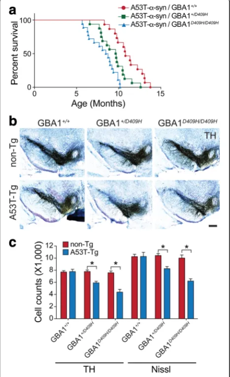
D409H GBA1 expression shortens lifespan and leads to dopaminergic degeneration in hA53Tα-synuclein Tg mice
The hA53T mutant α-synuclein Tg mice develop adult-onset phenotypes with rapidly progressive motor impair-ment that eventually leads to death [19]. To examine whether D409H GBA1 expression bearing decreased en-zyme activity affects the lifespan of hA53T α-synuclein Tg mice, littermates with the following genotypes were separated and aged: hA53T α-Syn, hA53T α-Syn;
GBA1+/D409H, hA53T α-Syn;GBA1D409H/D409H, and their
survival was monitored (Fig.3a). The hA53Tα-synuclein Tg mice lived an average of 10.8 months, as previously described [7]. The hA53T α-Syn;GBA1+/D409H lived an average of 9.7 months and the hA53T α-Syn;
GBA1D409H/D409H lived an average of 8.6 months,
indicating that the hA53T α-Syn;GBA1D409H/D409H significantly shortens lifespan of the hA53Tα-synuclein Tg mice by 2.2 months. Therefore, decreased enzyme activity due to D409H GBA1 expression has a boosting impact that expedites the onset and progression of the lethal phenotype induced by α-synuclein pathologies in the hA53Tα-synuclein Tg mice.
To determine whether the reduced GBA1 enzyme activity induces the loss of dopaminergic neurons in the hA53T α-synuclein mice, the number of TH
positive neurons in the SNpc was counted via an unbiased stereological analysis of the genotypes at 6 months of age (Fig. 3b and c). As previously de-scribed, at 6 months of age when the mice are asymptomatic, there was no obvious dopaminergic neurodegeneration in the hA53T α-synuclein Tg mice. In contrast, there was an approximately 24% loss of dopa-minergic neurons in the SNpc of the hA53T α-Syn;
GBA1+/D409H. Furthermore, there was 43% neuronal loss
in the SNpc of the hA53T α-Syn;GBA1D409H/D409H mutant mice.
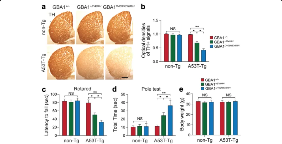
D409H GBA1 expression leads to reduction of the dopaminergic fiber density and alters behavioral deficits in the A53Tα-synuclein Tg mouse models
Since D409H GBA1 expression leads to dopaminergic neurodegeneration in the SNpc, next, tyrosine hydrox-ylase (TH)-immunopositive fiber density in the stri-atum was assessed (Fig. 4a). At 6 months of age when the mice were asymptomatic, there was no obvious dopaminergic terminal loss in the hA53T α-synuclein mice. However, there was approximately 32% TH fibers were lost in the striatum of the hA53T α-syn;
GBA1+/D409Hand 58% in the striatum of the hA53Tα
-Syn;GBA1D409H/D409H compared to the control group (Fig.4b).
To determine whether D409H GBA1 expression leading to decreased GBA1 enzyme activity leads to the abnormal behavior in the A53T α-synuclein, we performed a pole test, and rotarod analysis using a co-hort of 6 months of age of different genotypes (Fig. 4c and d). At 6 months of age, when the hA53T α -synu-clein mice were asymptomatic, there was no
significant behavioral impairment on the rotarod test. Average latency to fall in the accelerating rotarod was reduced in the hA53T α-Syn;GBA1+/D409H. The reduction in latency times was greater in the hA53T α-Syn;GBA1D409H/D409H mice at the 6 months of age (Fig.4c). We also conducted the pole test since it is a useful method for evaluating the mouse movement disorder caused by striatal dopamine depletion [26]. The pole test revealed that there was a moderate increase in the time to reach to the base of the pole in the hA53T α-Syn;GBA1+/D409H and a significantly greater increase in the hA53T α-Syn;GBA1D409H/D409H at 6 months of age (Fig. 4d). At 6 months of age, however, there was no significant difference in body weight (Fig.4e).
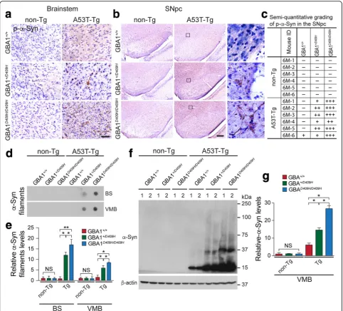
D409H GBA1 expression accelerates the accumulation of insolubleα-synuclein species in the brainstem and SNpc of A53Tα-synuclein Tg mice
As the accumulation of insoluble high molecular weight species ofα-synuclein is a prominent indicator of pathology in the A53Tα-synuclein mice [19], it was assessed via vari-ous techniques such as immunohistochemistry, dot blot, and immunoblot analysis (Fig. 5). Immunohistochemistry was conducted at 6 months of age when the mice were asymptomatic. At this time point, there was no obvious accumulation of α-synuclein phosphorylated at serine 129, which is closely associated withα-synuclein aggregation, in the SNpc and brainstem of the hA53Tα-Syn mice. In con-trast, immunohistochemistry revealed that some accumula-tion of α-synuclein phosphorylated at serine 129 in the
brainstem and SNpc of the hA53Tα-Syn;GBA1+/D409Hand significant accumulation in the brainstem and SNpc of the hA53Tα-Syn;GBA1D409H/D409Hat 6 months of age (Fig.5a, b, and c). None of the aggregates was detected in the brainstem and SNpc of non-Tg mice. Dot blot (Fig. 5d, and e) and immunoblot analysis (Fig. 5f and g) also demonstrated that the detergent-insoluble high mo-lecular weight species of α-synuclein accumulated in the ventral midbrain of the hA53T α-Syn;GBA1+/D409H and the accumulation was significantly increased in the ventral midbrain of the hA53T α-Syn;GBA1D409H/D409H at 6 months of age (Fig.5d,e,f, andg).
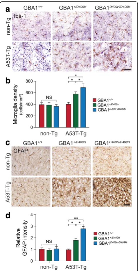
D409H GBA1 expression shows early neuroinflammation in the A53Tα-synuclein Tg mice
At 6 months of age there was no significant accumula-tion of Iba-1 and GFAP in the SNpc of A53T α -synuclein mice. For microglia activation, characterized by the increased expression of Iba-1, serves as an indir-ect indicator of neuronal abnormality in the A53T α -synuclein Tg mice. Thus, the enhanced expression of Iba1 was determined by immunohistochemistry. There was an increased Iba-1 immunoreactivity in the SNpc of hA53T α-Syn;GBA1+/D409H. The immunoreactivity was dramatically increased in the SNpc of the hA53Tα-Syn;
GBA1D409H/D409Hat 6 months of age (Fig.6aandb). As
the accumulation of glial fibrillary acidic protein (GFAP) is a prominent pathological indicator in the A53T α -synuclein Tg mice, its accumulation was also assessed via immunohistochemistry. There was an increased GFAP immunoreactivity in the SNpc of hA53T α-Syn;
GBA1+/D409H. The accumulation was further increased
in the SNpc of the hA53T α-Syn;GBA1D409H/D409H at 6 months of age (Fig.6candd).
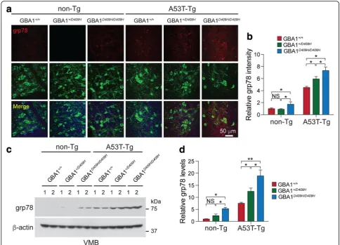
D409H GBA1 expression triggers ER stress early in the A53Tα-synuclein Tg mice
In symptomatic A53T α-synuclein Tg mice there was an accumulation of indicators of ER stress in a number of brain regions, including the brainstem and spinal cord [5]. 78 kDa glucose-regulated protein (grp78/BiP), an indica-tor of ER stress, was analyzed via immunohistochemisty and Immunoblot analysis using grp78 antibody (Fig. 7). Strikingly, immunohistochemistry demonstrated that there was a moderate increase of grp78 protein level in the SNpc tissues of the hA53T α-Syn;GBA1+/D409H and the upregulation of grp78 was significantly increased in the SNpc tissues of the hA53Tα-Syn;GBA1D409H/D409Hat 6 months of age (Fig.7aandc). Immunoblot analysis also demonstrated that the grp78 protein accumulated in the ventral midbrain of the hA53T α-Syn;GBA1+/D409H and the accumulation was further promoted in the ventral midbrain of the hA53T α-Syn;GBA1D409H/D409H at 6 months of age (Fig.7candd).
Discussion
The hypothesis that GBA1 could affectα-synuclein deg-radation and pathology has been tested in several animal models [8,10,30,37,44]. These models have allowed us to elucidate the relationship among GBA1, α-synuclein, and PD. However, these animal models do not fully represent the clinical observations seen in the GBA1-associated Parkinsonism such as earlier PD onset and DA neuronal loss. To gain further insights into the mechanisms by whichGBA1mutations increase the risk for PD and lead to the development of GBA1-assiciated parkinsonism, we crossbred GBA1 mice carrying D409H
Fig. 3D409H GBA1 expression shortens lifespan and leads to dopaminergic degeneration in the hA53Tα-synuclein transgenic mice.a,Survival was monitored from littermates with the following genotypes: hA53Ta-Syn/GBA1+/+(n= 15), hA53Tα-Syn/GBA1+/D409H(n = 16), hA53Tα-Syn/GBA1D409H/D409H(n= 18) mice. GBA1 mutation induces the lethal phenotype and TH-positive neuronal loss in the SNpc of A53T mutantα-synuclein transgenic mice.bandc,The number of TH-positive neurons in the SNpc was counted using stereological analysis with the indicated genotypes at 6 months of age. The scale bar is 200
knock-in mutation with human A53T α-synuclein Tg mice exhibiting neurological abnormalities, accumula-tion of α-synuclein aggregates, increased ER stress, neu-roinflammation, and shortened lifespan [5, 19]. Using this mouse model, we assessed the impact of D409H GBA1 mutation on the major phenotypes of the A53T α-synuclein Tg mice with disease onset and further examined the cardinal features seen in the GBA1-associated Parkinsonism through biochemical and im-munohistochemical analyses. Importantly, our findings reveal that the expression of D409H GBA1 mutation resulted in the loss of DA neurons, accelerates disease onset, exacerbates neuroinflammation and ER stress more extensively than the degree seen in the hA53Tα -synuclein Tg mice.
It has been reported that mutations inGBA1result in the production of misfolded GBA1, increased GBA1 ubiquitination, and premature degradation leading to quantitative loss in the protein levels [22,46]. We found the reduction of GBA1 protein expression and GBA1 enzyme activity in the brains of GBA1+/D409H and
GBA1D409H/D409H, which is similar to previous findings
that GBA1 deficiency due to expression of L444P mutation or heterozygous GBA1-null mutations results in GBA1 enzyme activity deficiency [30]. Importantly, there was even lower GBA1 enzyme activity in the
brains of hA53T α-Syn;GBA1+/D409Hand hA53T α-Syn;
GBA1D409H/D409H, which differs from the earlier finding
that heterozygous GBA1-null mutations in the A53T α-synuclein Tg mice does not lead to GBA1 enzyme deficiency [44]. Although the reason for this discrepancy is unclear, it is conceivable that the GBA1 enzyme deficiency due to D409H mutation may affects the disease onset in A53T α-synuclein Tg mice differently from heterozygous GBA1-null mutations. For instance, the buildup of the misfolded GBA1 D409H mutant pro-tein [29] and α-synuclein aggregates trigger ER stress [5], which would form a positive feedback loop to fur-ther impair GBA1 enzyme activity and consequently contribute toα-synuclein pathology and loss of DA neu-rons in the model. Since it is known that GBA1 enzyme activity was lowest in the SN of PD patients [1, 2, 11, 31], a further investigation will be required to determine GBA1 enzyme activity in different brain regions in this mouse model. In addition, it is possible that GBA1 D409H mutation may affect the lysosomal dysfunction in hA53Tα-synuclein Tg, thereby hampering the autophagy/ lysosomal degradation of pathological α-synuclein [34]. Future study will be required to test this possibility in our animal model.
In our model, the relationship between GBA1 enzyme activity and α-synuclein accumulation revealed that
GBA1 deficiency due to D409H mutation was associated with the increased levels of human and mouse α -synulcein proteins as well as the enhanced levels of high molecular weight α-synuclein aggregates in the ventral midbrain regions. Consistent with previous findings, our observations confirm that decreased GBA1 en-zyme activity due to GBA1mutations or null leads to increased α-synuclein levels in other models [8, 10,
30] and PD postmortem brains [11, 31]. Since the levels ofα-synuclein are greater in the brain of hA53T α-Syn;GBA1D409H/D409H compared to hA53T α-Syn;
GBA1+/D409H, the accumulation of α-synuclein is
dependent on the levels of GBA1 enzyme activity. On the other hand, our study revealed that phosphoserine 129 (pSer) α-synuclein immunoreactivity and high molecular weight α-synuclein species were detected in
the ventral midbrain of hA53T α-Syn;GBA1+/D409H and hA53T α-Syn;GBA1D409H/D409H mice at 6 months of age. Moreover, intensities of both pSer α-synuclein immunoreactivity and high molecular weight α-synuclein species were correlated with the enzyme activity levels of GBA1 in the hA53T α-Syn;GBA1+/D409H and hA53T
α-Syn;GBA1D409H/D409H mice. However, the pSer immunoreactivity and high molecular weight α -synuclein species were not present in the ventral mid-brain of the hA53Tα-Syn Tg mice at 6 months of age. Although the underlying mechanism of how D409H expression results in increased pathologic α-synuclein aggregates at the early time point is not clear, it is likely that additional α-synuclein accumulation trig-gered by GBA1 deficiency due to D409H expression pushes forward the levels of α-synuclein protein to reach quickly the threshold required for pathologic α -synuclein aggregates in the model at 6 months of age, eliciting overt DA neurodegeneration loss in the SNpc and PD related motor deficits in the same model. Based on our current observations, the hA53T α-Syn Tg mouse model may provide a valuable resource to uncover mechanisms of how PD-associated gene mu-tations can impact PD pathogenesis.
Importantly, we found the loss of nigrostriatal DA neu-rons in the SNpc of the hA53T α-Syn;GBA1+/D409H and hA53T α-Syn;GBA1D409H/D409H, which were not detected in the A53T α-synuclein Tg mice, GBA1+/D409H, and
GBA1D409H/D409H at 6 months of age. This result has not
been reported in the previous studies [10, 37, 44]. One possible explanation for this is due to the accumulation of pathologicα-synuclein aggregates, which may be sufficient to lead to the loss of nigrostriatal DA neurons in the SNpc at the time point. Another explanation might be behind neuroinflammation that contributes to neurodegeneration in neurodegenerative disorders including PD [20, 35, 38]. Neuroinflammation was present in the SNpc of hA53Tα -Syn;GBA1+/D409Hand hA53Tα-Syn;GBA1D409H/D409H, but not observed in A53Tα-synuclein Tg mice, GBA1+/D409H, and GBA1D409H/D409H at 6 months of age. The last explanation for this might be ER stress that contributes to neurodegeneration in neurodegenerative disorders including PD [14, 15, 28]. We also observed significantly changed levels of ER stress in the SNpc of hA53T α-Syn;
GBA1+/D409H and hA53T α-Syn;GBA1D409H/D409H at
6 month of age, which were not detected in A53T α-synuclein Tg mice, GBA1+/D409H, and GBA1D409H/D409H at the time point.
Although the penetrance of D409H GBA1 mutation is relatively lower than other mutations such as N370S and L444P GBA1 mutations [40], our current findings sug-gest that GBA1 deficiency due to D409H GBA1 muta-tion alone is not sufficient to cause PD but addimuta-tional factors, such as environmental factors or increased levels of α-synuclein could increase the penetrance through rendering the levels of α-synuclein accumulation close to the threshold required for α-synuclein aggregation. Our previous finding that GBA1 deficiency due to L444P GBA1 heterozygous mutation renders DA neu-rons more susceptible to MPTP intoxication [47] further
Fig. 6D409H GBA1 expression shows the activation of pathology-associated microglia and astrocyte activation in the SNpc of A53T
supports this hypothesis. Our study does not provide a detailed explanation on how D409H GBA1mutation contributes to severe neurodegeneration with the loss of DA neurons in the A53T α-synuclein Tg mice. Further studies need to be undertaken to account for the DA neurodegeneration. Also, it would be interesting to study how the regulation of formation of α-synuclein tetra-mers and other related multitetra-mers as well as the changes in the status of glycosphingolipids (GSLs) in the model is regulated [17].
Conclusions
In conclusion, our results indicate that GBA1 defi-ciency due to D409H GBA1 mutation that contributes to α-synuclein accumulation exacerbates neuronal vul-nerability in neurodegenerative processes triggered by A53T α-synuclein expression in vivo. The model that
recapitulates the cardinal PD phenotypes including loss of DA neurons, LB pathology, and motor deficits can be a useful tool to study in depth the possible mechanisms underlying neurodegeneration due to
GBA1 mutations and to test the efficacy of potential treatment against GBA1-associated PD and Dementia with Lewy bodies (DLB).
Acknowledgements
The authors acknowledge the joint participation by the Adrienne Helis Malvin Medical Research Foundation and the Diana Helis Henry Medical Research Foundation through its direct engagement in the continuous active conduct of medical research in conjunction with The Johns Hopkins Hospital and the Johns Hopkins University School of Medicine and the Foundation’s Parkinson’s Disease Program M-2014, H-1, H-2013. Funding
Availability of data and materials
All data generated or analyzed during the current study are included in this published article. All raw data and material are available on request.
Authors’contributions
DK and HSK designed research; DK, HH, SC, SHK, SL, JHP and S K performed research; DK analyzed data; DK and HSK wrote the paper. All authors read and approved the final manuscript.
Ethics approval and consent to participate
All experimental procedures were in accordance with the guidelines of Laboratory Animal Manual of National Institute of Health Guide for the Care and Use of Animals. All animal experiments were complied with the current laws of the United States. They were approved by the Johns Hopkins Medical Institute Animal Care and Use Committee.
Competing interests
The authors declare that they have no competing interests.
Publisher’s Note
Springer Nature remains neutral with regard to jurisdictional claims in published maps and institutional affiliations.
Author details
1Neuroregeneration and Stem Cell Programs, Institute for Cell Engineering,
The Johns Hopkins University School of Medicine, Baltimore, MD 21205, USA.
2
Department of Neurology, The Johns Hopkins University School of Medicine, Baltimore, MD 21205, USA.3Diana Helis Henry Medical Research
Foundation, New Orleans, Louisiana 70130, USA.4Adrienne Helis Malvin
Medical Research Foundation, New Orleans, Louisiana 70130, USA.
Received: 14 March 2018 Accepted: 19 April 2018
References
1. Alcalay RN, Levy OA, Waters CC, Fahn S, Ford B, Kuo SH, Mazzoni P, Pauciulo MW, Nichols WC, Gan-Or Z et al (2015) Glucocerebrosidase activity in Parkinson's disease with and without GBA mutations. Brain 138:2648–2658.
https://doi.org/10.1093/brain/awv179
2. Chiasserini D, Paciotti S, Eusebi P, Persichetti E, Tasegian A, Kurzawa-Akanbi M, Chinnery PF, Morris CM, Calabresi P, Parnetti L et al (2015) Selective loss of glucocerebrosidase activity in sporadic Parkinson's disease and dementia with Lewy bodies. Mol Neurodegener 10:15. https://doi.org/10.1186/s13024-015-0010-2
3. Choi S, Kim D, Kam TI, Yun S, Kim S, Park H, Hwang H, Pletnikova O, Troncoso JC, Dawson VL et al (2015) Lysosomal enzyme Glucocerebrosidase protects against Abeta1-42 oligomer-induced neurotoxicity. PLoS One 10: e0143854.https://doi.org/10.1371/journal.pone.0143854
4. Clark LN, Kartsaklis LA, Wolf Gilbert R, Dorado B, Ross BM, Kisselev S, Verbitsky M, Mejia-Santana H, Cote LJ, Andrews H et al (2009) Association of glucocerebrosidase mutations with dementia with lewy bodies. Arch Neurol 66:578–583.https://doi.org/10.1001/archneurol.2009.54
5. Colla E, Coune P, Liu Y, Pletnikova O, Troncoso JC, Iwatsubo T, Schneider BL, Lee MK (2012) Endoplasmic reticulum stress is important for the
manifestations of alpha-synucleinopathy in vivo. J Neurosci 32:3306–3320.
https://doi.org/10.1523/JNEUROSCI.5367-11.2012
6. Colla E, Jensen PH, Pletnikova O, Troncoso JC, Glabe C, Lee MK (2012) Accumulation of toxic alpha-synuclein oligomer within endoplasmic reticulum occurs in alpha-synucleinopathy in vivo. J Neurosci 32:3301–3305.
https://doi.org/10.1523/JNEUROSCI.5368-11.2012
7. Daher JP, Pletnikova O, Biskup S, Musso A, Gellhaar S, Galter D, Troncoso JC, Lee MK, Dawson TM, Dawson VL et al (2012) Neurodegenerative phenotypes in an A53T alpha-synuclein transgenic mouse model are independent of LRRK2. Hum Mol Genet 21:2420–2431.https://doi.org/10. 1093/hmg/dds057
8. Davis MY, Trinh K, Thomas RE, Yu S, Germanos AA, Whitley BN, Sardi SP, Montine TJ, Pallanck LJ (2016) Glucocerebrosidase deficiency in Drosophila results in alpha-Synuclein-independent protein aggregation and neurodegeneration. PLoS Genet 12:e1005944.https://doi.org/10.1371/ journal.pgen.1005944
9. DePaolo J, Goker-Alpan O, Samaddar T, Lopez G, Sidransky E (2009) The association between mutations in the lysosomal protein glucocerebrosidase and parkinsonism. Mov Disord 24:1571–1578.https://doi.org/10.1002/mds.22538
10. Fishbein I, Kuo YM, Giasson BI, Nussbaum RL (2014) Augmentation of phenotype in a transgenic Parkinson mouse heterozygous for a Gaucher mutation. Brain 137:3235–3247.https://doi.org/10.1093/brain/awu291
11. Gegg ME, Burke D, Heales SJ, Cooper JM, Hardy J, Wood NW, Schapira AH (2012) Glucocerebrosidase deficiency in substantia nigra of parkinson disease brains. Ann Neurol 72:455–463.https://doi.org/10.1002/ana.23614
12. Gegg ME, Schapira AHV (2018) The role of glucocerebrosidase in Parkinson disease pathogenesis. FEBS J.https://doi.org/10.1111/febs.14393
13. Goedert M (2001) Alpha-synuclein and neurodegenerative diseases. Nat Rev Neurosci 2:492–501.https://doi.org/10.1038/35081564
14. Hetz C, Mollereau B (2014) Disturbance of endoplasmic reticulum proteostasis in neurodegenerative diseases. Nat Rev Neurosci 15:233–249.
https://doi.org/10.1038/nrn3689
15. Hetz C, Saxena S (2017) ER stress and the unfolded protein response in neurodegeneration. Nat Rev Neurol 13:477–491.https://doi.org/10.1038/ nrneurol.2017.99
16. Kim C, Lv G, Lee JS, Jung BC, Masuda-Suzukake M, Hong CS, Valera E, Lee HJ, Paik SR, Hasegawa M et al (2016) Exposure to bacterial endotoxin generates a distinct strain of alpha-synuclein fibril. Sci Rep 6:30891.https:// doi.org/10.1038/srep30891
17. Kim S, Yun SP, Lee S, Umanah GE, Bandaru VVR, Yin X, Rhee P, Karuppagounder SS, Kwon SH, Lee H et al (2018) GBA1 deficiency negatively affects physiological alpha-synuclein tetramers and related multimers. Proc Natl Acad Sci U S A 115:798–803.https://doi.org/10.1073/ pnas.1700465115
18. Kitatani K, Sheldon K, Rajagopalan V, Anelli V, Jenkins RW, Sun Y, Grabowski GA, Obeid LM, Hannun YA (2009) Involvement of acid beta-glucosidase 1 in the salvage pathway of ceramide formation. J Biol Chem 284:12972–12978.
https://doi.org/10.1074/jbc.M802790200
19. Lee MK, Stirling W, Xu Y, Xu X, Qui D, Mandir AS, Dawson TM, Copeland NG, Jenkins NA, Price DL (2002) Human alpha-synuclein-harboring familial Parkinson's disease-linked ala-53–> Thr mutation causes neurodegenerative disease with alpha-synuclein aggregation in transgenic mice. Proc Natl Acad Sci U S A 99:8968–8973.https://doi.org/10.1073/pnas.132197599
20. Liddelow SA, Guttenplan KA, Clarke LE, Bennett FC, Bohlen CJ, Schirmer L, Bennett ML, Munch AE, Chung WS, Peterson TC et al (2017) Neurotoxic reactive astrocytes are induced by activated microglia. Nature 541:481–487.
https://doi.org/10.1038/nature21029
21. Lopez G, Sidransky E (2013) Predicting parkinsonism: new opportunities from Gaucher disease. Mol Genet Metab 109:235–236.https://doi.org/10. 1016/j.ymgme.2013.04.012
22. Lu J, Chiang J, Iyer RR, Thompson E, Kaneski CR, Xu DS, Yang C, Chen M, Hodes RJ, Lonser RR et al (2010) Decreased glucocerebrosidase activity in Gaucher disease parallels quantitative enzyme loss due to abnormal interaction with TCP1 and c-Cbl. Proc Natl Acad Sci U S A 107:21665–21670.
https://doi.org/10.1073/pnas.1014376107
23. Luk KC, Kehm V, Carroll J, Zhang B, O'Brien P, Trojanowski JQ, Lee VM (2012) Pathological alpha-synuclein transmission initiates Parkinson-like
neurodegeneration in nontransgenic mice. Science 338:949–953.https://doi. org/10.1126/science.1227157
24. Martin LJ, Pan Y, Price AC, Sterling W, Copeland NG, Jenkins NA, Price DL, Lee MK (2006) Parkinson's disease alpha-synuclein transgenic mice develop neuronal mitochondrial degeneration and cell death. J Neurosci 26:41–50.
https://doi.org/10.1523/JNEUROSCI.4308-05.2006
25. Mata IF, Samii A, Schneer SH, Roberts JW, Griffith A, Leis BC, Schellenberg GD, Sidransky E, Bird TD, Leverenz JB et al (2008) Glucocerebrosidase gene mutations: a risk factor for Lewy body disorders. Arch Neurol 65:379–382.
https://doi.org/10.1001/archneurol.2007.68
26. Matsuura K, Kabuto H, Makino H, Ogawa N (1997) Pole test is a useful method for evaluating the mouse movement disorder caused by striatal dopamine depletion. J Neurosci Methods 73:45–48
27. Mazzulli JR, Xu YH, Sun Y, Knight AL, McLean PJ, Caldwell GA, Sidransky E, Grabowski GA, Krainc D (2011) Gaucher disease glucocerebrosidase and alpha-synuclein form a bidirectional pathogenic loop in synucleinopathies. Cell 146:37–52.https://doi.org/10.1016/j.cell.2011.06.001
29. Migdalska-Richards A, Schapira AH (2016) The relationship between glucocerebrosidase mutations and Parkinson disease. J Neurochem 139(Suppl 1):77–90.https://doi.org/10.1111/jnc.13385
30. Migdalska-Richards A, Wegrzynowicz M, Rusconi R, Deangeli G, Di Monte DA, Spillantini MG, Schapira AHV (2017) The L444P Gba1 mutation enhances alpha-synuclein induced loss of nigral dopaminergic neurons in mice. Brain 140:2706–2721.https://doi.org/10.1093/brain/awx221
31. Murphy KE, Gysbers AM, Abbott SK, Tayebi N, Kim WS, Sidransky E, Cooper A, Garner B, Halliday GM (2014) Reduced glucocerebrosidase is associated with increased alpha-synuclein in sporadic Parkinson's disease. Brain 137: 834–848.https://doi.org/10.1093/brain/awt367
32. Nalls MA, Duran R, Lopez G, Kurzawa-Akanbi M, McKeith IG, Chinnery PF, Morris CM, Theuns J, Crosiers D, Cras P et al (2013) A multicenter study of glucocerebrosidase mutations in dementia with Lewy bodies. JAMA Neurol 70:727–735.https://doi.org/10.1001/jamaneurol.2013.1925
33. Nussbaum RL (2003) Alzheimer's disease and Parkinson's disease (vol 348, pg 1356, 2003). New Engl J Med 348:2588–2588
34. Pitcairn C, Wani WY, Mazzulli JR (2018) Dysregulation of the autophagic-lysosomal pathway in Gaucher and Parkinson's disease. Neurobiol Dis.
https://doi.org/10.1016/j.nbd.2018.03.008
35. Poewe W, Seppi K, Tanner CM, Halliday GM, Brundin P, Volkmann J, Schrag AE, Lang AE (2017) Parkinson disease. Nat Rev Dis Primers 3:17013.https:// doi.org/10.1038/nrdp.2017.13
36. Rocha EM, Smith GA, Park E, Cao H, Brown E, Hallett P, Isacson O (2015) Progressive decline of glucocerebrosidase in aging and Parkinson's disease. Ann Clin Transl Neurol 2:433–438.https://doi.org/10.1002/acn3.177
37. Rockenstein E, Clarke J, Viel C, Panarello N, Treleaven CM, Kim C, Spencer B, Adame A, Park H, Dodge JC et al (2016) Glucocerebrosidase modulates cognitive and motor activities in murine models of Parkinson's disease. Hum Mol Genet 25:2645–2660.https://doi.org/10.1093/hmg/ddw124
38. Salter MW, Stevens B (2017) Microglia emerge as central players in brain disease. Nat Med 23:1018–1027.https://doi.org/10.1038/nm.4397
39. Savitt JM, Dawson VL, Dawson TM (2006) Diagnosis and treatment of Parkinson disease: molecules to medicine. J Clin Invest 116:1744–1754.
https://doi.org/10.1172/JCI29178
40. Schapira AH (2015) Glucocerebrosidase and Parkinson disease: recent advances. Mol Cell Neurosci 66:37–42.https://doi.org/10.1016/j.mcn.2015.03.013
41. Schondorf DC, Aureli M, McAllister FE, Hindley CJ, Mayer F, Schmid B, Sardi SP, Valsecchi M, Hoffmann S, Schwarz LK et al (2014) iPSC-derived neurons from GBA1-associated Parkinson's disease patients show autophagic defects and impaired calcium homeostasis. Nat Commun 5:4028.https://doi.org/10. 1038/ncomms5028
42. Sidransky E, Lopez G (2012) The link between the GBA gene and parkinsonism. Lancet Neurol 11:986–998. https://doi.org/10.1016/S1474-4422(12)70190-4
43. Spillantini MG, Schmidt ML, Lee VM, Trojanowski JQ, Jakes R, Goedert M (1997) Alpha-synuclein in Lewy bodies. Nature 388:839–840.https://doi.org/ 10.1038/42166
44. Tayebi N, Parisiadou L, Berhe B, Gonzalez AN, Serra-Vinardell J, Tamargo RJ, Maniwang E, Sorrentino Z, Fujiwara H, Grey RJ et al (2017)
Glucocerebrosidase haploinsufficiency in A53T alpha-synuclein mice impacts disease onset and course. Mol Genet Metab 122:198–208.https://doi.org/10. 1016/j.ymgme.2017.11.001
45. Xu YH, Quinn B, Witte D, Grabowski GA (2003) Viable mouse models of acid beta-glucosidase deficiency: the defect in Gaucher disease. Am J Pathol 163: 2093–2101
46. Yang C, Wang H, Zhu D, Hong CS, Dmitriev P, Zhang C, Li Y, Ikejiri B, Brady RO, Zhuang Z (2015) Mutant glucocerebrosidase in Gaucher disease recruits Hsp27 to the Hsp90 chaperone complex for proteasomal degradation. Proc Natl Acad Sci U S A 112:1137–1142.https://doi.org/10.1073/pnas. 1424288112