See discussions, stats, and author profiles for this publication at: https://www.researchgate.net/publication/234164196
Short-time effects of coumarin along the maize
primary root axes
Conference Paper · May 2009
CITATIONS
10
READS
28
4 authors:
Some of the authors of this publication are also working on these related projects:
SIR-2014 (RBSI14L9CE)-MEDANAT - Mediterranean species as source of bioactive natural compounds: a physiological, molecular and metabolomic approach.View project
Morpho-physiological and Molecular Approaches To Improve Nutrient Use EfficiencyView project Antonio Lupini
Mediterranean University of Reggio Calabria
47 PUBLICATIONS 221 CITATIONS
SEE PROFILE
Agostino Sorgonà
Mediterranean University of Reggio Calabria
84 PUBLICATIONS 738 CITATIONS
SEE PROFILE
Fabrizio Araniti
Mediterranean University of Reggio Calabria
45 PUBLICATIONS 174 CITATIONS
SEE PROFILE
Maria Rosa Abenavoli
Mediterranean University of Reggio Calabria
113 PUBLICATIONS 743 CITATIONS
SEE PROFILE
ReSeaRch PaPeR ReSeaRch PaPeR
Plant Signaling & Behavior 5:11, 1-6; November 2010; © 2010 Landes Bioscience
*Correspondence to: Maria Rosa Abenavoli; Email: [email protected] Submitted: 07/14/10; Accepted: 07/14/10
Previously published online: www.landesbioscience.com/journals/psb/article/13021 DOI:
Introduction
Coumarin is a simple allelopathic compound found in many plants,1 widely distributed in both natural plant communities and
crops,2 where it plays an important role in plant-plant interactions
and biocommunication.3,4 Like other allelochemicals, such as
phenolic compounds released from living plants into the environ-ment,5 coumarin can influence many physiological and
biochem-ical processes: root growth,6-9 nitrate uptake and metabolism,10,11
respiration and photosynthesis12 and germination.13-15 The effect
of coumarin is species-specific7,15 and concentration-dependent,
often stimulatory at low and inhibitory at high concentrations.9,14
Coumarin has been widely studied for allelopathic effects on root growth, organ considered the primary target of this compound.16
Earlier investigations indicated clearly that coumarin changed root cell polarity of growth causing an inhibition of longitudinal root cell elongation accompanied by a simultaneous stimulation of radial expansion.6,17 These effects were also observed in alfalfa
grass, where the thickness of seminal roots was enlarged abnor-mally because of an inhibition of the longitudinal root growth.8
Recently, Abenavoli et al.15 demonstrated a selective and
species-specific effect of coumarin on the root growth of individual roots: 100 μM coumarin inhibited primary root length and stimulated lateral root formation in Arabidopsis. By contrast, in maize seed-lings, coumarin did not inhibit growth of the primary root rel-ative to seminal and nodal roots.9 However, these effects were
observed after long-term coumarin exposure (48 h), while little
The short-term effects of coumarin on three different maize primary root zones, transition zone (TZ, 3 mm) and two non-growing zones (NGZ1 and NGZ2 at 20 and 50 mm, respectively), were studied in order to investigate the effect of the allelochemical on maize root elongation rate (ReR). The ReR, plasma membrane (pm) h+-aTPase activity, quantitative
ph changes and cell membrane potentials were evaluated. The results showed that coumarin caused at the TZ (1) an increased ReR; (2) an enhancement of pm h+-aTPase activity and proton extrusion; and (3) a transient depolarization
followed by a hyperpolarization of cell membrane potential. These observations were not evident in the NGZ1 and NGZ2 of the maize root. coumarin-treatment in the NGZ1 did not change ReR, but caused a membrane depolarization, while the NGZ2 was mostly insensitive to the allelochemical. These data suggested that the primary maize root was sensitive to coumarin within a 20 mm section from the root tip, but the more distal NGZ2 was not involved in coumarin-elicited physiological responses.
Short-term effects of coumarin along the maize
primary root axis
antonio Lupini,1 agostino Sorgonà,1 anthony J. Miller2 and Maria Rosa abenavoli1,*
1Dipartimento di Biotecnologie per il Monitoraggio agro-alimentare ed ambientale (BIOMaa); Università Mediterranea di Reggio calabria; Salita Melissari; Reggio calabria,
Rc Italy; 2centre for Soil and ecosystem Function; Rothamsted Research; harpenden, hertfordshire UK
Key words: coumarin, membrane potential, pmH+-ATPase, proton efflux, root elongation rate
This manuscr
ipt has been published online, pr
ior to pr
inting. O
nce the issue is complete and page numbers hav
e been assigned, the citation will change accor
dingly
.
information is available for the short-term effects of coumarin on the morphological and physiological responses of maize roots. This knowledge may be more useful for a better understanding of the allelochemicals mode of action. Furthermore, plant root axes are characterized by having different zones with diverse anatomi-cal, morphological and physiological traits which can respond differently to nutrient, water and allelochemicals which are het-erogeneously distributed in soils. Particularly, the transition zone (TZ), localized 1.7–3.4 mm from the tip, has been character-ized as having special cell physiological properties which allow the root to respond to a wide range of environmental signals.22-24
Furthermore, coumarin induced swelling in the root apex, simi-lar to that caused by mechanical impedance in the TZ behind the meristem, has been reported.22
The differential short-term responses of three different pri-mary root zones of maize, TZ, non-growing zones 1, 2 (NGZ1 and NGZ2) to localized coumarin treatment have been investi-gated. In particular, the ability of coumarin to locally influence the cell plasma membrane, an early event of the allelochemical action,25 along the maize root axis has been studied. To address
these both questions, the effect of coumarin on pm H+-ATPase
activity, proton efflux and cell plasma-membrane electrical potential difference in each zone of the primary maize root was evaluated and the results are reported in this paper.
coumarin exposure, with lesser stimulation caused by 50 μM and none with a 25 μM coumarin treatments (Fig. 2). In NGZ1 and NGZ2, H+-ATPase activity was not significantly modified
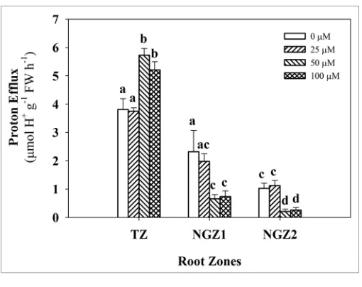
by treatment at all three coumarin concentrations (Fig. 2). For the TZ, the proton efflux was significantly stimulated by 50 and 100 μM coumarin treatments by 50.3 and 36.6% respectively, compared to the control, while this increase was not observed after 25 μM coumarin exposure (Fig. 3). By contrast, in the more distal root zones (NGZ1 and NGZ2), the proton efflux was significantly inhibited by 50 or 100 μM coumarin treatments by 71–79% and 68–74%, respectively, while 25 μM coumarin showed a similar behavior to the control (Fig. 3).
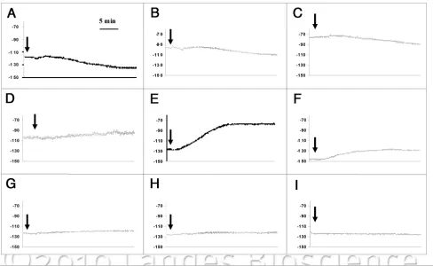
Cell membrane potentials, before, during and after the cou-marin treatment were recorded (Em), in order to detect the ini-tial cell responses within the various root zones of intact primary maize roots. After insertion of a microelectrode into the mature epidermal cells and the stabilization of Em (<10 min) in a nutrient solution without the coumarin, the Em was significantly different among the three root zones (Fig. 4). Then, the bathing medium was changed to one containing coumarin at different final con-centrations and the effect on the Em was evaluated within each of the root zones. In the TZ, all the coumarin concentrations caused an immediate and transient depolarization (less negative electrical potential), followed by a more negative hyperpolariza-tion (Fig. 4A–C). While the depolarization response was not significantly different among the various coumarin treatments, the hyperpolarization phase varied in relation to the allelochemi-cal concentrations (Table 1). Indeed, 50 μM coumarin caused a hyperpolarization of about 21 ± 1.8 mV, which was statistically different from 25 μM (9 ± 3.6 mV), but similar to that induced by 100 μM coumarin (14 ± 2 mV) (Table 1). In the NGZ1, the coumarin addition only elicited a depolarization with no subse-quent hyperpolarization (Fig. 4D–F). At the highest coumarin concentrations (50 and 100 μM), the depolarization of the cell membrane potential (26.3 ± 4.1 and 21.7 ± 2.3 mV, respectively) was significantly different from that measured with a 25 μM coumarin treatment (10 ± 0.6 mV) (Table 1). By contrast, in the NGZ2, coumarin did not cause any significant effects on the cell plasma membrane potential (Fig. 4G–I and Table 1).
Discussion
Coumarin treatments in the TZ, NGZ1 and NGZ2 of maize primary roots elicited differing morpho-physiological responses. More specifically in the TZ, but not at greater distances (NGZ1 and NGZ2), coumarin caused (1) an increased RER; (2) an higher H+-ATPase activity; (3) an enhancement of proton extrusion; and
(4) a transient depolarization followed by a sharp hyperpolariza-tion of membrane potential. This coumarin-induced pattern in TZ was similar to that induced by auxin in oat and maize coleop-tiles26,27 and may suggest an auxin-like behavior or/and an
inter-action with the auxin signalling pathways for this allelochemical. A possible interference of coumarin on auxin metabolism was already reported in Petunia hybrida.28 From these results, a
mech-anism action of coumarin in the TZ of maize primary root, based on the classic acid growth theory, could be proposed. Coumarin A different response pattern of RER to 0, 25, 50 and 100 μM
coumarin treatments within the TZ, NGZ1 and NGZ2 of intact root maize seedlings was clearly evident (Fig. 1). In the TZ, a significant increase of RER was induced by 50 and 100 μM cou-marin (0.0221 and 0.0258 mm min-1, respectively) compared to
the control (0.0133 mm min-1) and 25 μM coumarin treatment.
By contrast, in the NGZ1 and NGZ2, RER was not significantly affected by all coumarin concentration treatments (Fig. 1).
A similar response pattern within the TZ, NGZ1 and NGZ2 was observed for pm H+-ATPase activity when exposed
to coumarin. Indeed, in the TZ, H+-ATPase activity markedly
increased by 64% with respect to the control after 100 μM Figure 1. effect of coumarin supply (30 min) in agar medium on root
elongation rate of TZ (3 mm), NGZ1 (20 mm) and NGZ2 (50 mm) of pri-mary maize root. Means with different letters are significantly different (p < 0.05, Tukey’s test) with regard to root zones.
Figure 2. h+-aTPase activity (nmol P
also evoked for other environmental cues such as chilling,35 water
stress36 and nutritional signals such as nitrate37 and phosphate.38
An ecological role could be attributed to this localized root tip response to coumarin. The root tip is the zone first encountering and interacting with the soil environment in which, in addition to nutrient and water resources, allelochemicals such as cou-marin could be present. Indeed, plant residues, litter decomposi-tion in the top soil and rain leaching from foliage may provide sources of coumarin.38 Furthermore, root exudates also contain
coumarin providing a mechanism to sense resource competi-tion from other plants.40 Finally, coumarin concentrations in the
soil will depend on microbial activity, but the ranges used for experimental measurements seem feasible for those occurring in nature.39 Hence, during exploration for nutrients and water, the
root could be exposed to a locally allelochemical-enriched soil which is sensed by a root tip zone of environmental perception and signal transduction.
In conclusion, these results suggested several important con-siderations: (1) the TZ of primary maize root is the most sensitive to coumarin; (2) the morpho-physiological responses of the more apical root zones to the coumarin showed an auxin-like pattern. Further studies are necessary to better understand if the change in bioelectric pattern of membranes induced by coumarin could be due to an H+-coupled transport of the allelochemical as
sug-gested for monocarboxylic and benzoic acids29 or to alterations
in the flux of ions as reported for monoterpenes41,42 and then a
subsequent direct or indirect action in the TZ auxin perception system.
Materials and Methods
Plant material and growth condition. Maize (Zea mays L., cv Cecilia, Pioneer, Italia) seeds, previously immersed in deionized water for 48 h, were germinated over aerated 0.5 mM CaSO4 interacts with the plasma membrane causing an immediate
tran-sient depolarization and then subsequently, directly or indirectly, stimulated the pm H+-ATPase activity resulting in an increased
H+ efflux with consequently more negative hyperpolarization of
the plasma membrane. The increase of the proton release deter-mined an acidification of the apoplast thereby facilitating root growth rate as confirmed by the increased root elongation rate in the TZ after coumarin treatments (Fig. 1).
In contrast to the TZ, coumarin-response pattern in the NGZ1 displayed (1) an unchanged RER and pm H+-ATPase
activity; (2) an inhibition of proton extrusion; and (3) a sharp depolarization of membrane potential. Therefore, the only common effect induced by coumarin in both root zones is to the depolarization of the membrane, possibly suggesting that, although coumarin is electrically neutral, its uptake occurred via H+-coupled mechanism. This cotransport mechanism,
pro-posed by Pang et al.29 for undissociated phenolic acids, could be
responsible for the substantial membrane depolarization occur-ring after coumarin treatment. How can we explain the other contrasting responses to coumarin between the TZ and NGZ1 of maize primary root? Assuming that coumarin could exhibit an auxin-like behavior or/and interact with the auxin signalling pathways, probably a lower auxin sensitivity or concentration in the NGZ1 could be limiting the coumarin mediated responses in this root zone. Indeed, an asymmetrical auxin distribution along root axis of Arabidopsis thaliana (0 to 3 mm, 3 to 10 mm and 10 to 20 mm) with the highest concentrations in the root tip/ meristem/elongation zones and lowest toward the basal region was observed.30 A lower auxin content in the NGZ1 could not be
adequate to reach the level required for the auxin-induced activa-tion of the pm H+-ATPase, which in turn led to decreasing H+
efflux resulting in change in the RER. For example, an adequate auxin budget plays a central role in controlling lateral root initia-tion in Arabidopsis thaliana.31
Finally, the NGZ2 was the region of the maize primary roots less affected by coumarin treatment since no change in the root elongation rate, pm H+-ATPase activity and plasma membrane
potential was measured, although a decrease in the H+ efflux was
observed. As cells mature along the developing root axis, differ-ential changes in gene expression may explain these differences.33
The presence of lignified sclerenchymatous fibres32 and suberized
endo- and exodermal cells34 in the more mature root regions of
maize may limit the coumarin-plasma membrane interactions. Overall, these short-term coumarin treatment experiments confirm the plasma membrane transport activity as an early tar-get for the allelochemicals. Further, the experiments indicated the presence of a threshold concentration dividing the stimulatory from non-effect for the allelochemical. Indeed, 25 μM coumarin did not produce any physiological responses along primary maize root while 50 and 100 μM coumarin, as observed in a previous study on root anatomy, morphology and physiology in a durum wheat cultivar showed a stimulatory effect on root elongation rate.10 Finally, the results showed that the transition zone was the
region of the maize primary root most responsive to the coumarin treatments. The important “sensing zone” role of the root tip was
Figure 3. effect of coumarin on proton efflux from the TZ (3 mm), NGZ1
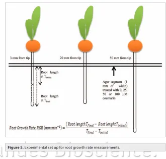
intact maize seedlings was determined as previously described by Sivaguru and Horst,43 with minor modifications. Briefly, agar
dissolved in 0.5 mM CaSO4 (0.75% w/v) was layered in Petri dishes (120 x 120 mm) and divided in three segments. Either zero, 25, 50 or 100 μM coumarin was added to the cooled agar solution only into the middle agar segment (1 mm width, Fig. 5). Primary root of intact maize seedlings was then vertically placed in the agar plates so that the various root zones, TZ, NGZ1 or NGZ2 were in contact with the coumarin-treated agar seg-ments. Then an image of individual root segment was captured after 0 and 30 min of coumarin exposure using a digital camera (Olympus C-5050). The length of each root zone was measured using the WinRHIZO pro STD 1600 software (Instruments Régent Inc., Canada) and, RER was calculated as the increase solution, in controlled conditions (continuous darkness; 24°C
and 70% RH). After 72 h, homogeneous seedlings were trans-ferred into hydroponic culture containing 1 l of aerated one-fourth strength Hoagland solution (NS, nutrient solution). The pH was adjusted to 6.0 with 0.1 N KOH. The seedlings were maintained in a growth chamber at 24 ± 1°C with a 14 h pho-toperiod, at a photon flux density of 300 μmolm-2s-1 at plant
height and 70% RH for seven days until the experimental mea-surements. All reagents used were of the highest analytical grade and were purchased from Sigma Chemical Co., (St. Louis, MO, USA).
Root elongation rate. Root elongation rate (RER) of three different zones (TZ, NGZ1 and NGZ2 at 3, 20 and 50 mm of distance from the root tip, respectively) of primary root of
Figure 4. electrophysiological traces of membrane potential of the TZ (3 mm), NGZ1 (20 mm) and NGZ2 (50 mm) of primary maize root exposed to 25
μM (a, D and G, respectively), 50 μM (B, e and h, respectively) or 100 μM coumarin (c, F and I, respectively). The data displayed the curves from repre-sentative membrane potential recording of five similar experimental results for 30 minute of exposure. arrow indicates the starting time of coumarin perfusion.
Table 1. Depolarization (DeP) and hyperpolarization (hYP) of root membrane potential along primary maize root by different concentrations of
coumarin
Coumarin (µM) Distance from tip (mm)
3 20 50
DEP HYP DEP HYP DEP HYP
25 4 ± 1a 9 ± 3.6a 10 ± 0.59a 0 0 0
50 3 ± 0.54a 21 ± 1.82b 26.33 ± 4.1b 0 0 0
100 3 ± 0.57a 14 ± 2ab 21.66 ± 2.3b 0 0 0
compartments each of which incorporated three distinct zones (TZ, NGZ1 and NGZ2) isolated by silicone grease. Each root zone compartment contained 0.5 mM CaSO4 pH 6.0 solutions, with or without (control) 25, 50 or 100 μM coumarin added. The pH of each compartmental solution was monitored for 30 min using a pH electrode (4.5 mm diameter tip, Thermo Scientific, Auchtermuchty, Scotland, model CMAW711). The extrusion rate was expressed as μmolH+g-1 fresh weight h-1 and
was calculated from the measured change in pH.
Membrane potential measurements. All electrophysiological experiments were performed on intact, 100 mm long, primary roots. The membrane electrical potential of the outermost layer of cells was measured at TZ, NGZ1 and NGZ2 using a standard glass microelectrode technique. Single-barrelled microelectrodes were prepared using filamented borosilicate glass as described pre-viously.50,51 The microelectrode was backfilled with 200 mM KCl
solution using a 70 mm long Microfil needle (World Precision Instruments Inc., Stevenage, UK). For the electrode impalement the primary root of intact maize seedlings, 7 day-old, was placed in a Plexiglass chamber and perfused with a solution contain-ing 0.5 mM CaSO4, 2 μM KNO3, 1 mM MES-NaOH (pH 6) and 25, 50 or 100 μM coumarin. Impalements with microelec-trodes were always made in mature epidermal cells and measured the voltage difference (mV), between the inside of the cell and the external bathing solution. The values from -70 to -140 were considered to define a successful cell microelectrode impalement and measurement. The initial impalement of an epidermal cell could be confirmed visually and by the accompanying jump in the voltage recorded after which it was not possible to see the precise location of the tip. Before the coumarin treatment, a time of the root length during the 30 min of
couma-rin treatment (Fig. 5). Care was taken to ensure that the maize roots were positioned vertically so that gravity-induced curvature did not inter-fere with the measurements.
H+-ATPase assay. Isolation of plasma mem-brane vesicles. Plasma memmem-brane vesicles were
isolated from primary root zones of maize seedlings using a small-scale procedure from Giannini et al.44 modified by Santi et al.45
Treated and control maize root zones (1–1.5 g) were homogenized in extraction buffer (250 mM sucrose, 10% (v/v) glycerol, 10 mM glycerol-1-phosphate, 2 mM MgSO4, 2 mM EDTA, 2 mM EGTA, 2 mM ATP, 2 mM DTT, 5.7% (w/v) choline chloride and 25 mM BTP buffered to pH 7.6 with MES and 1 mM PMSF and 20 mg/ml chimostatin freshly added before homogenization), filtered and centri-fuged twice at 12,700 g for 3 and 25 min, at 4°C. The suspension was layered over a 25/38% discontinuous sucrose gradient (10 mM DL-α -glycerol-1-phosphate, 2 mM MgSO4, 2 mM EGTA, 2 mM ATP, 1 mM PMSF, 2 mM DTT, 20 mg/ml chimostatin, 5.7% choline chloride, 5 mM BTP buffered at pH 7.4 with MES) and
centrifuged at 12,700 g for 60 min at 4°C. The vesicles, banding at the 25/38% interface layers, were collected and centrifuged at 14,000 g for 45 min at 4°C. The pellets, resuspended in a medium (20% glycerol (v/v), 2 mM EGTA, 2 mM EDTA, 0.5 mM ATP, 1 mM PMSF, 2 mM DTT, 20 mg/ml chimostatin, 5.7% choline chloride, 5 mM BTP buffered at pH 7 with MES), were immedi-ately frozen in liquid N2 and stored at -80°C until use.
Protein assay. Total soluble protein was estimated according to
the Bradford46 using bovine serum albumin as standard.
ATPase activity. ATP-hydrolyzing activity was determined
by measuring the release of inorganic phosphate, as described by Forbusch47 at 38°C. Assays were performed at 38°C in a 0.6
mL assay medium containing 50 mM BTP-MES, pH 6.5, 5 mM MgSO4, 5 mM ATP, 0.6 mM Na2MoO4, 100 mM KNO3, 1.5 mM NaN3, 0.01 % (w/v) Brij58, with or without 100 μM vana-date (V2O5), an inhibitor of P-type H+-ATPase.47 Sodium azide
and KNO3 were used as selective inhibitors of mitochondria and tonoplast H+-ATPase, respectively. The difference between these
two activities was attributed to the pmH+-ATPase. The reaction
was initiated by the addition 0.5–1.5 μg of membrane protein and was stopped after 30 min with a solution containing: 0.6 M HCl, 3% (w/v) SDS, 3% (w/v) ascorbic acid and 0.5% (w/v) ammo-nium molybdate at 2°C. The enrichment degree in plasma mem-brane of vesicles was determined in the presence of 0.1 mM V2O5, 1 mM NaN3 and 150 mM KNO3, selective inhibitors of plasma membrane, tonoplast and mitocondrial ATPase, respectively.
Proton efflux assay. H+ efflux was measured from the change
in pH of an unbuffered solution bathing the root (modified from Glass et al.48). Primary root of intact maize seedlings
was positioned in a chamber that was partitioned into three
ANOVA (coumarin concentration) with a completely random-ized design with 5 replicates, while RER data were representative of 10 replicates. The Tukey’s test was used for comparing the means within each root zones. Statistical analysis was run using Systat v. 8.0 software package (SPSS Inc.,).
interval (10 min) of stable cell electrical membrane potential was recorded (data not shown).
Statistical analysis. Data were firstly checked for deviations from normality and homogeneity of variances. The Em, proton efflux, pm H+-ATPase activity were analyzed using one-way
References
1. Zobel AM, Brown SA. Coumarins in the interaction between the plant and its environment. Allelopathy Journal 1995; 2:9-22.
2. Bruneton J. Pharmacognosy, Phytochemistry, Medicinal Plants. Hampshire, UK: Intercept Ltd., 1999; 263-277.
3. Bertin C, Yang X, Weston LA. The role of root exudates and allelochemicals in the rhizosphere. Plant and Soil 2003; 256:67-83.
4. Bais HP, Park SW, Weir TL, Callaway RM, Vivanco JM. How plants communicate using the underground information superhighway. Trends in Plant Science 2004; 9:26-32.
5. Rice EL. 1984; Allelopathy. Academic Press Inc., Orlando.
6. Svensson SB. The effect of coumarin on root growth and root histology. Physiologia Plantarum 1971; 24:446-70.
7. Kupidlowska E, Kowalec M, Sulkowski G, Zobel AM. The effect of coumarin on root elongation and ultra-structure of meristematic cell protoplast. Annals Botany 1994; 73:525-30.
8. Chon SU, Choi SKC, Jung S, Jang HG, Pyo BS, Kim SM. Effects of alfalfa leaf extracts and phenolic allelochemicals on early seedling growth and root mor-phology of alfalfa and barnyard grass. Crop protection 2002; 21:1077-82.
9. Abenavoli MR, Sorgonà A, Albano S, Cacco G. Coumarin differentially affects the morphology of dif-ferent root types of maize seedlings. J Chem Ecol 2004; 30:1871-83.
10. Abenavoli MR, De Santis C, Sidari M, Sorgonà A, Badiani M, Cacco G. Influence of coumarin on the net nitrate uptake in durum wheat. New Phytol 2001; 150:619-27.
11. Abenavoli MR, Sorgonà A, Sidari M, Badiani M, Cacco G. Coumarin inhibits the growth of carrot (Daucus
carota L. cv. Saint Valery) cells in suspension culture. J
Plant Physiol 2003; 160:227-37.
12. Moreland ED, Novitzky WP. 1987; Effects of acid cou-marins and flavonoids on isolated chloroplast and mito-chondria. In: Waller GR, Ed. Allelochemicals: Role in Agriculture and Forestry. American Society Sympsium Series. Washington 1987; American Chemical Society. 13. Aliotta G, Cafiero G, Fiorentino A, Strumia S.
Inhibition of radish germination and root growth by coumarin and phenylpropanoids. J Chem Ecol 1993; 19:175-83.
14. Abenavoli MR, Cacco G, Sorgonà A, Marabottini R, Paolacci AR, Ciaffi M, et al. The inhibitory effects of coumarin on the germination of durum wheat (Triticum turgidum ssp. durum, CV. Simeto) seeds. J Chem Ecol 2006; 32:489-506.
15. Pergo ER, Abrahim D, da Silva PCS, Kern KA, da Silva LJ, Voll E, et al. Bidens pilosa L. Exhibits high sensitiv-ity to coumarin in comparison with three other weed species. J Chem Ecol 2008; 34:499-507.
16. Schreiner O, Reed HS. The toxic action of certain organic plant constituents. Botanical Gazette 1908; 45:43-102.
17. Avers CJ, Goodwin RH. Studies on root. IV. Effects of coumarin and scopoletin on the standard root growth pattern of Phleum pratense. Am J Bot 1956; 43:612-20.
18. Abenavoli MR, Nicolò A, Lupini A, Oliva S, Sorgonà A. Effects of different allelochemicals on root morphol-ogy of Arabidopsis thaliana. Allelopathy Journal 2008; 22:245-52.
19. Glass ADM, Dunlop J. Influence of phenolic acids on ion uptake. IV. Depolarization of membrane potentials. Plant Physiol 1974; 54:855-8.
20. Hejl AM, Koster KL. The allelochemical sorgoleone inhibits root H+-ATPase and water uptake. J Chem Ecol 2004; 30:2181-91.
21. Ahn SJ, Rengel Z, Matsumoto H. Aluminium-induced plasma membrane surface potential and H+-ATPase activity in near-isogenic wheat lines differing in toler-ance to aluminium. New Phytologist 2004; 162:71-9. 22. Baluška F, Barlow PW, Kubica S. Importance of the
post-mitotic isodiametric growth (PIG) region for growth and development of root. Plant and Soil 1994; 167:31-41.
23. Baluška F, Barlow PW, Hauskrecht M, Kubica S, Parker JS, Volkmann D. Microtubule arrays in maize root cells. Interplay between the cytoskeleton, nuclear organization and post-mitotic cellular growth patterns. New Phytologist 1995; 130:177-92.
24. Baluška F, Barlow PW, Volkmann D. Complete disin-tegration of the microtubular cytoskeleton precedes its auxin-mediated reconstruction in postmitotic maize root cells. Plant Cell Physiol 1996; 37:1013-21. 25. Einhellig FA. Mechanisms and modes of action of
alle-lochemicals. In: Putnam AP, Teng CS, Ed. The science of allelopathy. John Wiley & Sons, New York 1986; 170-88.
26. Bates GW, Goldsmith MHM. Rapid response of the plasma-membrane potential in oat coleoptiles to auxin and other weak acids. Planta 1983; 159:231-7. 27. Karcz W, Burdach Z. A comparison of the effects of
IAA and 4-Cl-IAA on growth, proton secretion and membrane potential in maize coleoptile segments. J Exp Bot 2002; 53:1089-98.
28. Abenavoli MR, Sorgonà A, Muscolo A. Morphophysiological changes in tissue culture of
Petunia hybrida in response to the allelochemical
cou-marin. Allelopathy J 2001; 8:171-7.
29. Pang J, Cuin T, Shabala L, Zhou M, Mendham N, Shabala S. Effect of secondary metabolites associ-ated with anaerobic soil conditions on ion fluxes and electrophysiology in barley roots. Plant Physiol 2007; 145:266-76.
30. Casimiro I, Marchant A, Bhalerao RP, Beekman T, Dhooge S, Swarup R, et al. Auxin transport promotes Arabidopsis lateral root initiation. Plant Cell 2001; 13:843-52.
31. Lucas M, Godin C, Jay-Allemand C, Laplaze L. Auxin fluxes in the root apex co-regulate gravitropism and lateral root initiation. J Exp Bot 2008; 59:55-66. 32. Colmer TD, Bloom AJ. A comparison of NH4+ and
NO3- net fluxes along roots of rice and maize. Plant Cell Environ 1998; 21:240-6.
33. Birnbaum K, Shasha DE, Wang JY, Jung JW, Lambert GM, Galbraith DW, et al. A gene expression map of the Arabidopsis root. Science 2003; 302:1956-60. 34. Ferguson IB, Clarkson DT. Simultaneous uptake and
translocation of magnesium and calcium in barley (Hordeum vulgare L.) roots. Planta 1976; 128:267-9. 35. Goulas E, Dily FL, Ourry A. Effects of a cold treatment
of the root system on white clover (Trifolium repens L.) morphogenesis and nitrogen reserve accumulation. J Plant Physiol 2003; 160:893-902.
36. Blake TJ, Ferrell WK. The association between soil and xylem water potential, leaf resistance and absci-sic acid content in droughted seedlings of Douglas-fir (Pseudotsuga menziesii). Physiol Plant 1977; 39:106-9.
37. Sorgonà A, CaccoG. Linking the physiological param-eters of nitrate uptake with root morphology and topology in wheat (Triticum durum Desf.) and in citrus rootstock (Citrus volkameriana Ten & Pasq). Canadian journal of botany-revue canadienne de botanique 2002; 80:494-503.
38. Goff MD, Koide RT. Investigation of the relationship between root tip and phosphorus uptake. In: Lynch JP, Deikman J, ed. Phosphorus in plant biology: regulatory roles in molecular, cellular, organismic and ecosystem processes. American Society of Plant Physiologist. Rockville Md 1998; 322-3.
39. Yamamoto Y. Movement of allelopathic compound coumarin from plant residue of sweet vernalgrass (Anthoxanthum odoratum L.) to soil. Japanese Society of Grassland Science 2009; 55:36-40.
40. Narasimhan K, Basheer C, Bajic VB, Swarup S. Enhancement of plant-microbe interactions using a rhizosphere metabolomics-driven approach and its application in the removal of polychlorinated biphe-nyls. Plant Physiol 2003; 132:146-53.
41. Maffei M, Camusso W, Sacco S. Effect of Mentha x
pip-erita essential oil and monoterpenes on cucumber root
membrane potential. Phytochemistry 2001; 58:703-7. 42. Sanders D, Bethke P. Membrane transport. In: Gruissem
W, Jones R ed. Biochemistry and molecular biol-ogy of plants. American Society of Plant Physiologists, Rockville 2000; 110-59.
43. Sivaguru M, Horst WJ. The distal part of the transition zone is the most aluminium-sensitive apical root zone of maize. Plant Physiol 1998; 116:155-63.
44. Giannini JL, Ruiz-Cristin J, Briskin DP. A small scale procedure for the isolation of transport compe-tent vesicles from plant tissues. Anal Biochem 1988; 174:561-7.
45. Santi S, Locci G, Pinton R, Cesco S, Varanini Z. Plasma membrane H+-ATPase in maize roots induced for NO3- uptake. Plant Physiol 1995; 109:1277-83. 46. Bradford MM. A rapid and sensitive method of
quan-titation of microgram quantities of protein utilizing the principle of protein dye binding. Anal Biochem 1976; 72:248-54.
47. Forbush B. Assay of the Na+ K+-ATPase in plasma membrane preparations: increasing the permeability of membrane vesicles using sodium dodecyl sulfate buff-ered with bovine serum albumin. Anal Biochem 1983; 128:159-63.
48. Sze H. H+-translocating ATPases: advances using mem-brane vesicles. Ann Rev Plant Physiol 1985; 36:175-208.
49. Glass ADM, Siddiqui MY, Giles KI. Correlations between potassium uptake and hydrogen efflux in barley varieties. A potential screening method for the isolation of nutrient efficient lines. Plant Physiol 1981; 68:457-9.
50. Miller AJ. Ion-selective microelectrodes for measure-ment of intracellular ion concentrations. Methods Cell Biology 1996; 49:275-91.