E
WAS
AWICKA, A
NNAD
ŁUGOSZ, D
OROTAP
RZYBYLSKAThe Effect of Cyclosporin A on Free Radical
Processes and Interactions with Antioxidants
Badanie wpływu cyklosporyny A na procesy wolnorodnikowe
i jej interakcji z antyoksydantami
Department of Toxicology, Silesian Piasts University of Medicine in Wrocław, Poland
Adv Clin Exp Med 2006, 15, 3, 435–444 ISSN 1230−025X
ORIGINAL PAPERS
Abstract
Background. Previous study of a group of patients treated with CsA after renal transplantation showed a statisti− cally elevated concentration of lipid peroxidation products, measured as the MDA + 4−HNE level in blood plasma. Objectives. Evaluating whether CsA is responsible for the increase in MDA concentration or hydroxyl radical ge− neration by an in vitromethod. Moreover, the authors investigated whether two important lipophilic antioxidants, coenzyme Q10 and vitamin E, could be considered as protective or reparative agents in free radical processes cau−
sed by CsA.
Material and Methods. Mitochondria isolated from human placenta were used as the experimental model. MDA content was measured by the TBARS method, the •OH level by deoxirybose degradation, and the protein concen−
tration by the Lowry method. The influence of CsA in doses of 30–750 ng/ml on MDA and •OH concentration was
investigated. Also evaluated were the protective (antioxidant given before CsA) and reparative (antioxidant given after CsA) effects of CoQ10 and vitamin E in oxidative stress caused by CsA. Coenzyme Q10 was used in two con−
centrations, 1.5 and 3.0 µg/ml, and vitamin E in the three concentrations of 1.5; 5.0, and 15.0 µg/ml.
Results. The examinations showed that CsA in doses of 450–750 ng/ml increased MDA concentration in mitochon− dria in a statistically significant way. A significant positive linear correlation, with a Pearson’s coefficient of r= = 0.6030 (p= 0.000), between the dose of CsA and MDA level was obtained. Elevated •OH formation was caused
by the highest applied dose of CsA, 750 ng/ml. Coenzyme Q10at a dose of 3.0 µg/ml protected against increased
lipid peroxidation caused by CsA with high significance, but it did not repair. The elevated •OH formation after
750 ng/ml of CsA was not decreased by CoQ10at both concentrations used, unlike vitamin E, which in the three
experimental concentrations (1.5, 5.0, and 15 µg/ml) acted as a protective and reparative agent, decreasing OH for− mation (p < 0.01). Vitamin E inhibited lipid peroxidation in both protective and reparative ways, and it was observed that the highest dose of vitamin E (15 µg/ml) was the most effective in protection.
ConclusionCyclosporin at high doses can induce free radicals processes, evaluated as an elevated MDA concen− tration and •OH generation. The investigations with CoQ
10and vitamin E point out the possibility of reducing the
negative effects of CsA by these antioxidants. Moreover, the usefulness of the mitochondrial model from human placenta for in vitroanalysis was estimated (Adv Clin Exp Med 2006, 15, 3, 435–443).
Key words: cyclosporin A, malondialdehyde, hydroxyl radical, coenzyme Q10, vitamin E.
Streszczenie
Wprowadzenie. Wcześniejsze badania wykonane w Katedrze Toksykologii w grupie pacjentów po przeszczepie nerek, leczonych cyklosporyną A, wykazały statystycznie istotne zwiększenie stężenia produktów peroksydacji li− pidów, mierzone stężeniem MDA + 4−HNE w osoczu.
Cel pracy. Wyjaśnienie mechanizmu badanych zjawisk przez ocenę in vitrowpływu CsA na peroksydację lipido− wą, w tym na stężenie MDA oraz wytwarzanie rodnika hydroksylowego. Doświadczenia miały na celu ocenę włas− ności antyoksydacyjnych koenzymu Q10 oraz witaminy E w odniesieniu do stresu oksydacyjnego wywołanego
przez CsA.
Materiał i metody. Modelem do badań były mitochondria izolowane z łożysk ludzkich. Oznaczanie stężenia MDA wykonano za pomocą metody TBARS, liczbę rodników •OH przez degradację dezoksyrybozy, a stężenie białka metodą Lowry’ego. Badano wpływ CsA w dawkach 30–750 ng/ml na MDA oraz •OH w mitochondriach. Ocenio−
Cyclosporin A (CsA) is a natural polypeptide with wide clinically use as a strong immunosup− pressive agent in organ transplantation. Among its other effects, it prevents transplant rejection by in− fluencing the course of many inflammatory pro− cesses through decreasing the synthesis and secre− tion of proinflammatory cytokines. Cyclosporin therapy has revolutionized organ transplantation; however, it is not devoid of side−effects [1, 2]. Some recent scientific reports point to the role of CsA in inducing oxidative stress. Authors’ own previous research, conducted on a group of pa− tients treated with CsA after renal transplantation, showed statistically significant enhanced lipid pe− roxidation, measured as malondialdehyde with 4−hydroxylnonenal (MDA + 4−HNE) concentra− tion, compared with controls [3].
The aim of the present study was to determine the influence of CsA on free radical processes, mea− sured as MDA concentration and hydroxyl radical generation, and to estimate the usefulness of antio− xidants in decreasing the lipid peroxidation indu− ced by CsA. The investigations also estimated the interaction of CsA with the antioxidants coenzyme Q10 (CoQ10) and vitamin E. Authors’ earlier rese−
arch demonstrated that CoQ10 did not inhibit
enhanced lipid peroxidation after four weeks of application to patients after renal transplantation; however, it advantageously lowered atherogenici− ty indicators [3]. Therefore the question arose whether coenzyme Q10does not scavenge the free
radicals generated by CsA, which would explain observed lack of its influence on MDA level in pa− tients after renal transplantation, or whether CoQ10
supplementation should last longer, as some CoQ10
researchers suggested. Therefore an evaluation of the antioxidative abilities of CoQ10inin vitrosta−
tes of enhanced by CsA lipid peroxidation appear− ed to be sensible. The mitochondrial model from human placenta was used in the experiments.
Besides CoQ10, the antioxidative effect of vita−
min E on conditions of oxidative stress induced by CsA was examined. Coenzyme Q10is synthesized
in all tissues and cells of organisms in amounts to fulfill its antioxidative role in conditions of correct homeostasis. Under conditions of illness which handicap the function of tissue and organs, for example after renal transplantation, the local bio− synthesis of CoQ10can be scarce. Coenzyme Q10in
the hydrochinone form plays an important antioxi− dative role. It deactivates radicals, including toco− pheryl radicals, and prevents lipid peroxidation during its initiation as well as propagation. Vita− min E, however, is an important hydrophobic an− tioxidant acting in the second phase of the process, scavenging free radicals and interrupting the chain reaction during propagation [4].
Material and Methods
The examinations were conducted on an in vi− tro model using mitochondria isolated from hu− man placenta from natural deliveries. Mitochon− dria were isolated by homogenizing the material in 5 mM Tris−HCl buffer, pH 7.4, containing 0.23 M mannitol, 0.07 M sucrose, 1 mM EDTA, and 0.2% bovine serum albumin (BSA). The homoge− nate was centrifuged at 1000 ×g (4°C) and then recentrifuged under the same conditions at 12,000 ×g. The mitochondrial supernatant was collected and washed three times in 50 mM Tris−HCl buffer, pH 7.4, containing 0.575% KCl at 4°C. The resul− ting mitochondria were stored at –80°C for no lon− ger than 3 months [5, 6]. The degree of lipid pero− xidation was measured by the MDA concentration [7]. Hydroxyl radical generation was estimated using the deoxirybose degradation method [8].
Used in the experiments were: cyclosporin A (Calbiochem) and Sandimmun, cyclosporinum
no zarówno działanie ochronne koenzymu Q10oraz witaminy E (podając antyoksydant przed CsA), jak i napraw−
cze (podając antyoksydant po CsA) na peroksydację lipidową indukowaną CsA.
Wyniki.Nastąpiło statystycznie istotne zwiększenie stężenia MDA po cyklosporynie w dawkach 450–750 ng/ml z dodatnią korelacją liniową dawka–odpowiedź (r = 0,6030; p = 0,000), a generację rodnika •OH indukowała CsA o stężeniu 750 ng/ml. Oznaczając działanie CoQ10, stwierdzono, że antyoksydant w dawce 3,0 µg/ml działał
ochronnie, zapobiegając lipidowej peroksydacji wywołanej przez CsA, mierzonej stężeniem MDA; nie wykazywał natomiast działania naprawczego i ochronnego na zwiększoną generację rodników •OH po ekspozycji mitochon− driów na CsA. Witamina E, zastosowana w badaniach w trzech stężeniach: 1,5; 5,0 oraz 15,0 µg/ml wykazywała istotne statystycznie działanie ochronne i naprawcze na zwiększoną generację rodników •OH po CsA (p < 0,01), a najsilniejsze działanie obniżające MDA wykazywała witamina E w stężeniu 15,0 µg/ml.
Wnioski. Cyklosporyna A w dużych stężeniach (> 450 ng/ml) może indukować wolnorodnikowe uszkodzenia li− pidów, oceniane podwyższonym stężeniem MDA oraz ••OH. Wyniki badań dotyczących oceny własności ochron− nych i naprawczych koenzymu Q10 oraz witaminy E wskazują, że suplementacja pacjentów po przeszczepie nerek
koenzymem Q10 oraz witaminą E może korzystnie hamować nasilenie peroksydacji lipidów w warunkach ekspo−
zycji na cyklosporynę A. Wykazano ponadto użyteczność modelu mitochondrialnego do badań (Adv Clin Exp Med 2006, 15, 3, 435–443).
(Novartis) and the antioxidants CoQ10 (Q10
904944, JEMO PHARM) and vitamin E (D−alpha− tocopherol, ICN Biomedicals Inc.). The MDA and
•OH concentrations were expressed relative to the
level of mitochondrial protein. The protein con− centration in the mitochondria used in the experi− ments was 0.5 mg/ml. Mitochondrial proteins were measured by the Lowry method.
The Procedure
for Evaluating the Effects
of CsA on MDA Concentration
in Mitochondria
The mitochondrial suspension (1 ml) was sti− mulated by 30 µl of 1% t−BOOH at 37°C for 30 min. Then the samples were incubated at 37°C for 30 min. with 30 µl of CsA at concentrations from 30 ng/ml to 750 ng/ml. After incubation, 0.5 ml of 20% trichloroacetic acid (TCA), 1.5 ml of 0.67% thiobarbituric acid (TBA), and 30 µl of 1% buty− lhydroxytoluene (BHT) were added and the sam− ples were incubated at 85°C for 15 min. The MDA concentration was measured spectrophotometri− cally at 535 nm and expressed as nmol per mg mi− tochondrial protein and calculated using a molar absorption coefficient of 1.56 × 10⁵M–1cm–1. The
results were compared with the control (K) prepa− red in the same way as the experimental sample, but without the CsA.
The Procedure for Evaluating
the Effects of CsA on
••OH
Generation in Mitochondria
The mitochondrial suspension (0.5 ml) was in− cubated at 37°C for 15 min. with 15 µl of CsA at concentrations from 30 to 750 ng/ml, 0.5 ml of 20 mmol/l deoxyribose, and 15 µl of 1% t−BOOH. After incubation, the samples were centrifuged. Then 0.5 ml of 20% TCA, 1.5 ml of 0.67% TBA, and 30 µl of 1% BHT were added to 0.8 ml of the supernatant and the samples were incubated at 85°C for 15 min. After cooling and centrifuging, the hydroxyl radical level was measured spectro− photometrically at 532 nm and calculated using the molar coefficient of absorption of 1.56 × 10⁵
M–1cm–1 [8]. The results were compared with the
control (K1) prepared in the same way, but witho− ut the CsA. For each dose of CsA, 10–14 samples were made.
The Procedure for Evaluating
the Antioxidative Effects
of CoQ
10and Vitamin E
in Mitochondria Exposed to CsA
The antioxidative effects of coenzyme Q10 (in
two doses: 1.5 and 3.0 µg/ml) and vitamin E (in three doses: 1.5, 5.0, and 15.0 µg/ml) on MDA concentration and hydroxyl radical generation in mitochondria exposed to CsA were examined. Investigated was whether coenzyme Q10 and vita−
min E could be considered as a protective (antiox− idant given to mitochondria before the CsA) or reparative (antioxidant given to mitochondria after CsA) agents in oxidative stress caused by CsA. Coenzyme Q10 or vitamin E were given to the mito−
chondrial suspension in a quantity of 30 µl when their influence on MDA concentration was mea− sured and in a quantity 15 µl when the antioxidant effect on hydroxyl radical generation was estimated. For each dose of CsA, 10–14 samples were made.
Results of the study were statistically analyzed using the Student’st−test. The Pearson coefficient was used to describe correlation. Statistical evalu− ation of the results was performed using the pro− gram Statistics 6.0. Differences with p< 0.05 were regarded as statistically significant.
Results
The concentration of CsA in the blood of a pa− tient after renal transplantation depends on the time of measurement. It reaches average values of 642–714 ± 422 ng/ml in the second hour after ap− plication [1, 9]. Desirable, though, are therapeutic levels (Co) of 100–250 ng/ml. Levels below 100 ng/ml are associated with the risk of rejection and levels above 250 ng/ml correlate with nephrotoxi− city and hepatotoxicity [10]. Evaluating the effect of CsA at concentrations of 30–750 ng/ml on lipid peroxidation, statistically significant increases in MDA concentration in mitochondria compared with the control K (4.11 nmol/mg protein) were obtained only at doses of 450–750 ng/ml (4.49, 5.13, and 5.76 nmol/mg protein, respectively) (Ta− ble 1). Estimating the dose−response dependence, significant positive linear correlation, with the Pe− arson’s coefficient r= 0.6030 (p= 0.0000), between CsA dose and MDA level was obtained. Thus CsA in concentrations > 450 ng/ml enhanced lipid pe− roxidation, with significant growth observed for the concentrations of 600 and 750 ng/ml (p = 0.006 and p= 0.0002).
T
able 1.
The ef
fect of CsA
on MDA
concentration compared with the control (K)
T
a
bela 1.
Wpływ CsA
na stężenie MDA
w
porównaniu z
grupą kontrolną K
Control group
Examined samples
(Grupa (Badane
próby)
kontrolna) KS
Dose of CsA
30 75 90 150 300 450 600 750
(Dawka CsA) ng/ml MDA
concentration 4.1 1 ± 0.86 3.93 ± 0.40 3.88 ± 0.48 3.78 ± 0.45 3.89 ± 0.40 4.38 ± 1.04 4.49 ± 0.95 5.13 ± 0.95 5.76 ± 1.14
(Stężenie MDA) nmol/mg protein Statistical significance K−S
p = 0.9301
p = 0.2546 p = 0.21892 p = 0.7256 p = 0.2925 p = 0.0486 p = 0.0066 p = 0.00028
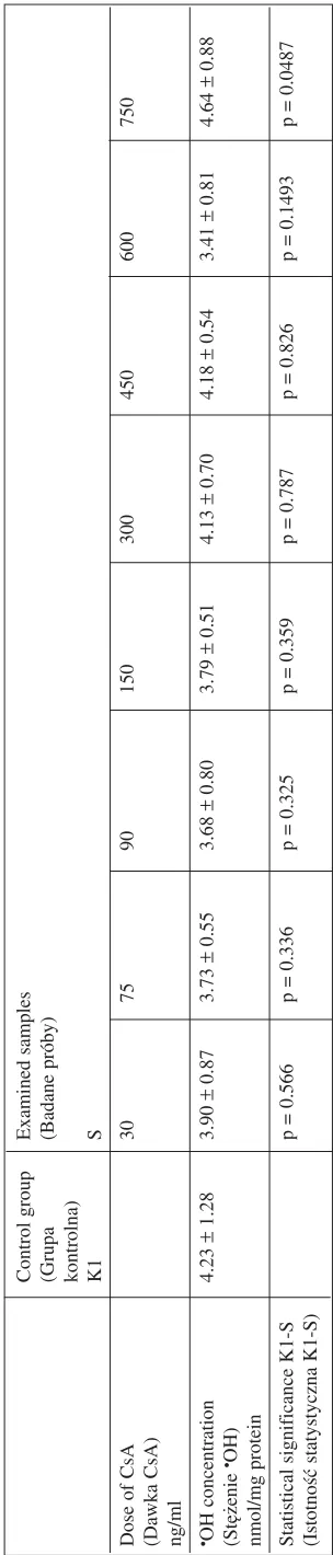
(Istotność statystyczna K−S) Table 2.
The ef
fect of CsA
on
•OH generation compared with the control (K1)
T
abela 2.
Wpływ CsA
na wytwarzanie
•OH w
porównaniu z
grupą kontrolną K1
Control group Examined samples (Grupa (Badane próby) kontrolna) K1 S
Dose of CsA
30 75 90 150 300 450 600 750
(Dawka CsA) ng/ml •OH concentration
4.23 ± 1.28 3.90 ± 0.87 3.73 ± 0.55 3.68 ± 0.80 3.79 ± 0.51 4.13 ± 0.70 4.18 ± 0.54 3.41 ± 0.81 4.64 ± 0.88 (Stężenie •OH)
nmol/mg protein Statistical significance K1−S
p = 0.566 p = 0.336 p = 0.325 p = 0.359 p = 0.787 p = 0.826 p = 0.1493 p = 0.0487
processes, i.e. ascertain whether it affects •OH ge− neration. The results indicate that only the highest concentration of CsA (750 ng/ml) induced a stati− stically significant difference in •OH generation (4.64 nmol/mg protein) compared with the control. Thus the mechanism related to •OH generation could be of no essential importance in the influ− ence of routine CsA doses on lipid peroxidation.
Evaluating the antioxidative effect of CoQ10
and its interaction with CsA, two doses of antioxi− dant (1.5 and 3.0 µg/ml) were used. In previous in− vestigations concerning the protective and repara− tive effects of CoQ10on lipid peroxidation induced
by various xenobiotics, these two doses were shown to be beneficial and effective [11]. The pro− tective and reparative influence of CoQ10was esti−
mated in mitochondria exposed to CsA. The pro− tective effect was measured when CoQ10 was ad−
ded to the mitochondria before CsA, and the reparative when CoQ10 was added to the sample
after CsA (30 minutes after CsA action).
Only the lower dose of CoQ10 (1.5 µg/ml) sho−
wed a weak effect, decreasing the lipid peroxida− tion induced by only two CsA concentrations (300 and 750 ng/ml) of the four tested (Fig. 1). Howe− ver, the lack of such an effect by the higher dose of CoQ10 (3.0 µg/ml) and the very low statistical
significance of the results for the 1.5 ug/ml dose (p≈0.04) suggest that CoQ10in concentrations of
1.5–3.0 µg/ml is not effective as a reparative agent in lowering CsA−induced lipid peroxidation. There was no interaction between CoQ10and CsA at con−
centrations of 75 ng/ml and 450 ng/ml or higher. The next experiment indicates that CoQ10 can be
a strong protective agent against lipid peroxidation generated by CsA. A significant drop in MDA concentration was observed when CoQ10was used
before CsA, e.g. with p = 0.0009 for the concen− tration of 450 ng/ml (Fig. 2). However, in order to obtain such a result, the 3.0−µg/ml concentration of CoQ10 was needed because the lower concentra−
tion (1.5 µg/ml) did not cause such an effect. To a certain degree, the study carried out in the
in vitromodel explains why there was no decrease
in lipid peroxidation in patients after renal trans− plantation who were given CoQ10 for 4 weeks in
authors’ earlier experiment. It appears that in order to obtain the expected effect, treatment with CoQ10
supplementation should be used in the earliest phase of transplantation, because CoQ10 is more
a protective than a reparative agent against CsA li− pid peroxidation. The results obtained do not indi− cate interaction between CsA and CoQ10 in influ−
encing lipid peroxidation.
In the next stage of experiment, the combined influence of CsA and CoQ10 on •OH generation
was estimated. At a concentration of 1.5 µg/ml, co− enzyme Q10 did not affect •OH generation. There
was also no dose effect of CsA until the 600 ng/ml concentration on •OH formation. The combined effect did not show any interaction in •OH level. Coenzyme Q10added before or after CsA did not
influence •OH generation.
In the experiment with vitamin E, CsA in four doses (300, 450, 600, and 750 ng/ml) and vitamin E in three doses (1.5, 5.0, and 15.0 µg/ml) were used. The protective effect (vitamin E given before CsA) and the reparative effect (vitamin E given after CsA) were evaluated. The 1.5−µg/ml dose of vitamin E ac− ted protectively against lipid peroxidation at two CsA doses and reparative at one (Fig. 3). Vitamin E at a concentration of 5.0 µg/ml acted protectively at three CsA doses (450, 600, and 750 ng/ml), but re− parative only at one CsA dose (750 ng/ml) (Fig. 4).
0 2 4 6 8
75 300 450 750 ng/ml
dose of CsA dawka CsA
CsA protective ochronne
repairing naprawcze MDA concentration (nmol/mg protein) stężenie MDA (nmol/mg białka)
Fig. 1.The protective and reparative effect of CoQ10 at
a dose of 1.5 µg/ml on MDA concentration after expo− sure to CsA
* significantly lower than the CsA sample, p < 0.05 Ryc. 1.Wpływ ochronny i naprawczy CoQ10 w dawce
1,5 µg/ml na poziom MDA po ekspozycji na CsA * Wyniki statystycznie istotnie mniejsze od prób z CsA
0 2 4 6 8
75 300 450 750 ng/ml
dose of CsA dawka CsA
CsA protective ochronne
repairing naprawcze MDA concentration (nmol/mg protein) stężenie MDA (nmol/mg białka)
Fig. 2.The protective and reparative effect of CoQ10 at
a dose of 3.0 µg/ml on MDA concentration after expo− sure to CsA
* significantly lower than the CsA sample, p < 0.05 Ryc. 2.Wpływ ochronny i naprawczy CoQ10 w dawce
The most effective was the highest dose of vitamin E (15.0 µg/ml). It statistically significantly lowered MDA in mitochondria exposed to CsA. Protective action was observed towards three CsA concentra− tions (300, 600, and 750 ng/ml) and reparative ac− tion towards two (300 and 750 ng/ml) (Fig. 5).
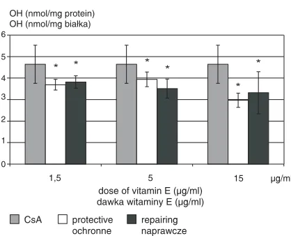
Vitamin E was specially effective in influen− cing •OH generation after exposure to CsA at a dose of 750 ng/ml. It acted in all the three doses applied (1.5, 5.0, and 15.0 µg/ml). Vitamin E statistically significantly lowered the •OH level, acting equally protective and reparative (Fig. 6).
In summary, although vitamin E decreased CsA−induced lipid peroxidation in a way compara− ble to CoQ10, its high efficiency in scavenging •OH
radicals indicates that it could be a better repara− tive agent than CoQ10. Coenzyme Q10 seems be
better in protective action against lipids peroxida− tion caused by CsA.
Discussion
One mechanism of the toxic effect of xenobi− otics is the initiation of free radical reactions, through their influence on the oxygen metabolite chain or the biotransformation of xenobiotics to free radicals. Some xenobiotics can accept an elec− tron from reductase P−450, giving rise to radicals.
300 450 600 750 ng/ml
dose of CsA dawka CsA
CsA protective ochronne
repairing naprawcze 0
2 4 6 8
MDA concentration (nmol/mg protein) stężenie MDA (nmol/mg białka)
1 3 5 7
Fig. 3. The protective and reparative effect of vitamin E at a dose of 1.5 µg/ml on MDA concentration after exposure to CsA
* significantly different from the CsA sample, p< 0.05 Ryc. 3.Wpływ ochronny i naprawczy witaminy E w dawce 1,5 µg/ml na poziom MDA po ekspozycji na CsA
* Wyniki statystycznie istotnie różne od prób z CsA
300 450 600 750 ng/ml
dose of CsA dawka CsA
CsA protective ochronne
repairing naprawcze MDA concentration (nmol/mg protein) stężenie MDA (nmol/mg białka)
*
* * * *
0 1 3 6 8
5 7
4
2
Fig. 4.The protective and reparative effect of vitamin E at a dose of 5.0 µg/ml on MDA concentration after exposure to CsA
* significantly different from the CsA sample, p< 0.05 Ryc. 4.Wpływ ochronny i naprawczy witaminy E w dawce 5,0 µg/ml na poziom MDA po ekspozycji na CsA
* Wyniki statystycznie istotnie różne od prób z CsA
300 450 600 750 ng/ml
dose of CsA dawka CsA
CsA protective ochronne
repairing naprawcze 0
6 8
4
2
* *
* * * *
MDA concentration (nmol/mg protein) stężenie MDA (nmol/mg białka)
Fig. 5.The protective and reparative effect of vitamin E at a dose of 15 µg/ml on MDA concentration after exposure to CsA
* significantly different from the CsA sample, p< 0.05 Ryc. 5.Wpływ ochronny i naprawczy witaminy E w dawce 15 µg/ml na poziom MDA po ekspozycji na CsA
* Wyniki statystycznie istotnie różne od prób z CsA
* * * *
* *
1,5 5 15 µg/ml
dose of vitamin E ( g/ml) dawka witaminy E ( g/ml)
µ µ
CsA protective ochronne
repairing naprawcze OH (nmol/mg protein)
OH (nmol/mg białka)
0 6
5
4
3
2
1
Fig. 6.The protective and reparative effect of vitamin E at doses of 1.5, 5.0, and 15.0 µg/ml on •OH generation after exposure to CsA
* significantly lower than the CsA sample, p< 0.05 Ryc. 6.Wpływ ochronny i naprawczy witaminy E w dawkach 1,5; 5,0 oraz 15 µg/ml na generację rodni− ka hydroksylowego po ekspozycji na CsA
These radicals easily transfer the extra electron to molecular oxygen, forming a superoxide anion radical (O2) and regenerating the parent xenobiot−
ic, which is ready to gain a new electron. Through this “redox cycling”, one electron−acceptor xeno− biotic molecule can generate many O2molecules.
Superoxide radicals can be transformed into hydroxyl radicals. Free radicals are able to damage endogenous cell structure, such as nucleic acids, proteins, and lipids [12].
In this study the influence of CsA on free radi− cal damage to lipids, known as lipid peroxidation, was estimated. Mitochondria isolated from human placenta were exposed to different concentration of CsA. The selection of doses was dictated by the ther− apeutic concentrations of CsA in the blood of pa− tients directly after renal transplantation (Co) or two hours after CsA application (C2). The concentra− tions used were within the range of 30 to 750 ng/ml. Analysis of the results indicates that CsA has limited influence on lipid peroxidation. Statistical− ly significant increases in MDA concentration were observed after CsA doses of 450–750 ng/ml. Similarly, Chen et al. [10] noticed that CsA in a therapeutic concentration (500 ng/ml) shows an oxidizing effect, measured as lipid peroxidation in human spleen cells in vitro. In children after liver transplantation, CsA concentrations in plasma of 70–120 ng/ml did not cause statistically significant increases in MDA levels compared with controls [13]. Singh et al. [14] even reported that low doses of CsA (3 mg/kg b.w. intravenously) protected rat kidney from lipid peroxidation induced by ische− mic reperfusion. Also, significant decreases in thiobarbituric−reactive substance (TBARS) con− centrations and increases in the activities of the an− tioxidative enzymes SOD, GR, catalase, and redu− ced glutathione were noted. Cyclosporin partially counteracts lipid peroxidation induced by adria− mycin in the kidneys and blood of rats, probably by stimulation of superoxide dismutase [15]. In re− search on rats it was observed that lipid peroxida− tion depends on CsA dose and cannot be measured below a certain CsA concentration. In research on patients with rheumatoid arthritis treated with low doses of CsA it was observed that 2.5–3.5 mg/kg b.w. of CsA did not increase MDA and F2−isopro− stanes concentrations in the urine [16]. Thus the influence of CsA on lipid peroxidation is different, depending on the factor initiating peroxidation and the investigative model.
In most studies, the CsA doses examined were approximately 100–250 ng/ml. Research on the in− fluence of CsA doses over 250 ng/ml on lipid pe− roxidation also seemed useful. These doses are present in the blood at two−hour (C2) post−dose CsA levels [1, 9].
The present study shows that CsA doses of 450–750 ng/ml statistically significantly increase lipid peroxidation, measured as MDA concentra− tion. Because CsA in concentrations of 450–600 ng/ml did not increase the •OH level in a statisti− cally significant manner, it seems that the mecha− nism of CsA action on lipid peroxidation is not connected with •OH generation. Perhaps other ra− dicals, e.g. superoxide radicals, are more involved in the action of CsA. The superoxide radical can be transformed into the •OH radical, which is able to initiate the peroxidation of lipids. The metabo− lism of CsA mainly takes place in the wall of inte− stine and in the liver with the participation of the isoenzyme CYP 3A4. This pathway can be a source of superoxide radical or hydrogen peroxide induced by CsA.
Research performed in vitroand in vivoindi− cate that though CsA is capable of generating free radicals, it cannot be assumed that they are sup− plied directly by the drug. Therefore, the unsolved problem remains as to where and how the radicals are generated. It seems most probable that the ba− sic radical induced by CsA is superoxide radical. This is because the radicals generated by CsA ori− ginate from arachidonic acid conversion by lipo− xygenases induced by CsA [2]. However, in the urine of rats treated with CsA, the presence of electron spin resonance (ESR) signals dependent on CsA were identified. Further analysis revealed that these signals are from •OH radicals [17, 18]. The explanation is that the superoxide anion which is linked to CsA metabolism may be trapped by ni− tric oxide, leading to peroxynitrite formation and inactivation of prostacyclin synthase via the nitra− tion of tyrosine residue. This could be the reason why only •OH radicals were detected [2].
From clinical point of view, minimizing side− effects after CsA treatment is both essential and obligatory. Excessive oxidative stress requires the use of antioxidants which protect cells from free radical damage. The hydrophobic character of CsA may suggest that the most potent protector from its harmful effect on lipid membranes can be the main hydrophobic antioxidant, vitamin E as well as CoQ10. It was observed that vitamin E lo−
The coenzyme Q10is the sole lipid−soluble an−
tioxidant which can be synthesized in the body and regenerated from the oxidized form. Coenzyme Q10
participates in vitamin E regeneration by deactiva− tion of tocopheryl radicals. Previous research has shown the efficiency of CoQ10 and vitamin E as
agents which protect from the free radical damage caused by certain xenobiotics [6, 11, 19]. However, four week’s of supplementation with CoQ10in pa−
tients after renal transplantation did not successful− ly reduce enhanced lipid peroxidation as demon− strated by the MDA+ 4−HNE level [3].
In the present in vitrostudy both CoQ10and vi−
tamin E exerted equal antioxidative activity in lo− wering the adverse influence of CsA on lipid pero− xidation. The mechanism of this activity may result from direct CoQ−reduced from which reaction with superoxide radical generated by CsA or from the ac− tion of CoQ10as a free radical scavenger (e.g. lipid
peroxides). The 3−µg/ml dose of CoQ10 showed
good protective effect on the lipid peroxidation caused by CsA (MDA level). The efficiency of CoQ10 supplementation in conditions of oxidative
stress after transplantation is the object of research. Kucharska [20] pointed out decreasing levels of CoQ10in the blood of patients after heart transplan−
tation. Genova [21] noted that oxidative stress increased endogenous CoQ10 consumption. CoQ10
supplementation can fulfill protective functions. There are not many reports about the antioxidative function of CoQ10 on exposure to xenobiotics.
Beyer [4] and Takahashi [22] reported that CoQ10
protects liver cells of rats from free radical damage caused by carbon tetrachloride. Present results indi− cate that in conditions of oxidative stress induced by CsA, supplementation with CoQ10could sometimes
be advantageous. The mechanism of CoQ10 action
seems to be connected with superoxide radical inac− tivation, because authors’ earlier examinations did not show a CoQ10effect on •OH radical generation
[23]. It is important that a lack of CoQ10−CsA inter−
action on MDA and •OH levels was observed. The results encourage examining the application of CoQ in an earlier phase of transplantation.
The mechanism of vitamin E action in redu− cing lipid peroxidation induced by CsA seems to be different from that of CoQ10. Vitamin E showed
antioxidative properties in in vitro experiments through blocking the oxidative action of H2O2and
CsA [10]. Andres et al. [24], in research on rat he− patocyte cultures, observed the activity of vitamin E as a free radical scavenger as well as its effect on the endogenous antioxidative defense system, e.g. increases in the activities of the antioxidative en− zymes superoxide dismutase (SOD), glutathione peroxidase (GPx), and catalase. Therefore, vita− min E can reduce oxidative stress by inhibiting li−
pid peroxidation in the propagation stage. It could also influence antioxidative enzyme activity. It has been established that CsA reduced the pool of glu− thatione in the liver and kidneys and caused a sti− mulation of lipid peroxidation [10, 25].
Own study pointed out that a reparative effect was observed more often after vitamin E applica− tion than after CoQ10 treatment. Perhaps this is
a result of the different influences of vitamin E and CoQ10 on •OH radicals. It showed that
CoQ10 did not effect •OH generation, while vita−
min E lowered •OH level statistically significant− ly, independently of the time of application (equal protective and repairing effect). No interaction of vitamin E with the influence of CsA on MDA and
•OH radicals was observed, except for a single
case. Parra et al. [26], who served vitamin E to rats in water for 15 days before 10−day CsA− intoxication, noticed that the supplementation of rats with vitamin E stopped such adverse effects of CsA as the synthesis of tromboxane A, super− oxide radicals, MDA, and H2O2. Vitamin E served
to rats tree times a week in a dose of 50 mg/kg b.w. for 3 weeks before CsA administration inhibited the acute renal failure induced by CsA. This was probably done through free radical scavenging and prevention of tromboxane synthesis. The pro− tective effect of vitamin E appeared effective in reducing lipid peroxidation and enhancing the concentration of thiol groups [27]. In present experiments the dose of 15.0 µg/ml of vitamin E produced the most potent effect, and at three of the examined CsA concentrations, i.e. 300, 600, and 750 ng/ml, it exerted statistically significant protective and reparative effects. This influence of vitamin E on hydroxyl radicals was indepen− dent of the time of vitamin E treatment. It acted equally well as an •OH radical scavenger both before and after CsA administration. As is well known, the antioxidative action of tocopherols relies on participation in the second line of defense against reactive oxygen species. Toco− pherols are good scavengers of secondarily gener− ated organic radicals and are active in terminating the lipid peroxidation process.
The authors conclude that 1) cyclosporin caus− es an increase in lipid peroxidation in mitochon− dria only at high doses (450–750 ng/ml), 2) cyclosporin in a concentration of 750 ng/ml enhances hydroxyl radical generation, 3) coen− zyme Q10appears more a protective (given before
References
[1] Kavukcu S, Soylu A, Turkmen M, Kasap B, Gumustekin M, Gulay H:Two−hour post−dose cyclosporin A levels in adolescent renal transplant recipients in the late post−transplant period. Pediatr Nephrol 2004, 19, 667–671. [2] Paolini M, Biagi GL, Cantelli−Forti G:Cyclosporin A and free radical generation. Trends Pharmacol Sci 2001,
22(1), 14–15.
[3] Długosz A, Kuźniar J, Sawicka E, Marchewka Z, Lembas−Bogaczyk J, Sajewicz W, Boratyńska E:Oxidative stress and coenzyme Q10supplementation in renal transplant recipients. Int Urol Nephrol 2004, 36, 253–258.
[4] Beyer RE:The participation of coenzyme Q in free radical production and antioxidation, Free Rad Biol Med 1990, 8, 545–565.
[5] Radi R, Sims S, Cassina A, Turrens JR:Role of catalase and cytochrome c in hydroxyperoxide−dependent lipid peroxidation and chemiluminescence in rat heart and kidney mitochondria. Free Rad Biol Med 1993, 15, 653–659. [6] Długosz A, Piotrowska D: Lipid peroxidation stimulated by Solvesso, Bawanol and methanol and its counterac−
tion by antioxidants in human placental mitochondria. Toxicol in Vitro 2002, 16, 649–656. [7] Buege JA, Aust SD:Microsomal lipid peroxidation. Meth Enzymol 1978, 52, 302–310.
[8] Bartosz G: The second face of oxygen. Wydawnictwo Naukowe PWN, Warszawa 1995, 301–302.
[9] Pape L, Lehnhardt A, Latta K, Ehrich JH, Offner G:Cyclosporin A monitoring by 2−h levels: preliminary tar− get levels in stable pediatric kidney transplant recipients. Clin Transplant 2003, 17, 546–548.
[10] Chen Ch, Johnston TD, Reddy KS, Merrick JC, Mastrangelo M, Ranjan D:Cyclosporine directly causes oxi− dative stress and promotes Epstein−Barr virus transformation of human B cells. J Surg Res 2001, 100, 166–170. [11] Długosz A, Sawicka E, Marchewka Z:Styrene and ethylene glycol have a synergetic effect on lipid peroxida−
tion that is better protected than repaired by CoQ10.Toxicol in Vitro, 2005, 19(5), 581–588.
[12] Klassen CD: Toxicology. The basic science of poisons. International Edition New York 1996, 5thed., 35–74.
[13] Granot E, Shemesh L, Rivkin L, Kohen R:Plasma and low−density lipoprotein lipid peroxidation in cyclospo− rine A−treated children after liver transplant. Transplant Proc 1998, 30, 4057–4059.
[14] Singh D, Chander V, Chopra K:Cyclosporine protects against ischemia/reperfusion injury in rat kidneys. Toxi− cology 2005, 207(3), 339–347.
[15] Zima T:The influence of cyclosporin on lipid peroxidation and superoxide dismutase in adriamycin nephropathy in rats. Nephron 1997, 75(4), 464–468.
[16] Wang Ch, Salahudeen AK:Lipid peroxidation accompanies cyclosporine nephrotoxicity: Effect of vitamin E. Kidney Int 1995, 47, 927–934.
[17] Zhong Z, Arteel GE, Connor HD, Yin M, Frankenberg MV, Stachlewitz RF, Raleigh JA, Mason RP, Thur− mann RG:Cyclosporin A increases hypoxia and free radical production in rat kidneys: prevention by dietary gly− cine. Am J Physiol 1998, 275, 595–604.
[18] Zhong Z, Connor HD, Yin M, Moss N, Mason RP, Bunzendahl H, Forman DT, Thurman RG: Dietary gly− cine and renal denervation prevents cyclosporin A−induced hydroxyl radical production in rat kidney. Mol Phar− macol 1999, 56, 455–463.
[19] Długosz A, Sawicka E:Chemoprotective effect of coenzyme Q on lipids in the paint and lacquer industry wor− kers. Int J Occup Med Environ Health 1998, 11(2), 153–163.
[20] Kucharska J, Gvozdjakova A, Mizera S:Coenzyme Q10 depletion in rejection episodes in patients after heart transplantation. Bratis Med J 1996, 97, 603–606.
[21] Genova ML, Aurelio M, Formiggini G, Nardo B, Cuccomarino S, Turi P, Merlo Pich M, Lenaz G, Bovina C: Protective effect of endogenous coenzyme Q in rats subjected to partial hepatic ischemia and reperfusion. First Conference of the International Coenzyme Q10 Association, Boston 1998.
[22] Takahashi T, Sugimoto N, Takanata K, Okamoto T, Kishi T:Cellular antioxidant defence by a ubiquitol−rege− nerating system coupled with cytosolic NADPH−dependent ubiquinone reductase: protective effect against carbon tetrachloride−induced hepatotoxicity in the rat. Biol Pharm Bull 1996, 19, 1005–1012.
[23] Piotrowska D:Biological investigation of the oxidative capabilities of selected occupational exposure agents and an evaluation of the protective role of some antioxidants [Badanie biologicznych zdolności oksydacyjnych wybra− nych czynników narażenia zawodowego i ocena ochronnej roli niektórych antyoksydantów]. Doctoral disserta− tion, Akademia Medyczna, Wrocław 2004.
[24] Andres D, Cascales M:Novel mechanism of vitamin E protection against cyclosporine A cytotoxicity in cultu− red rat hepatocytes. Biochem Pharmacol 2002, 64(2), 267–276.
[25] Moran D, De Buitrago JM, Fernandez E, Galan AI, Munoz ME, Jimenez R:Inhibition of biliary glutathione secretion by cyclosporine A in the rat: Possible mechanisms and role in the cholestasis induced by the drug. J He− patol 1998, 29, 68–73.
[26] Parra T, De Arriba G, Arribas I, Lema GP, Rodriguez−Puyol D, Rodriguez−Puyol M:Cyclosporine A neph− rotoxicity: role of thromboxane and reactive oxygen species. J Lab Clin Med. 1998, 131(1), 63–70.
Address for correspondence:
Ewa Sawicka
Department of Toxicology
Silesian Piasts University of Medicine in Wrocław ul. Traugutta 57/59
50−417 Wrocław, Poland
E−mail: [email protected]
Conflict of interest: None declared.
Received: 30.06.2005 Revised: 30.11.2005 Accepted: 7.12.2006
Praca wpłynęła do Redakcji: 30.06.2005 r. Po recenzji: 30.11.2005 r.