Levent Gökgözoğlu
1, A–C, Mine İslimye
2, A, B, Hasan Onur Topçu
3, D–F,
Utku Özcan
3, E, FThe Effects of Total Abdominal Hysterectomy
on Ovarian Function – Serial Changes in Serum
Anti-Müllerian Hormone, FSH and Estradiol Levels
1 Giresun Bulancak State Hospital, Giresun, Turkey2 Department of Obstetrics and Gynecology, Balıkesir Univesity, Balıkesir, Turkey 3 Zekai Tahir Burak Women’s Health Education and Research Hospital, Ankara, Turkey
A – research concept and design; B – collection and/or assembly of data; C – data analysis and interpretation;
D – writing the article; E – critical revision of the article; F – final approval of article; G – other
Abstract
Objectives. The aim of this study was to evaluate ovarian function with longitudinal changes in serum levels of anti-mullerian hormone (AMH), follicle stimulating hormone (FSH) and estradiol (E2) after total abdominal hys-terectomy (TAH) with ovarian conservation.
Material and Methods. In this prospective longitudinal study, a total of 29 women, aged 39 to 48 years, suffering from uterine pathologies underwent TAH with ovarian conservation. Their serum AMH, FSH and E2 levels were measured at baseline, at the first month and the third month after TAH.
Results. There was a statistically significant decrease in AMH serum levels between the baseline and the first postoperative month; the values were 0.22 (0.16–1.49)ng/mL and 0.18 (0.04–0.52) ng/mL, respectively. However, significant differences were not seen for serum levels of FSH and E2 when baseline and one-month values were compared. In addition, no statistically significant differences were detected between the baseline and third-month serum AMH, FSH and E2 levels.
Conclusions. The study demonstrated that TAH affects ovarian function temporarily (Adv Clin Exp Med 2014, 23, 5, 821–825).
Key words: anti-mullerian hormone, hysterectomy, ovarian conservation.
Adv Clin Exp Med 2014, 23, 5, 821–825 ISSN 1899–5276
ORIGINAL PAPERS
© Copyright by Wroclaw Medical University
Hysterectomy is one of the most frequent sur-gical procedures in women of reproductive age [1]. In women between 40 and 49 years old, if the cas-es do not have an ovarian pathology, the decision whether to perform a hysterectomy with bilater-al oophorectomy or without oophorectomy is still a big problem for gynecologists.
More than half of all women who have hys-terectomies are younger than 44 years old [2]. Hysterectomy with ovarian conservation may be performed on women in their early forties. De-spite this protective approach, there are still con-cerns about the effects of hysterectomy on the ovaries. The effects of hysterectomy on the ova-ries are open to dispute. Several studies have been
published about the ovarian reserve after hysterec-tomy; among the effects reported were acute per-manent decrease in the ovarian reserve [3], de-creases in the ovarian reserve [4], and no change in the ovarian reserve [5].
Anti-mullerian hormone (AMH) is a constant and valuable marker in the evaluation of the ovar-ian reserve and it does not change with the men-strual cycle [6–8]. Follicle stimulating hormone (FSH) and estradiol (E2) are also important mark-ers in the evaluation of the ovarian reserve.
Material and Methods
This prospective longitudinal study was con-ducted at Zekai Tahir Burak Women’s Health Ed-ucation and Research Hospital in Ankara, Tur-key. This is a tertiary reference research hospital; the participants were mostly referred from outside medical centers. A total of 29 women between 39 and 48 years old and suffering from uterine pathol-ogies were included in the study. TAH with bilat-eral ovarian and tubal conservation using surgical sutures was performed on these women for be-nign gynecologic diseases. The inclusion criteria for the study were regular (21–35 days) menstru-al cycles; normmenstru-al ovarian reserve, as indicated by basal FSH levels < 10 IU/L; no history of ovarian surgery or ovarian abnormalities; no history of in-fertility, ovulatory dysfunction or other endocrine disorders. The study was approved by the institu-tion’s ethics committee, and written informed con-sent was obtained from all the participants.
After taking a complete history, including an ob-stetrical and gynecological history, the patients were examined. The data were recorded including age, gra-vidity, parity, indication of hysterectomy, pre-opera-tive FSH, E2, AMH levels on day 3 of the menstrual cycle, and post-operative levels of these hormones.
The baseline blood collection was performed in the morning of day 3 of the menstrual cycle before the surgery. All the operations were per-formed in the follicular phase of the menstrual cycle. After the hysterectomy, due to the absence of menstrual flow, the patients were instructed to recognize their early follicular phase by consulting their past menstrual diary and noting a rapid de-crease of self-reported “fluid retention”, indicat-ed by a feeling of bloating, indicat-edema and/or noctu-ria during the expected menses [9]. The ovanoctu-rian reserve was re-evaluated when the early follicular phase was confirmed by a serum progesterone (P) level < 1 ng/mL in conjunction with an ultrasound evaluation showing the absence of a dominant fol-licle > 10 mm in either of the ovaries.
After the blood collection, the serum was stored at –70°C until the measurement of the AMH levels. The serum levels of AMH were determined using a commercially available enzyme-linked immuno-sorbent assay kit (Diagnostic Systems Laborato-ries, Webster, TX USA). FSH and serum E2 were measured by electrochemiluminescence immu-noassay (ECLIA, Roche). The statistical analysis was performed using SPSS software (version 11.5, SPSS, Chicago, IL USA). The continuous variables were analyzed by the Shapiro-Wilk test. Descrip-tive statistics for continuous variables were shown as mean with standard deviation or median, and categorical variables were given as case numbers
and percentiles. The Friedman test was used to in-vestigate the longitudinal significant change in se-rum FSH, estradiol and AMH levels. Where an important difference was detected, the Wilcoxon signed-rank test was performed. Spearman’s cor-relation test was used to detect significant correla-tions between continuous variables. The Bonferro-ni correction was used to counteract Type 1 errors in the multiple comparisons, A p value < 0.05 was considered statistically significant.
Results
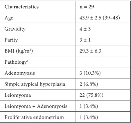
The characteristics and post-operative his-topathology results of the cases are presented in Table 1. The mean age of the women in the study was 43.9 ± 2.5; there was only one woman aged 48 and she had no premenopausal symptoms. Table 2 shows the baseline, post-operative first month and third month’s serum FSH (mIU/mL), E2 (pg/mL) and AMH (ng/mL) levels. There was no statistical-ly significant difference between the baseline and follow-up serum FSH and E2 levels; however, the differences between the baseline and post-opera-tive first month’s serum AMH levels were statisti-cally significant (p = 0.009).
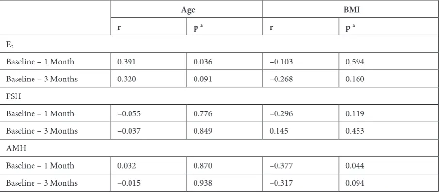
The correlation coefficient and materiality lev-els of BMI and age in relation to the baseline, post-operative first and third month changes in serum FSH, E2 and AMH levels are shown in Table 3. There was no statistically significant correlation between age, BMI and the changes in serum lev-els of FSH, E2 and AMH. The Bonferroni correc-tion was used and p < 0.017 was accepted as statis-tically significant.
Table 1. The demographic and clinical characteristics of the patients
Characteristics n = 29
Age 43.9 ± 2.5 (39–48)
Gravidity 4 ± 3
Parity 3 ± 1
BMI (kg/m2) 29.3 ± 6.3
Pathologya
Adenomyosis 3 (10.3%) Simple atypical hyperplasia 2 (6.8%) Leiomyoma 22 (75.8%) Leiomyoma + Adenomyosis 1 (3.4%) Proliferative endometrium 1 (3.4%)
Discussion
In this longitudinal prospective study, ovari-an function was evaluated in 29 women aged 39 to 48 years and subjected to TAH for benign diseases of the uterus. The serum levels of AMH, FSH and E2 were measured pre-operatively and in the first and third post-operative months, and transient changes in serum AMH levels were detected. Sub-sequently, all of the hormone levels reached nor-mal ranges by the third month.
AMH is a promising screening test for ovar-ian function. Low threshold values of AMH have a good specificity for poor ovarian reserve. FSH and E2 serum levels were also used to evaluate ovarian reserve. However, fluctuating levels of FSH and E2 may result in abnormal evaluations of ovarian function [3].
Conservation of the ovaries protects women from bone resorption in the premenopausal and postmenopausal period [10, 11]. Surgically in-duced menopause may also increase the risks of cardiovascular diseases and of psychosexual and cognitive dysfunction [12–14].
In some past studies, a decline in ovarian func-tion after hysterectomy was demonstrated; howev-er, some of those studies were designed retrospec-tively [15, 16]or were based on a questionnaire-style survey about climacteric complaints [17, 18].
Souza et al. [5] evaluated ovarian histology and function before and after TAH in 25 patients with symptomatic uterine fibroids. They did not find any change in the serum levels of hormones. His-tologically; however, they demonstrated a statisti-cally significant decrease in ovarian reserve.
In a rat model study, Ozdamar et al. report-ed ovarian histological changes after hysterectomy such as an increased number of atretic and cystic follicles and a decline in normal follicle count on the ovaries [19]. In another experimental rat mod-el study, Tapisiz et al. found histological chang-es in ovarichang-es that may adversely affect ovarian reserve [20].
In a recent prospective cohort study, Moor-man et al. evaluated ovarian function after hyster-ectomy and found risk ratios for ovarian failure to be 2.93 (95% CI 1.57–5.49) in women who un-derwent unilateral oophorectomy, and 1.74 (95% Table 2. Longitudinal changes in serum FSH, E2 and AMH levels
Baseline First Month Third Month p a
FSH (mIU/mL) 6.0 (4.3–7.9) 6.6 (4.6–9.7) 5.2 (4.4–6.5) 0.485 E2 (pg/mL) 107 (60–206.7) 102.4 (45.8–197.4) 127.8 (51.1–216.1) 0.519
AMH (ng/mL) 0.22 (0.16–1.49)b 0.18 (0.04–0.52)b 0.17 (0.04–0.69) 0.036
a – Friedman test; b – the difference between baseline and first-month AMH was found significant by using the Wilcoxon signed rank test (p = 0.009).
Table 3. Coefficient of correlation and materiality levels of BMI and age relative to the baseline, first and third post-opera-tive month changes in serum FSH, E2 and AMH levels
Age BMI
r p a r p a
E2
Baseline – 1 Month 0.391 0.036 –0.103 0.594
Baseline – 3 Months 0.320 0.091 –0.268 0.160
FSH
Baseline – 1 Month –0.055 0.776 –0.296 0.119
Baseline – 3 Months –0.037 0.849 0.145 0.453
AMH
Baseline – 1 Month 0.032 0.870 –0.377 0.044
Baseline – 3 Months –0.015 0.938 –0.317 0.094
CI 1.14–2.65) in women in whom both ovaries re-mained [21]. Farquar et al. investigated the asso-ciation between menopause and hysterectomy, and demonstrated that the onset of menopause in women who underwent hysterectomy retain-ing both ovaries was nearly four years earlier when compared to women with intact uteri and also that the onset of menopause in women who retained one ovary more than four years earlier than wom-en who retained both ovaries [22].
After hysterectomy, evaluation of ovarian ar-terial blood flow may be helpful to predict ovari-an function [23, 24]. Lee et al. [25] designed a pro-spective study to determine ovarian function after hysterectomy that evaluates both ovarian arterial blood flow indices and serum AMH levels. They stated that there is no association between hyster-ectomy and a decline in ovarian function for up to 3 months post-operatively. In another study, de-spite a decline in the ovarian blood flow in women who underwent hysterectomy, total antral follicle count and total ovarian volume were similar, and those results suggest changes in ovarian function after hysterectomy [26].
AMH is a valuable marker for assessing ovarian function. In a recent study, Atabekoglu et al. [27] investigated serum AMH levels during a 24-month follow-up period in women who had undergone TAH. They demonstrated a 30% loss of ovarian reserve in hysterectomized women when
compared with controls; however, these findings did not reach statistical significance.
In a prospective randomized trial, serum AMH levels were evaluated in subjects who had undergone uterine artery embolization and hysterectomy [28]. The mean AMH levels were significantly decreased at 6 weeks, but a recovery of AMH levels occurred during the 24-month post-operative follow-up peri-od. Those data are similar to the results of the current study, in which a significant difference was found in serum AMH levels when the baseline and first post-operative month were compares, but subsequently the difference decreased by the third month.
In this study, to prevent the probable adverse effects of ovarian diseases on ovarian functions, only cases with uterine pathologies were includ-ed in the study. The authors think that removal of the uterine arteries, which are an important blood supply to the ovaries, causes an acute decrease in serum AMH levels. In the recovery period, ovari-an arteries, which are the main blood supply to the ovaries, may compensate for the loss of the uterine arteries in the ovarian blood supply.
In the current study, transient changes in se-rum levels of AMH were found in the first post-operative month, which improved by the third post-operative month. The authors therefore con-clude that ovarian function is affected after TAH, but those effects are not permanent and recovery takes only a few months.
References
[1] Baskett TF: Hysterectomy: evolution and trends. Best Pract Res Clin Obstet Gynaecol 2005, 19, 295–305.
[2] Keshavarz HHS, Burney A, Marchbanks P: Hysterectomy surveillance – United States, 1994–1999. Surveillance Summaries MMWR 2002, 1–7.
[3] Vuorento T, Maenpaa J, Huhtaniemi I: Follow-up of ovarian endocrine function in premenopausal women after hysterectomy by daily measurements of salivary progesterone. Clin Endocrinol (Oxf) 1992, 36, 505–510.
[4] Siddle N, Sarrel P, Whitehead M: The effect of hysterectomy on the age at ovarian failure: identification of a sub-group of women with premature loss of ovarian function and literature review. Fertil Steril 1987, 47, 94–100.
[5] Souza AZ, Fonseca AM, Izzo VM, Clauzet RM, Salvatore CA: Ovarian histology and function after total abdomi-nal hysterectomy. Obstet Gynecol 1986, 68, 847–849.
[6] La Marca A, Volpe A: Anti-Müllerian hormone (AMH) in female reproduction: is measurement of circulating AMH a useful tool? Clin Endocrinol 2006, 64, 603–610.
[7] Hehenkamp WJ, Looman CW, Themmen AP, de Jong FH, Te Velde ER, Broekmans FJ: Anti-Mullerian hor-mone levels in the spontaneous menstrual cycle do not show substantial fluctuation. J Clin Endocrinol Metab 2006, 91, 4057–4063.
[8] La Marca A, Stabile G, Artenisio AC, Volpe A: Serum anti-Müllerian hormone throughout the human menstrual cycle. Hum Reprod 2006, 21, 3103–3107.
[9] Prior JC: Exercise-associated menstrual disturbances. In: Reproductive Endocrinology, Surgery and Technology. Eds.: Adashi EY, Rock JA, Rosenwaks Z. Raven Press, New York 1996, 1077–1091.
[10] Judd HL, Judd GE, Lucas WE, Yen SS: Endocrine function of the postmenopausal ovary: concentration of andro-gens and estroandro-gens in ovarian and peripheral vein blood. J Clin Endocrinol Metab 1974, 39, 1020–1024.
[11] Fogle RH, Stanczyk FZ, Zhang X, Paulson RJ: Ovarian androgen production in postmenopausal women. J Clin Endocrinol Metab 2007, 92, 3040–3043.
[12] Falkeborn M, Schairer C, Naessen T, Persson I: Risk of myocardial infarction after oophorectomy and hysterec-tomy. J Clin Epidemiol 2000, 53, 832–837.
[14] Rivera CM, Grossardt BR, Rhodes DJ, Brown Jr RD, Roger VL, Melton III LJ: Increased cardiovascular mortal-ity after early bilateral oophorectomy. Menopause 2009, 16, 15–23.
[15] Siddle N, Sarrel P, Whitehead M: The effect of hysterectomy on the age at ovarian failure: identification of a sub-group of women with premature loss of ovarian function and literature review. Fertil Steril 1987, 47, 94–100.
[16] Kaiser R, Kusche M, Würz H: Hormone levels in women after hysterectomy. Arch Gynecol Obstet 1989, 244, 169–173.
[17] Oldenhave A, Jaszmann LJ, Everaerd WT, Haspels AA: Hysterectomized women with ovarian conserva-tion report more severe climacteric complaints than do normal climacteric women of similar age. Am J Obstet Gynecol 1993, 168, 765–771.
[18] Riedel HH, Lehmann-Willenbrock E, Semm K: Ovarian failure phenomena after hysterectomy. J Reprod Med 1986, 31, 597–600.
[19] Ozdamar S, Ulger H, Sorkun HC, Muderris I: Effects of hysterectomy on ovarian morphology and serum FSH level in rats. Maturitas 2005 16, 52, 60–64.
[20] Tapisiz OL, Gungor T, Aytan H, Zergeroğlu S, Mulazimoglu B, Bilge U, Leyla Mollamahmutoglu L: Does hys-terectomy affect ovarian function? Histopathologic evaluation and serum FSH, inhibin A, and inhibin B levels in an experimental rat model. Eur J Obstet Gynecol Reprod Biol 2008, 140, 61–66.
[21] Moorman PG, Myers ER, Schildkraut JM, Iversen ES, Wang F, Warren N: Effect of Hysterectomy With Ovarian Preservation on Ovarian Function. Obstet Gynecol 2011, 118, 1271–1279.
[22] Farquhar CM, Sadler L, Harvey SA, Stewart AW: The association of hysterectomy and menopause: a prospective cohort study. BJOG 2005, 112, 956–962.
[23] Janson PO, Jansson I: The acute effect of hysterectomy on ovarian blood flow. Am J Obstet Gynecol 1977, 127, 349–352.
[24] Chalmers C: Does hysterectomy in a premenopausal woman affect ovarian function? Med Hypotheses 1996, 46, 573–575.
[25] Lee DY, Park HJ, Kim BG, Bae DS, Yoon BK, Choi D: Change in the ovarian environment after hysterectomy as assessed by ovarian arterial blood flow indices and serum anti-Mullerian hormone levels. Eur J Obstet Gynecol Reprod Biol 2010, 151, 82–85.
[26] van Rooij IA, Broekmans FJ, Te Velde ER: Serum anti-Mullerian hormone levels: a novel measure of ovarian reserve. Hum Reprod 2002, 17, 3065–3071.
[27] Atabekoglu C, Taskın S, Kahraman K, Gemici A, Taskın EA, Ozmen B, Berker B, Sonmezer M: The effect of total abdominal hysterectomy on serum anti-Mullerian hormone levels: a pilot study. Climacteric 2012, 15, 393–397.
[28] Hehenkamp WJ, Volkers NA, Broekmans FJ: Loss of ovarian reserve after uterine artery embolization: a random-ized comparison with hysterectomy. Hum Reprod 2007, 22, 1996–2005.
Address for correspondence:
Hasan Onur Topçu
Zekai Tahir Burak Women’s Health Education and Research Hospital Talatpasa Avenue, Hamamönü
Ankara Turkey
Tel: +90 532 635 95 38
E-mail: [email protected] Conflict of interest: None declared Received: 29.08.2013