Dominika Klimkiewicz-Blok
1, A–F, Jerzy Florjański
1, C, E, F,
Jerzy Zalewski
2, A, C, E, F, Radosław Blok
3, C–EAnalysis of the Concentrations of Interleukin 18
in Amniotic Fluid in the Second and the Third
Trimesters of Pregnancy
Analiza stężeń IL-18 w płynie owodniowym
w II i III trymestrze ciąży
1 Second Department of Gynecology and Obstetrics, Wroclaw Medical University, Poland
2 Department of Gynecology and Obstetrics, Division of Public Health, Wroclaw Medical University, Poland 3 First Department of Gynecology and Obstetrics, Wroclaw Medical University, Poland
A – research concept and design; B – collection and/or assembly of data; C – data analysis and interpretation;
D – writing the article; E – critical revision of the article; F – final approval of article; G – other
Abstract
Background. Interleukin 18 (IL-18) is a glycoprotein produced by macrophages. IL-18 influences different popu-lations of T lymphocytes and NK cells and stimulates the production of INF-gamma by these cells. IL-18 induces both Th1 and Th2 response. That is why IL-18 is a unique cytokine.
Objectives. The aim of work was to examine the concentration of interleukin 18 in amniotic fluid in the 2nd and the 3rd trimesters of physiological pregnancies.
Material and Methods. 74 pregnant women were qualified to take part in the studies. The amniotic fluid samples by amniocentesis were taken from the patients. Two groups were distinguished among the examined patients: group I – 45 pregnant women qualified for genetic amniocentesis between the 15th and 19th week of pregnancy. All findings of the cytogenetic tests were normal. Group II: 29 pregnant women in their 3rd trimester were qualified for diagnostic amniocentesis in order to determine the biological maturity of the fetuses. The concentration of IL-18 was marked with the immunoenzymatic method ELISA with the use of the kit produced by the MBL company. Method sensitivity was < 12.5 pg/mL.
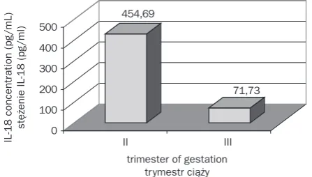
Results. In the 2nd trimester of pregnancy the average concentration of IL-18 in the amniotic fluid was 454.69 pg/mL and in the 3rd trimester was 71.73 pg/mL. The obtained data proved that the average concentration of IL-18 in the 2nd trimester was significantly higher than in the 3rd trimester. The obtained differences in the findings were statistically significant (p < 0.05).
Conclusions. The presence and high levels of IL-18 in the amniotic fluid in the 2nd trimester of pregnancy indi-cate an early process of initiation of immunological mechanisms by the fetus. An average concentration of IL-18 in the amniotic fluid was significantly higher in the 2nd trimester of pregnancy than in the 3rd trimester, which may indicate theinfluential role of IL-18 on the development of the immune response in the fetus in this period of gestation (Adv Clin Exp Med 2013, 22, 5, 699–703).
Key words: interleukin 18, amniotic fluid.
Streszczenie
Wprowadzenie. Interleukina 18 (IL-18) to glikoproteina wytwarzana głównie przez makrofagi. IL-18 oddziałuje na różne populacje limfocytów T i komórki NK oraz stymuluje wytwarzanie IFN-gamma przez te komórki. IL-18 indukuje zarówno odpowiedź typu komórkowego, jak i humoralnego. Pod tym względem IL-18 jest cytokiną uni-katową.
Cel pracy. Badanie stężeń IL-18 w płynie owodniowym w II i III trymestrze ciąż fizjologicznych.
Adv Clin Exp Med 2013, 22, 5, 699–703 ISSN 1899–5276
ORIGINAL PAPERS
Biological properties of IL-18 confirm the spe-cial function of this cytokine in the activation of physiological and pathological immunological mechanisms during pregnancy. IL-18 was identi-fied by Okamura et al. in 1995 as a factor inducing INF-gamma synthesis. Moreover, IL-18 was con-firmed to be related to Th1 response [1].
IL-18 is supposed to be released out of the cells which are influenced by apoptosis (as a re-sult of viruses contamination or influence of bac-terial toxins). For immunological resistance cells, IL-18 is one of the first signs of the beginning of infection [2–4]. The main source of IL-18 are mac-rophages. Moreover, IL-18 is produced by kerati-nocytes, most epithelial cells (pulmonal alveoli, in-testinal epithelial cells) and osteoblasts [5–8].
Functionally, IL-18 is close to IL-12. IL-18 in-fluences mainly different T lymphocyte popula-tions and NK cells. Moreover, IL-18 stimulates the production of interferon (IFN)-gamma by these cells – especially strong in the presence of IL-12. IL-18 strengthens the cytotoxicity of TCD8+ lympho-cytes, NK cells and CD4+ lymphocytes [1, 9–11].
It is interesting that IL-18 stimulates also se-cretion Th2 cytokines – IL-4, IL-3 by CD4+ lym-phocytes, NK cells and basophiles. Therefore, IL-18 induces both Th1 and Th2 response. That is why IL-18 is a unique cytokine [12, 13].
IL-18 plays a major role in the menstrual cy-cle and in the development of pregnancy. It has been shown that the level of IL-18 and its recep-tor increases rapidly in ovarium thecal cells, if it is stimulated by hormones (the maximal value in periovulation phase). IL-18 is necessary for proper ovary function [14].
Gutman’s et al. research confirmed that IL-18 takes part in the developmental process of ovula-tion and its influence on pathogenesis of ovaries hyperstimulation [15].
The role of IL-18 in early gestation was al-so confirmed by Zhang et al. research. The au-thors showed that IL-18 (through stimulation of IFN-gamma synthesis by decidual NK cells) takes
part in the process of vessels implantation and remodeling [16].
The cells of syncytiotrophoblast, chorioamni-on and fetal blood cells include the IL-18 receptor (IL-18R). The presence of receptor positive cells in decidua can be confirmed between the 6th and 10th week of gestation [17].
IL-18 also plays a role in pathogenesis arte-rial hypertension during pregnancy. Huang et al. showed higher values of IL-18 concentration in blood serum and placentas in pregnancies compli-cated by arterial hypertension [18].
However, reports about IL-18 values in blood serum during pregnancy were ambiguous. Accord-ing to the research conducted by Ida et al., the con-centration of IL-18 in blood serum during preg-nancy increasing gradually – since the 1st trimester until the 3rd trimester and even in the delivery [19]. But Kruse et al. reported that IL-18 concentration in blood serum in the 1st and 2nd trimester were lower than in non-pregnant women [20].
According to the research, IL-18 was present in the amniotic fluid and fetal membranes and its higher expression was connected with intrauterine infection, premature rupture of membranes and premature labor [21–24].
Material and Methods
The clinical material contained 74 samples of amniotic fluid. The samples were taken by way of amniocentesis from pregnant women. The materi-al was collected in the Clinic for Fetmateri-al Development Disorders of the Wroclaw Medical University dur-ing 2004–2006. The study obtained the acceptance of the Bioethical Committee of the Medical Uni-versity of Wroclaw.
The collected samples of amniotic fluid were divided into two groups. Group I – 45 pregnant women were qualified for genetic amniocentesis between the 15th and 19th week of pregnancy. The amniocentesis was carried out on the patients due
Materiał i metody. Do badań zakwalifikowano 74 ciężarne, od których pobrano płyn owodniowy za pomocą amniopunkcji. Wśród badanych pacjentek wyodrębniono 2 grupy: grupę I – 45 ciężarnych zakwalifikowanych do amniopunkcji genetycznej między 15 a 19 tyg.; wszystkie otrzymane wyniki badań cytogenetycznych były prawi-dłowe oraz grupę II – 29 ciężarnych w III trymestrze ciąży, które zakwalifikowano do amniopunkcji diagnostycznej, aby określić dojrzałość biologiczną płodów. Stężenia IL-18 oznaczano immunoenzymatycznie (ELISA), używając zestawu firmy MBL. Czułość metody wynosiła < 12,5 pg/ml.
Wyniki. W II trymestrze ciąży średnie stężenie IL-18 w wodach płodowych wyniosło 454,69 pg/ml, a w III try-mestrze 71,73 pg/ml. Średnie stężenie IL-18 w płynie owodniowym było istotnie większe w II trytry-mestrze ciąży. Uzyskane w wynikach różnice były istotne statystycznie (p < 0,05).
Wnioski. Obecność i duże stężenia IL-18 w wodach płodowych w II trymestrze ciąży świadczą o wczesnym procesie uruchomienia mechanizmów immunologicznych przez płód. Średnie stężenie IL-18 w płynie owodniowym jest istotnie większe w II trymestrze ciąży niż w III trymestrze, co może oznaczać, że IL-18 odgrywa istotną rolę w roz-woju odpowiedzi immunologicznej płodu w tym okresie ciąży (Adv Clin Exp Med 2013, 22, 5, 699–703).
to: the women’s age (over 35), incorrect results of biochemical tests of the mother’s blood and abnor-mal ultrasound screening.
The observed women were between 24 and 46 years of age (with an average of 38.5). The Genetic Laboratory of the Medical University of Wroclaw determined the kariotype of fetuses. The results of the fetal cytogenetic tests among the ex-amined fetuses were normal. Group II – 29 preg-nant women between the 36th and 40th week of pregnancy were qualified for diagnostic amnio-centesis with the aim of determining the biolog-ical maturity of the fetus. The observed women were between 22 and 38 years of age (with an av-erage of 28.5).
The amniotic fluid samples were centrifuged at a speed of 3,000 spins per min for 10 min and were then subjected to refrigeration at a tempera-ture of –82o Celsius. The concentration of IL-18
was marked with the ELISA method using a kit made by the MBL company. Method sensitivity was < 12.5 pg/mL. Absorbance value was read at a wavelength of 450 nm.
In all the analysed groups, Shapiro-Wilka test did not show abnormalities from normal distribu-tion. The calculations were performed with the use of the system STATISTICA 8.0, StatSoft, Inc. 2007. IL-18 concentration in the examined groups were compared with each other using the t-Student test for independent group. The value p < 0.05 proved to be statistically significant.
Results
Table 1 presents the comparison of IL-18 con-centration in the amniotic fluid of groups I and II – in the 2nd and 3rd trimesters of gestation. The average level of IL-18 in the amniotic fluid in the 2nd trimester of gestation was 454.69 pg/ mL, whereas in the third trimester it was 71.73 pg/ mL. These differences were found to be statistical-ly significant (p < 0.05) – Fig. 1.
Discussion
The number of researches appraising the lev-el of IL-18 in amniotic fluid was not large. In the available literature there was an analysis of IL-18 concentration in amniotic fluid in normal preg-nancies and pregpreg-nancies complicated by symp-toms of threatening preterm labour or premature rupture of fetal membranes [21–23].
Pacora et al. were the first who described the presence of IL-18 in amniotic fluid, in the umbili-cal blood and in the serum of pregnant women. The authors showed that IL-18 concentration in amni-otic fluid increased during pregnancy, which might show the maturing of the fetus’ immune system and the important role of IL-18 in a normal preg-nancy (2nd trimester: median 6.2 pg/mL; pregnan-cy in term: median 14.9 pg/mL; p < 0.0001) [23]. The results of our research were completely dif-ferent from these shown by Pacora et al., because the IL-18 level in amniotic fluid had a decreasing trend and amounts: median 416.33 pg/mL – 2nd
Table 1. The level of concentration of IL-18 in the amniotic fluid in first and second examined groups
Tabela 1. Stężenie IL-18 w wodach płodowych w badanych grupach
Groups
(Grupy) IL-18 (pg/mL) (Wartość p-value p)
average
(średnia) ± SD(odchylenie standardowe) min.–max.(minimum–maksimum)
Group I (Grupa I)
(n = 45) 454.69 228.21 129.66–1118.83 p < 0.05
Group II (Grupa II)
(n = 29) 71.73 84.24 22.58–420.91
Fig. 1. Comparison of average concentration IL- 18 in the amniotic fluid in the first and second examined groups
p < 0,05, ± SD Group I – 228.21, ± SD Group II – 84.24.
Ryc. 1. Porównanie średnich wartości stężeń IL-18 w wodach płodowych w I i II badanej grupie
p < 0,05, ± SD grupa I – 228,21, ± SD grupa II – 84,24.
454,69
71,73
0 100 200 300 400 500
IL−18 concentration (pg/mL)
stężenie IL−18 (pg/ml)
II III
trimester, median 44.66 pg/mL – 3rd trimester (p < 0.05). The differences concern also the results of IL-18 concentration which were repeatedly high-er in our research compared to the results shown by Pacora et al., both in pregnancies complicated by infections and in normal pregnancies.
Jacobson et al. also tested IL-18 concentra-tion in amniotic fluid in pregnancies complicated by preterm labour or the premature rupture of the fetal membrane and in normal pregnancies [21]. In pregnancies threatened with preterm labour the authors reported relation between increasing IL-18 level in amniotic fluid (median 1.01ng/mL; p = 0.046) and bacterial infection in amniotic flu-id and the time of delivery (labour during 7 days). The results proved that IL-18 was one of important elements of fetus immunological response. In preg-nancies carried to full term, the presence of IL-18 in amniotic fluid was confirmed in just 11% (3/28) of cases [21]. In our research IL-18 was present in all tested probes of amniotic fluid (n = 29) taken between 36–40 week of pregnancy, and its concen-tration was between 3.83 pg/mL – 420.91 pg/mL; median – 0.44 ng/mL.
As well Menon et al. confirmed the increase of IL-18 concentration in amniotic fluid, both in pregnancies complicated by preterm labour or premature rupture of fetal membrane [22]. In our
research, IL-18 median in amniotic fluid in preg-nancies carried to full term (38–40 weeks) was 0.46 ng/mL and was 2.9-fold higher than for preg-nancies carried to full term as presented by Menon et al. (median 0.16ng/mL) [22].
As well Lemancewicz and al. showed the pres-ence of IL-18 in amniotic fluid in pregnancies com-plicated by preterm labour (median 720 pg/mL) and in pregnancies carried to full term (median – 268 pg/mL) (200). In our research, IL-18 con-centration in amniotic fluid in the third trimester of pregnancy (36–40 week) were much lower (me-dian 44.66 pg/mL) comparing to the results de-scribed by Lemancewicz et al. [25].
The analysis of available literature and our re-search results demonstrates inconsistencies of the IL-18 level in amniotic fluid during pregnancy. Only Pacora’s et al. research estimated IL-18 con-centration in amniotic fluid in the second trimes-ter [23]. The high IL-18 concentration level in am-niotic fluid in the second trimester of pregnancy (15–19 week) obtained in our research could con-firm an important function of IL-18 in immuno-logical response during this time of pregnancy.
Biological properties of IL-18 confirmed the special role of this cytokin in the activation of phys-iological and pathological immunological mecha-nisms during pregnancy.
References
[1] Okamura H, Tsutsi H, Komatsu T, Yutsudo M, Hakura A, Tanimoto T, Torigoe K, Okura T, Nukada Y, Hattori K: Cloning of a new cytokine that induces IFN-gamma production by T cells. Nature 1995, 378, 88–91.
[2] Tsutsui H, Kayagaki N, Kuida K, Nakano H, Hayashi N, Takeda K, Matsui K, Kashiwamura S, Hada T, Akira S, Yagita H, Okamura H, Nakanishi K: Caspase-1-independent, Fas/Fas ligand-mediated IL-18 secretion from mac-rophages causes acute liver injury in mice. Immunity 1999, 11, 359–367.
[3] Csernok EM, Schmitt W, Bainton DF, Gross WL: Activated neutrophils express proteinase 3 on their plasma membrane in vitro and in vivo. Clin Exp Immunol 1994, 95, 244–250.
[4] Felderhoff-Mueser U, Schmidt OI, Oberholzer A, Bührer C, Stahel PF: IL-18: a key player in neuroinflammation and neurodegeneration? Trends Neurosciences 2005, 28, 487–493.
[5] Stoll S, Müller G, Kurimoto M, Saloga J, Tanimoto T, Yamauchi H, Okamura H, Knop J, Enk AH: Production of IL-18 (↓IFN-gamma-inducing factor↓) messenger RNA and functional protein by murine keratinocytes. J Immunol 1997, 159, 298–302.
[6] Takeuchi M, Nishizaki Y, Sano O, Ohta T, Ikeda M, Kurimoto M: Immunohistochemical and immuno-electron-microscopic detection of interferon gamma-inducing factor (interleukin-18) in mouse intestinal epithelial cells. Cell Tissue Res 1997, 289, 499–503.
[7] Cameron LA, Taha RA, Tsicopoulos A, Kurimoto M, Olivenstein R, Wallaert B, Minshall EM, Hamid QA:
Airway epithelium expresses interleukin-18. Eur Respir J 1999, 14, 553–559.
[8] Udagawa N, Horwood NJ, Elliott J, Mackay A, Owens J, Okamura H, Kurimoto M, Chambers TJ, Martin TJ, Gillespie MT: Interleukin-18 (interferon gamma inducing factor) is produced by osteoblasts and acts via granulo-cyte – macrophage colony stimulating factor and not via interferon – gamma to inhibit osteoclast formation. J Exp Med 1997, 185, 1005–1012.
[9] Yoshimoto T, Takeda K, Tanaka T, Ohkusu K, Kashiwamura S, Okamura H, Akira S, Nakanishi K: IL-12 up-regulates IL-18 receptor expression on T cells, Th1 cells and B cells: synergism with IL-18 for IFN gamma produc-tion. J Immunol 1998, 161, 3400–3407.
[10] Okamura H, Kashiwamura S, Tsutsui H, Yoshimoto T, Nakanishi K: Regulation of interferon-gamma produc-tion by IL-12 and IL-18. Curr Opin Immunol 1998, 10, 259–264.
[12] Nakanishi K, Yoshimoto T, Tsutsui H, Okamura H: Interleukin-18 is unique that stimulates both Th1 and Th2 responses depending on its cytokine milieu. Cytokine Growth Factor Rev 2001, 12, 53–72.
[13] Yoshimoto T, Mizutani H, Tsutsui H, Noben-Trauth N, Yamanaka K, Tanaka M, Izumi S, Okamura H, Paul WE, Nakanishi K: IL-18 induction of IgE: dependence on CD4+ T cells, IL-4 and STAT6. Nat Immunol 2000, 1, 132–137.
[14] Tsuji Y, Tamaoki TH, Hasegawa A, Kashiwamura S, Iemoto A, Ueda H, Muranaka J, Adachi S, Furuyama J, Okamura H, Koyama K: Expression of interleukin-18 and its receptor in mouse ovary. Am J Reprod Immunol 2001, 46, 349–357.
[15] Gutman G, Soussan-Gutman L, Malcov M, Lessing JB, Amit A, Azem F: Interleukin-18 is high in the serum of IVF pregnancies with ovarian hyperstimulation syndrome. AJRI 2004, 51, 381–384.
[16] Zhang JH, Borzychowski AM, Takeda K, Akira S, Croy BA: Analysis of cytokine regulators inducing interferon production by mouse uterine natural killer cells. Biol Reprod 2003, 69, 404–411.
[17] Tokmadzić VS, Tsuji Y, Bogović T, Laskarin G, Cupurdija K, Strbo N, Koyama K, Okamura H, Podack ER, Rukavina D: IL-18 is present at the maternal-fetal interface and enhances cytotoxic activity of decidual lympho-cytes. Am J Reprod Immunol 2002, 48, 191–200.
[18] Huang X, Huang H, Dong M, Yao Q, Wang H: Serum and placental interleuki-18 are elevated in preeclampsia. J Reprod Immunol 2005, 65, 77–87.
[19] Ida A, Tsuji Y, Muranaka J, Kanazawa R, Nakata Y, Adachi S, Okamura H, Koyama K: IL-18 in pregnancy; the elevation of IL-18 in maternal peripheral blood during labour and complicated pregnancies. J Reprod Immunol 2000, 47, 65–74.
[20] Kruse N, Greif M, Moriabadi NF, Marx L, Toyka KV, Rieckmann P: Variation in cytokine mRNA expression during normal pregnancy. Clin Exp Immunol 2000, 119, 317–322.
[21] Jacobsson B, Holst RM, Mattsby-Baltzer I, Nikolaitchouk N, Wennerholm UB, Hagberg H: Interleukin-18 in cervical mucus and amniotic fluid: relationship to microbial invasion of the amniotic fluid, intra-amniotic inflam-mation and pretem delivery. Int J Obstet Gynecol 2003, 110, 598–603.
[22] Menon R, Lombardii SJ, Fortunato SJ: IL-18, a product of choriodecidual cells, increase during premature rup-ture of membranes but fails to turn on the Fas-FasL- mediated apoptosis pathway. J Assist Reprod Gen 2001, 18, 276–280.
[23] Pacora P, Romero R, Maymon E, Gervasi MT, Gomez R, Edwin SS, Yoon BH: Participation of the novel cyto-kine interleukin 18 in the host response to intra-amniotic infection. Am J Obstet Gynecol 2000, 183, 1138–1143.
[24] Splíchalová A, Trebichavský I, Muneta Y, Mori Y, Splíchal I: Effect of bacterial virulence on IL-18 expression in the amnion infected with Echerichia coli. Am J Reprod Immunol 2005, 53, 255–260.
[25] Lemancewicz A, Urban R, Urban J, Skotnicki M, Kretowska M, Sierakowski S: Evaluation of interleukin con-centrations in amniotic fluid in preterm and term parturition and in oligohydramnion. Med Sci Monit 2001, 7, 924–927.
Address for correspondence:
Dominika Klimkiewicz-Blok
II Department of Gynecology and Obstetrics Wroclaw Medical University Borowska 213
50-528 Wrocław Poland
Tel.: +48 071 733 14 00 Fax: +48 071 733 14 09 E-mail: [email protected]
Conflict of interest: None declared