Akram Beheshti
1, A, C, F, Younes Shafigh
2, A, B, Hossien Parsa
2, E, F,
Amir A. Zangivand
1, DComparison of High-Frequency
and MIST Ultrasound Therapy
for the Healing of Venous Leg Ulcers
1 Department of Dermatology, Qazvin University of Medical Sciences, Qazvin, Iran 2 Department of General Surgery, Qazvin University of Medical Sciences, Qazvin, Iran
A – research concept and design; B – collection and/or assembly of data; C – data analysis and interpretation;
D – writing the article; E – critical revision of the article; F – final approval of article; G – other
Abstract
Background. Venous leg ulcers (VLUs) are a health problem in clinical care. Several options can be employed as adjuvant to standard treatment.
Objectives. We have aimed to analyze the effect of standard ulcer care alone with high-frequency ultrasound (HFU) and MIST ultrasound therapy on VLUs.
Material and Methods. Ninety patients with VLUs were assigned into the standard treatment, HFU and MIST ultrasound groups. All groups received the standard wound care. In the ultrasound groups, HFU and MIST ultra-sound therapy was administered to wounds 3 times per week until the wound healed. Time of complete wound healing was recorded. Wound size, pain, and edema were assessed at baseline and after 2 and 4 months. Also, patients were instructed to contact our clinic monthly, and recurrence of VLUs was recorded for 6 months after complete wound healing. The data was analyzed using a Student’s t-test, ANOVA, c2, or Fisher’s exact test. P < 0.05 was considered significant.
Results. Mean time duration of complete wound healing in the first, second and third groups was 8.13 (SD 1.40), 6.10 (SD 1.47) and 5.70 (SD 1.57) months, respectively (p < 0.0001). Size of ulcer, mean degree of pain and edema in ultrasound therapy was decreased after the 4-month visit in comparison to the standard-treatment group (p = 0.01, p < 0.0001 and p < 0.0001, respectively). Also, our results don’t show any significant differences between groups in the recurrence of VLUs during a 6-month follow up after complete wound healing (p = 0.37).
Conclusions. Our results in the present study show the significant effectiveness of ultrasound therapy in wound healing. Differences between the two ultrasound therapy groups were not statistically significant (Adv Clin Exp Med 2014, 23, 6, 969–975).
Key words: ultrasound therapy, venous leg ulcers.
Adv Clin Exp Med 2014, 23, 6, 969–975 ISSN 1899–5276
ORIGINAL PAPERS
© Copyright by Wroclaw Medical University
Venous leg ulcers (VLUs) are wounds with long healing time and frequent recurrence in elder-ly adult clinics and affect approximateelder-ly 1.1 to 1% of the world’s population [1, 2]. Several hypothe-ses may help explain the origin of VLUs including insufficient veins or valves (dysfunctional valves in the veins that allow backward blood recirculation due to incomplete valve closure) or impaired cle function which may lead to abnormal calf mus-cle pump function (elevated ambulatory venous pressure). These changes subsequently result in lo-cal venous dilatation and pooling, concomitantly
trapping leukocytes that may release proteolytic enzymes that destroy tissue. Venous pooling also induces inter endothelial pore widening and de-position of fibrin and other macromolecules that “trap” growth factors within them, rendering them unavailable for wound repair [3–6].
grafts, tissue engineered skin, growth factor thera-py and surgery [8].
Ultrasound has been used as a therapeutic mo-dality for nearly 50 years [9]. In recent years, ul-trasound therapy has been utilized for the man-agement of chronic wounds in some centers [10]. Although high frequency ultrasound (HFU) (1–3 MHz) has been used in clinical practice in most studies, and shown to promote healing of some injuries [11–13], it can cause burns or endo-thelial injury and usage of it is limited in medical practice. On the other hand, several experiments using ultrasound have shown that the application of low doses in the treatment of skin wounds are more effective in wound healing than high dose ultrasound [14].
Thus, noncontact ultrasound therapy is among the newer modalities. Operating at a markedly low-er frequency (40 kHz), it was approved for use in the wound care setting by the FDA in 2004 [15].
In our country, due to lifestyle modifications, ageing of the Iranian population and numerous chronic co-morbid conditions such as coronary heart disease, essential hypertension, diabetes mel-litus, obesity, immobility, peripheral arterial dis-ease, neuropathy etc., management of VLUs is es-timated to become a leading cost on our national health system.
To control the previously described abnormal-ities and decrease further costly medical or surgi-cal investigations, we need to identify accurate and appropriate strategies.
The focus of this study was to compare the ef-fect of standard ulcer care alone, with HFU and with noncontact ultrasound therapy. Therefore, in a com-parative study, we measured the mean time duration of complete wound healing, edema, pain, size of ul-cers and recurrence rate of VLUs in all groups.
Material and Methods
Protocol of the Study
From April 2011 to August 2012, 90 patients diagnosed with VLUs were enrolled in this study after obtaining informed consent. All chosen pa-tients received wound care in only one hospital- -based, outpatient wound program that was locat-ed at the vascular clinic of Shahid Rajaii Hospital (Qazvin, Iran).
The exclusion criterion were allergy to ultra-sound contact gel, pregnancy, or with any of the known contraindications to ultrasound includ-ing ankle or knee prosthesis or metal in the lower leg, suspected or confirmed local cancer or meta-static disease and neuropathy, no clinical evidence
of infections including active cellulites, suspicious thrombophlebitis and no history of antibiotic ther-apy at the time of enrollment. The original protocol also stated the study would not recruit people with peripheral arterial disease, diabetes or rheumatoid arthritis. VLUs that had the following characteris-tics were included for study: wound duration lon-ger than 4 weeks and no clinical improvement after using the clinic’s standard care (SOC) for healing during a 2 week period [16].
All patients were randomly assigned into the standard treatment group, HFU and MIST ultra-sound group. Randomization was performed by means of sealed opaque envelopes containing com-puter generated random numbers. In the first clin-ical visit, a baseline assessment of wound size, pain and edema was performed, and also treatments starting in this visit. All groups received compres-sion therapy as the standard of wound care. In the ultrasound groups, HFU therapy and MIST thera-py were administered to wounds 3 times per week until the wound healed.
Afterwards, monthly clinical visits were per-formed and the size of the ulcer, pain and edema were recorded at 2nd and 4th month after the initial
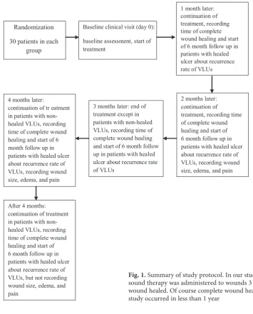
study. Also, the time duration of complete wound healing was recorded during the monthly visit in this study. After complete healing in each patient, a 6-month clinical follow up was performed for the patient and the rate of recurrence of VLUs calculat-ed (Fig. 1 summarizes the protocol of the study).
Ultrasound Therapy
HFU therapy was applied with a SoLo Thera-sonic 355 machine (EMS Physio, Wantage, UK). The ultrasound transducer head was sterilized with alcohol wipes. Ultrasound was then applied to the skin surrounding the reference ulcer, us-ing a water based contact gel recommended by the manufacturer, for 5–10 min by moving the trans-ducer head in a slow, controlled manner around the edges of the ulcer in overlapping circles to cov-er the skin evenly. Ulccov-ers of area < 5 cm2 received
ultrasound for 5 min, those of ≥ 10 cm2 received
10 minutes’ ultrasound. For ulcers between 5 cm2
and 10 cm2, treatment time in min equaled the
ul-cer area in cm² (ulul-cer of 7 cm2 area = 7 minutes’
treatment).
The MIST therapy system noncontact ultra-sound device delivers low-intensity (0.1–0.8 W/cm2),
low-frequency (40 kHz) ultrasound energy via at-omized, sterile saline mist to the wound bed with-out directly contacting the body or the wound.
a valve that controls the flow of saline to the trans-ducer surface. The product’s recommended treat-ment algorithm is based on longer treattreat-ment times for greater total ulcer area. At our facility, the pro-tocol is to treat wounds up to 4 cm2 with 4 min
of MIST therapy; larger wounds receive longer treatment times (max treatment time = 12 min for wounds larger than 10 cm2).
Data Collection and Variable
Definition
Pain was assessed by a numerical rating scale in which the patient was instructed to choose a num-ber from 0 for “without pain” to 20 for “unbearable pain” [17]. To assess leg edema [18], the examin-er pressed his fingexamin-ertip against a bony prominence for 5 s, and then removed it. A residual indenta-tion indicated pitting edema, which was graded on a scale of 1 (mild) to 4 (severe).
Ethical Approval
The study was approved by the ethics commit-tee of the university before its initiation, and the protocols used conformed to the ethical guidelines of the 1975 Helsinki Declaration.
Statistical Analysis
The statistical evaluation was performed by computer analysis with SPSS Software (Statisti-cal Package for the Social Sciences, version 11.0, SPSS Inc, Chicago, Ill, USA). The Student’s t test, ANOVA, c2, or Fisher’s exact test were used, where appropriate, for comparing clinical data be-tween all groups. Continuous data was recorded as mean ± standard deviation. P value less than 0.05 was considered significant.
Fig. 1. Summary of study protocol. In our study, HFU and MIST ultra-sound therapy was administered to wounds 3 times per week until wound healed. Of course complete wound healing in all patients in this study occurred in less than 1 year
Randomization
30 patients in each group
Baseline clinical visit (day 0):
baseline assessment, start of treatment
2 months later: continuation of
treatment, recording time of complete wound healing and start of 6 month follow up in patients with healed ulcer about recurrence rate of VLUs, recording wound size, edema, and pain 3 months later: end of
treatment except in patients with non-healed VLUs, recording time of complete wound healing and start of 6 month follow up in patients with healed ulcer about recurrence rate of VLUs
4 months later:
continuation of tr eatment in patients with non-healed VLUs, recording time of complete wound healing and start of 6 month follow up in patients with healed ulcer about recurrence rate of VLUs, recording wound size, edema, and pain
After 4 months: continuation of treatment in patients with non-healed VLUs, recording time of complete wound healing and start of 6 month follow up in patients with healed ulcer about recurrence rate of VLUs, but not recording wound size, edema, and pain
Results
Ninety patients diagnosed with VLUs (46 men and 44 women), aged 58.5 (SD 11.6) took part in this study. Inspections of background characteristics between study groups showed a generally good bal-ance of the demographic and clinical characteristics collected and mean age of patients, wound duration and mean initial size of ulcer, and even the distribu-tion of men and women was not significant.
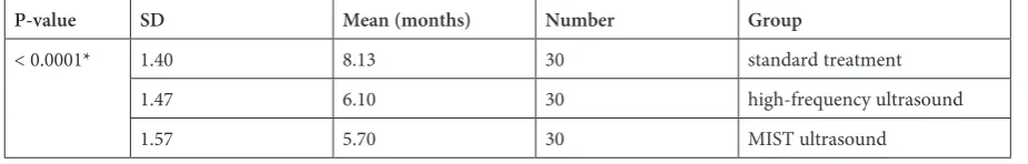
Mean time duration of complete wound heal-ing in the first, second and third groups was 8.13 (SD 1.40), 6.10 (SD 1.47) and 5.70 (SD 1.57) months, respectively (p < 0.0001; Table1). The results be-tween the duration of complete wound healing in the ultrasound treatment groups was not a statisti-cally significant difference (p = 0.22; Table 1).
Edema at the first clinical visit was mild to se-vere in all groups and after treatment had subsid-ed in all groups. In spite of the fact that subsid-edema was not statistically significantly different in all groups at baseline and 2-month visits, after 4 months the edema was more subsided in the 2nd and 3rd groups
in comparison to the standard treatment, and re-covery from edema was statistically significantly better in the ultrasound groups (p = 0.02; Table 2). Also, the assessment of leg edema between HFU
and MIST ultrasound therapy didn’t show any sig-nificant differences.
The mean degree of pain was recorded in all groups and changes in pain after 2 and 4 months were shown in this study. Our results indicate the mean degree of pain decreased more in the 2nd and
3rd groups and these decreases was statistically
sig-nificant (p < 0.0001; Table 3). There were not any significant differences between HFU and MIST ul-trasound therapy. The analysis of wound surface and size of ulcer showed the mean size of the ul-cer at the 1st clinical visit and 2 months after the
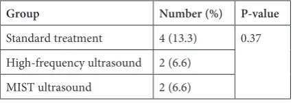
baseline clinical visit were not statistically signif-icantly different, but after 4 months our results showed significant differences (p = 0.01; Table 4). On the other hand, our results showed VLU recur-rence 6 months after complete wound healing was seen in 4 cases in the standard group, 2 cases in the HFU and 2 cases in the MIST ultrasound group (p = 0.37; Table 5).
Discussion
VLUs are one of the main burdens for pa-tients and healthcare service centers. Traditional wound healing intervention such as compression
Table 1. Mean time duration of complete wound healing
P-value SD Mean (months) Number Group
< 0.0001* 1.40 8.13 30 standard treatment
1.47 6.10 30 high-frequency ultrasound
1.57 5.70 30 MIST ultrasound
* p-value between high frequency ultrasound and MIST ultrasound was calculated at 0.22.
Table 2. Changes in edema
P-value Staging of edema Number of patients
in each group Time
4 plus 3 plus 2 plus 1 plus
0.31 7 8 5 10 standard treatment visit 1
3 8 7 12 high-frequency ultrasound
8 8 5 9 MIST ultrasound
0.64 5 6 7 12 standard treatment 2 months
after
3 4 9 14 high-frequency ultrasound
5 4 7 14 MIST ultrasound
0.02* 5 5 12 8 standard treatment 4 months
after
1 1 5 23 high-frequency ultrasound
4 2 3 21 MIST ultrasound
bandages are the mainstay and standard treatment for chronic venous ulcers. Today, several system-ic adjunctive treatments, for example applsystem-ication of ultrasound etc., may be used in conjunction with compression therapy. HFU has been used in clinical practice in musculoskeletal disorders, pri-marily by physical therapists, wound healing, and sports medicine with both thermal and mechanical
effects for many years. The therapeutic effect of ul-trasound therapy in the kilohertz (low frequency) range has been approved for use in the wound care setting in recent years.
The main effect of low frequency ultrasound is a mechanical property [19]. Also, it has been pro-posed that low frequency ultrasound in the KHz range may improve wound healing via the pro-duction, vibration, and movement of micron-sized bubbles in the coupling medium and tissue.
The results of the present study show thatthe size of the ulcers in ultrasound therapy were small-er in the visits aftsmall-er 4 months. Also, mean time du-ration of complete wound healing 4 months af-ter the initial study was very fast in comparison to standard treatment alone. However, these dif-ferences were not significantly different between HFU with MIST therapy.
Table 3. Changes in pain
P-value SD Mean (cm2) Number of patients Group Time
0.38 3.69 9.90 30 standard treatment visit 1
2.52 8.50 30 high-frequency ultrasound
5.23 9.43 30 MIST ultrasound
< 0.0001* 3.54 7.80 30 standard treatment 2 months after 2.30 4.93 30 high-frequency ultrasound
3.19 4.46 30 MIST ultrasound
< 0.0001* 2.09 6.56 30 standard treatment 4 months after 2.12 4.20 30 high-frequency ultrasound
2.70 4.20 30 MIST ultrasound
* p-value between high frequency ultrasound and MIST ultrasound therapy in visit 1, 2 months after and 4 months after was calculated at 0.38, 0.16 and 0.98, respectively.
Table 4. Changes in mean of ulcer size
P-value SD Mean (cm2) Number of patients Group Time
0.98 3.07 8.96 30 standard treatment visit 1
2.37 9.03 30 high-frequency ultrasound
2.27 9.10 30 MIST ultrasound
0.16 2.42 5.63 30 standard treatment 2 months after
2.35 4.76 30 high-frequency ultrasound
3.19 4.46 30 MIST ultrasound
0.01* 2.95 4.80 30 standard treatment 4 months after
2.16 3.70 30 high-frequency ultrasound
1.93 3.30 30 MIST ultrasound
* p-value between high frequency ultrasound and MIST ultrasound therapy in visit 1, 2 months after and 4 months after was calculated 0.91, 0.68 and 0.45, respectively.
Table 5. Recurrence of VLUs after 6-month follow up
Group Number (%) P-value
Standard treatment 4 (13.3) 0.37 High-frequency ultrasound 2 (6.6)
Our results emphasize similar results in oth-er studies. For example, in a randomized, con-trolled, double-blinded study, Ennis et al. exam-ined the effectiveness of MIST ultrasound therapy after 12 weeks of care for the healing of recalci-trant diabetic foot ulcers. The authors concluded, the proportion of wounds healed in the active ul-trasound therapy device group was significantly higher than that in the control group (40.7% vs. 14.3%, p = 0.0366, Fisher’s exact test) [20].
Also Ennis et al., in another non-comparative study, used MIST ultrasound during an 8-month period and ultimately concluded that 69% of the wounds were healed and median time to healing was 7 weeks when MIST ultrasound was used as a stand-alone therapy [19].
In another study, Kavros et al. assessed MIST ultrasound therapy in the treatment of non-healing leg and foot ulcers associated with chronic critical limb ischemia. The subjects included 35 patients who received MIST ultrasound therapy plus the standard of wound care for 12 weeks (treatment group) and 35 patients who received the standard of wound care alone (control group). The main out-come measurements showed a significantly higher percentage of patients treated with the treatment group achieved greater than 50% wound healing at 12 weeks than those treated with the standard of care alone (63% vs. 29%; p < 0.001) [21]. Also, this author et al. in another study indicated healing time reductions (9.8 ± 5.5 weeks vs. 5.5 ± 2.8 weeks (p < 0.0001)) and wound volume percent improve-ment (37.3% ± 18.6% vs. 94.9% ± 9.8% (p < 0.0001)) in comparing the clinic’s standard care with MIST ultrasound therapy [22].
Our results about changes in pain after 2 and 4 months of the initial of study also showed decreas-es in the ultrasound treatment groups in compari-son to standard treatment alone. Of course in this item, the differences were not significant between MIST and HFU therapy. In similar literature, Geh-ling and Samies reviewed and recorded pain scores of 15 consecutive patients (7 men and 8 women, age range of 28 to 88 years) with painful,
non-healing, lower-extremity wounds treated for 2 to 4 weeks with MIST ultrasound therapy. Mean pain scores decreased from 8.07 ± 1.91 pre-treatment to 1.67 ± 1.76 post-treatment (p = 0.0003) [23].
The recurrence rate of VLUs in our study was 13.3% in the standard treatment group and 6.6% in the ultrasound group. The recurrence rate of this disorder in previous studies was variable between 26% and 69% [24]. Our results were lower than similar studies but we believe the 6-month follow up is very short to truly decide about the potential of prevention of recurrence in patients treated with ultrasound therapy.
Overall, our results in the present study are similar to other published literature in the world and show the significant effectiveness of ultrasound therapy, especially MIST therapy, in wound healing as an adjuvant therapy. This method prepares the wound bed for healing by reducing the bioburden, enhancing angiogenesis, assisting in debridement of necrotic and devitalized tissues, and stimulating cellular activity. From another perspective, the tis-sue repair and wound healing process has 3 phases: inflammatory, proliferative and remodeling. Appli-cation of the MIST therapy system for tissue repair in the initial inflammatory stage could cause a pro-motion of it. However, we bear in mind that the MIST therapy system does not have only anti-in-flammatory properties. In the proliferative phase, it also affects ultrasound induced edema resolution, cellular element migration and division, accelerat-ed granulation tissue formation and stimulataccelerat-ed fi-broblasts for collagen production. At the last phase of wound healing, the scar tissue that is exposed to the MIST therapy system may be stronger and more elastic compared to normal scar tissue.
In conclusion, according to the very limited ef-fects identified in individuals in the MIST py group, which showed earlier response to thera-py based on wound area and volume reductions, it could give us a cost savings through a prominent reduction in therapeutic times. Additional work on cost-effective outcomes and planning are great-ly needed for the future.
References
[1] Moffatt C, Dorman M: Recurrence of leg ulcers within a community leg ulcer service. J Wound Care 1995, 4, 57–61.
[2] Grindlay A, MacLellan D: Inpatient management of leg ulcers: A costly option? Prim Intent 1997, 5, 24–26.
[3] Wienert V, Vanscheidt W, Rabe E: Leg ulcer due to venous insufficiency. In: Westerhof W. Leg ulcers: diagnosis and treatment. 1st ed. Amsterdam: Elsevier Science Publishers 1993, 83–113.
[4] Cherry GW, Hofman D, Cameron J, Poore SM: Bandaging in the treatment of venous ulcers. A European view. Ostomy Wound Manage 1996, 42, 13–18.
[5] Ryan TJ: The management of leg ulcers. 2nd ed. Oxford: Oxford University Press 1987, 1–101.
[6] Stücker M, Reich S, Robak-Pawelczyk B: Changes in venous refilling time from childhood to adulthood in sub-jects with apparently normal veins. J Vasc Surg 2005, 41, 296–302.
[8] Trent JT, Falabella A, Eaglstein WH, Kirsner RS: Venous ulcers: pathophysiology and treatment options. Ostomy Wound Manage 2005, 51, 38–56.
[9] Hill OR: Ultrasound biophysics: a perspective. Br J Cancer 1982, 82, 46–51.
[10] Moffatt C, Martin R, Smithdale R: Leg ulcer management. Blackwell Publishing Ltd 2007.
[11] Kibler WB, Duerler K: Electrical stimulation and application of heat.In: DeLee & Drez’s Orthopaedic Sports Medicine: Principles and Practice. Eds.: DeLee J, Drez D, Miller MD, Saunders, Philadelphia 2003, 2nd ed., 349–351, 356–359.
[12] Cameron MH: Thermal agents: cold and heat, ultrasound, and electrical currents. In: Physical Agents in Rehabilitation: From Research to Practice. Ed.: Cameron MH, Saunders, St. Louis 2003, 2nd ed., 133–259.
[13] Busse JW, Bhandari M, Kulkarni AV, Tunks E: The effect of low-intensity pulsed ultrasound therapy on time to fracture healing: a meta-analysis. CMAJ 2002, 166, 437–441.
[14] Ernst E: Ultrasound for cutaneous wound healing. Phlebology 1995, 10, 2–4.
[15] Unger PG: Low-frequency, noncontact, nonthermal ultrasound therapy: a review of the literature. Ostomy Wound Manage 2008, 54, 57–60.
[16] Ennis WJ, Meneses P: Comprehensive wound assessment and treatment system. In: Falabella A, Kirsner R, eds. Wound Healing. Boca Raton, FL: Taylor and Francis 2005, 59–68.
[17] Chibnall JT, Tait RC: Pain assessment in cognitively impaired and unimpaired older adults: a comparison of four scales. Pain2001, 92, 173–186.
[18] Brodovicz KG, McNaughton K, Uemura N, Meininger G, Girman CJ, Yale SH: Reliability and feasibility of methods to quantitatively assess peripheral edema. Clin Med Res 2009, 7, 21–31.
[19] Ennis WJ, Valdes W, Gainer M, Meneses P: Evaluation of clinical effectiveness of MIST ultrasound therapy for the healing of chronic wounds. Adv Skin Wound Care 2006, 19, 437–446.
[20] Ennis WJ, Foremann P, Mozen N, Massey J, Conner-Kerr T, Meneses P: Ultrasound therapy for recalcitrant diabetic foot ulcers: results of a randomized, double-blind, controlled, multicenter study. Ostomy Wound Manage 2005, 51, 24–39.
[21] Kavros SJ, Miller JL, Hanna SW: Treatment of ischemic wounds with noncontact, lowfrequency ultrasound: the Mayo clinic experience, 2004–2006 Adv Skin Wound Care 2007, 20, 221–226.
[22] Kavros SJ, Schenck EC: Use of noncontact low-frequency ultrasound in the treatment of chronic foot and leg ulcerations: A 51-patient analysis. J Am Podiatr Med Assoc 2007, 97, 95–101.
[23] Gehling ML, Samies JH: The effect of noncontact, low-intensity, low-frequency therapeutic ultrasound on lower-extremity chronic wound pain: A retrospective chart review. Ostomy Wound Manage 2007, 53, 44–50.
[24] Nelson E, Cullum N, Jones J: Venous leg ulcers. British Medical Journal (Clinical Research Edition) 2006, 172, 1447–1452.
Address for correspondence:
Hossien Parsa
Clinical Research Development Unit Qazvin University of Medical Sciences Qazvin
Iran
Tel.: +98 281 334 74 96
E-mail: [email protected]
Conflict of interest: None declared