Received: June 20, 2016. Accepted: October 6, 2016. Pre-published: October 14, 2016.
©2017 Ferrata Storti Foundation
Check the online version for the most updated information on this article, online supplements, and information on authorship & disclosures: www.haematologica.org/content/102/3/445
Material published in Haematologica is covered by copyright. All rights are reserved to the Ferrata Storti Foundation. Use of published material is allowed under the following terms and conditions:
https://creativecommons.org/licenses/by-nc/4.0/legalcode. Copies of published material are allowed for personal or inter-nal use. Sharing published material for non-commercial pur-poses is subject to the following conditions:
https://creativecommons.org/licenses/by-nc/4.0/legalcode, sect. 3. Reproducing and sharing published material for com-mercial purposes is not allowed without permission in writing from the publisher.
Correspondence:
[email protected]Ferrata Storti Foundation
EUROPEAN HEMATOLOGY ASSOCIATION
Haematologica
2017
Volume 102(3):445-453
doi:10.3324/haematol.2016.151209
I
n the bone marrow, endothelial cells are a major component of the
hematopoietic stem cell vascular niche and are a first line of defense
against inflammatory stress and infection. The primary response of an
organism to infection involves the synthesis of immune-modulatory
cytokines, including interferon alpha. In the bone marrow, interferon
alpha induces rapid cell cycle entry of hematopoietic stem cells
in vivo
.
However, the effect of interferon alpha on bone marrow endothelial cells
has not been described. Here, we demonstrate that acute interferon
alpha treatment leads to rapid stimulation of bone marrow endothelial
cells
in vivo
, resulting in increased bone marrow vascularity and vascular
leakage. We find that activation of bone marrow endothelial cells
involves the expression of key inflammatory and endothelial
cell-stimu-latory markers. This interferon alpha-mediated activation of bone
mar-row endothelial cells is dependent in part on vascular endothelial gmar-rowth
factor signaling in bone marrow hematopoietic cell types, including
hematopoietic stem cells. Thus, this implies a role for hematopoietic
stem cells in remodeling of the bone marrow niche
in vivo
following
inflammatory stress. These data increase our current understanding of
the relationship between hematopoietic stem cells and the bone marrow
niche under inflammatory stress and also clarify the response of bone
marrow niche endothelial cells to acute interferon alpha treatment
in vivo.
IFN
α
-mediated remodeling of endothelial cells
in the bone marrow niche
Áine M. Prendergast,1,2 Andrea Kuck,1,2Mieke van Essen,2Simon Haas,1,2
Sandra Blaszkiewicz1,2 and Marieke A. G. Essers1,2
1Heidelberg Institute for Stem Cell Technology and Experimental Medicine and 2Hematopoietic Stem Cells and Stress Group, Division of Stem Cells and Cancer,
Deutsches Krebsforschungszentrum, Heidelberg, Germany
ABSTRACT
Introduction
Tissue vasculature serves as a barrier between sites of inflammation or infection and immune cells.1Endothelial cells (ECs) are a diverse cell population which line
the vasculature. These cells form a cell monolayer and are interconnected by junc-tion molecules, including VE-cadherin and ESAM. This regulatory monolayer is ensheathed by pericytes and forms a selective, semi-permeable barrier that regu-lates tissue fluid homeostasis and migration of blood cells through the vessel wall.2
Thus, ECs are primary responders to inflammation and infection. During an inflam-matory response, ECs proliferate in order to maintain vessel integrity.3 Immune cell
loss, as well as interactions between immune cells and ECs, facilitates the emigra-tion of circulating cells across the EC barrier to sites of inflammaemigra-tion. This process can, in turn, lead to EC activation.4,5 Production of pro-inflammatory factors and
the interaction between stimulatory cytokines and chemokines is a critical step in the inflammatory response. Interferon α (IFNα) is one of the most prominent immune-modulatory cytokines which is produced to facilitate the response to inflammation. Vascular endothelial growth factor (VEGF) regulates ECs during both homeostasis and inflammation. VEGF regulation is central to vascular dynamics, promoting EC survival, proliferation and migration.4,6
In the bone marrow (BM) microenvironment, the vascular system consists of a network of sinusoids, arterioles, and transition zones. Subtypes of BM vessels are
heterogenous in both properties and functions.7,8 BM ECs
form a critical part of the hematopoietic stem cell (HSC) vascular niche. ECs have well-defined roles in HSC func-tion and maintenance, retaining HSCs in culture and con-tributing to the creation of HSC niches.9-13In vivoablation
of ECs leads to hematopoietic failure.14 In response to
infection, hematopoietic cells mediate an altered expres-sion of adherens molecules on the surface of ECs.13,15 This
suggests that HSCs may also directly affect ECs. However, in contrast to the defined roles for ECs on HSCs, the effect of HSCs on ECs in the BM niche remains unclear. In addi-tion, little is known about the influence of inflammatory stress on ECs in the BM, or the interaction between IFNα and VEGF.
The stimulatory effect of IFNαon HSCs in vivois not reflected in vitro. In vitro, IFNα has an inhibitory effect which leads to inhibition of HSC proliferation.16This
sug-gests a role for the BM niche, including ECs, in inflamma-tion-induced HSC stimulation. However, how crosstalk between HSCs and ECs in the BM is regulated under inflammatory stress remains unknown. To understand how inflammatory stress impacts on ECs in the BM niche, we investigated how BM ECs respond to IFNαin vivoand how the interaction between HSCs and ECs is regulated. We found that IFNαtreatment of mice led to a rapid stim-ulation of BM ECs in vivo. IFNαstimulation of ECs was both direct and indirect. VEGF signaling, mediated by other BM hematopoietic cell types including HSCs, was a central mediator of the observed EC stimulation. This novel communication between activated hematopoietic cells and ECs in the BM suggests an acute 'emergency' response of the BM niche to primary inflammatory signal-ing from the hematopoietic system.
Methods
Animals
Eight- to 12-week old female wild-type (WT) mice [C57Bl/6J (CD45.2), Harlan Laboratories or B6.SJL-Ptprca Pepcb/BoyJ (CD45.1), Charles River Laboratories] and IFNAR–/–mice17 were
maintained in individually ventilated cages in the Deutsches Krebsforschungszentrum animal facility. All animal protocols were approved by the Animal Care and Use Committees of the German Regierungspräsidium Karlsruhe für Tierschutz und Arzneimittelüberwachung.
In vivo
treatments
Mice were injected intraperitoneally (i.p.) with PBS, 5 mg/kg polyinosinic-polycytidylic acid (pI:C) (Invitrogen), subcutaneously (s.c) with 5x10⁶U/kg recombinant mouse IFNα(Miltenyi Biotech)
or intravenously (i.v.) with 2.5 mg/kg Avastin (Roche).
In vivo
vascular labeling
In vivolabeling was carried out as described by Kunisaki et al.18
Evans blue assay
Evans blue assay was carried out as described by Radu et al.19
Isolation of BM Cells and flow cytometry
Mice were sacrificed and BM cells were subsequently prepared and analyzed as described in Haas et al.20 In addition, ECs were
stained using antibodies against CD4, CD8 CD11b, B220a, Gr-1 and TER119 as indicated,20 and CD45 (30-F11), CD31 (390),
VE-Cadherin (VECD1), VEGFR2 (Avas12), ESAM (1G8) (Biolegend), and Laminin (Sigma). Cells were stained with anti-VEGF antibody according to the manufacturer's instructions (Abcam).
Figure 1. Interferon α(IFNα) treatment leads to increased bone marrow (BM) vascularity and vascular permeability. (A) Representative sections of murine femurs, with metaphysis and diaphysis regions indicated, from wild-type (WT) C57Bl/6 mice treated with either PBS or the IFN mimetic, pI:C, (5 mg/kg for 24 h). 8 μm sec-tions of femurs were stained with Laminin (green) and mounted in DAPI containing mountant (blue). Scale bar represents 100 mm. (B) Quantification of Laminin positive vasculature in BM sections. Corrected total cell fluorescence is represented as Arbitrary Units (AU). (C) Laminin expression on ECs (Lin–CD45–CD31+) from
WT mice treated with either PBS, pI:C (5 mg/kg for 24 h) or IFNα(5x10⁶U/kg for 24 h) was quantified by flow cytometry. (D) Graph representing the vessel diameter
in BM from WT mice treated with either PBS or pI:C (5 mg/kg for 24 h) quantified following in vivo labeling with Alexa 633. (E) Evans blue assay to determine vessel leakiness in WT and IFNAR–/–mice treated with PBS (0 h) or pI:C (5 mg/kg for 24 h). Absorbance was measured at 620 nm. Data are representative of 3 or more
independent experiments. Data are presented as mean±Standard Error of Mean (SEM) (n≥3). Statistical analysis was performed using unpaired Student t-test (two-tailed). ns: not significant, *P<0.001, **P<0.0001.
A B C
D E
*
* **
**
Vascular endothelial growth factor ELISA
ELISA was carried out on BM supernatant from one crushed tibia and femur per mouse according to the manufacturer's instructions (BD Bioscience).
BrdU incorporation assay
Mice were injected i.p. with BrdU (18 mg/kg, Sigma) 16 h prior to harvesting the BM. BM cells were stained as described and BrdU staining was carried out using the BD PharmigenTMBrdU
Flow Kit according to the manufacturer’s instructions.
Bone marrow transplantations
3x10⁶BM cells were diluted in 200 ml PBS and i.v. injected into
lethally irradiated (2x500 rad) WT or IFNAR–/–mice.
Immunofluorescence of bone sections
8 mm bone sections of frozen femurs were prepared using the Kawamoto tape method.21 In brief, sections were stained
overnight using anti-VEGFR2 (Avas12) and anti-Laminin antibod-ies, and subsequently with Alexa Fluor 488 secondary antibody (Jackson) for 1 h at room temperature. Images were acquired using an LSM710 microscope and were prepared using FIJI software. Corrected total cell fluorescence was calculated as: integrated den-sity - (area of selected cell x mean fluorescence of background readings).
Statistical analysis
GraphPad Prism® 6.0 was used for statistical analysis and graphical representation of data. Statistical analysis was
per-Figure 2. Bone marrow (BM) endothelial cells (EC) are stimulated following interferon α(IFNα) treatment in vivo. (A) FACS analysis of percentage of BrdU positive ECs (Lin–CD45–CD31+) from wild-type (WT) or IFNAR–/–mice treated with either phosphate-buffered saline (PBS) (0 h) or IFNα(5x10⁶U/kg for up to 24 h) and BrdU
(18 mg/kg, 16 h). (B) Percentage of Lin- CD45- CD31+BM cells in BM from WT mice treated with either PBS, the interferon mimetic, pI:C, (5 mg/kg for 24 h), or IFNα
(5x10⁶U/kg for 24 h). (C and D) FACS analysis of the expression of ESAM, VE-Cadherin or Laminin on ECs (Lin- CD45- CD31+) from (C) WT mice treated with either
PBS or IFNα(5x10⁶U/kg for 24 h) or (D) IFNAR–/–mice treated with either PBS or pI:C (5 mg/kg for 24 h). (E and F) FACS analysis of the expression of ESAM,
VE-Cadherin or Laminin on ECs (Lin–CD45–CD31+) from WT mice treated with either (E) pI:C (0-5 mg/kg for 24 h) or (F) IFNα(0-5x10⁶U/kg for 24 h). (G) FACS analysis
of the expression of Laminin on ECs (Lin–CD45–CD31+) from WT mice treated with either PBS (0 h) or pI:C (5 mg/kg for 0-120 h). (H) FACS analysis of the expression
of VE-Cadherin and ESAM on ECs (Lin–CD45–CD31+) from WT mice treated with either PBS (0 h) or pI:C (5 mg/kg for 0-120 h). Data are representative of 3 or more
(A-C) or 2 or more (D-H) independent experiments. Data are presented as mean±Standard Error of Mean (SEM) (n≥3). Statistical analysis was performed using unpaired Student t-test (two-tailed). ns: not significant, *P<0.05, **P<0.01, ***P<0.001, ****P<0.0001.
Figure 3. Bone marrow (BM) endothelial cells (EC) are not activated by multiple rounds of treatment with the interferon mimetic, pI:C. (A) Experimental design. 1x: treatment with PBS or interferon, pI:C, for 24 h. 8x: treatment with PBS or pI:C every second day for 16 days. Mice were sacrificed 24 h after final treatment (on day 17). (B) FACS analysis of the expression of ESAM, VE-Cadherin and Laminin on ECs (Lin–CD45–CD31+) from wild-type (WT) mice treated with either 1 round or 8
rounds of PBS or pI:C (5 mg/kg). (C) Data are representative of 2 or more independent experiments and data are presented as mean±Standard Error of Mean (SEM) (n≥3). Statistical analysis was performed using unpaired Student t-test (two-tailed). ns (not significant), *P<0.01, **P<0.0001
A B C D
E F G H
formed using unpaired Student t-test (two-tailed). All data are expressed as mean±Standard Error of Mean (SEM) unless other-wise indicated. Statistical significance is indicated in the individual figures.
Results
Acute inflammatory stress induces increased BM
vascularity and vessel permeability
To monitor the response of the BM vasculature to inflammatory stress, we treated WT C57Bl/6 mice with a single dose of the IFNαmimetic, pI:C, to mimic an acute inflammatory response. After 24 h, there was a significant increase in BM vasculature in both the diaphysis and metaphysis regions of the BM in pI:C-treated WT mice in comparison to mice treated with PBS, as visualized and quantified by anti-Laminin staining in frozen BM sections (Figure 1A and B). Increased expression of Laminin on ECs upon injection of either pI:C or IFNαwas confirmed by FACS analysis (Figure 1C). To quantify the IFNα-mediated increase in vasculature, BM vessels were directly labeled in vivoby i.v. injection of Alexa Fluor 633 phalloidin18 (Figure
1D). Quantification of BM vessel diameter based on Alexa 633 labeling showed that the BM vasculature became enlarged 24 h following pI:C treatment. The integrity of the BM vasculature was quantified using an Evans blue assay, as previously described.19 Evans blue staining in the
BM of PBS-treated mice showed basal efflux of macromol-ecules over the EC vasculature under homeostasis (0 h, Figure 1E). However, 24 h after pI:C treatment, BM Evans blue staining increased 2-fold in WT mice, but not in mice lacking the IFNαreceptor (IFNAR–/–) (Figure 1E). This
indi-cated that increased vessel leakage was the result of IFNα
signaling. Taken together, the observed increase in BM vascularity, Laminin expression on ECs and compromised vessel integrity suggests that acute inflammatory signaling stimulates the vasculature within the BM.
Acute inflammatory stress induces transient BM EC
proliferation and activation
in vivo
To investigate whether the observed vascular expansion was due to an increased activation of ECs, we next ana-lyzed the proliferative and activation status of ECs follow-ing IFNα treatment. BrdU incorporation was increased after 4 h in BM ECs (Lin–CD45–CD31+) from mice treated
with IFNαin comparison to PBS-treated mice (Figure 2A and B). This suggested an increase in cells which were in S-phase of the cell cycle. IFNαtreatment of IFNAR–/– mice
confirmed that the increased BrdU incorporation was due to IFN signaling. To determine the activation status of BM ECs, we analyzed the expression of the key cellular junc-tion proteins ESAM, VE-cadherin and Laminin.22
Twenty-four hours after either IFNα or pI:C treatment of mice, expression of ESAM, VE-cadherin and Laminin were up-regulated on the surface of WT BM ECs (Figure 2C) but not on IFNAR–/–BM ECs (Figure 2D). Indeed, increased BM
EC activation was detectable even from low-dose treat-ment. Exposure of mice to low-dose pI:C (0.5 mg/kg) (Figure 2E) or IFNα (0.1 Units/kg) (Figure 2F) led to increased expression of activation markers. These data indicated that BM ECs were activated by IFNα or pI:C treatment in an IFNα-dependent manner, and that BM ECs were activated even in response to low doses of ment. When mice were allowed to recover after treat-ment, upregulation of Laminin (Figure 2G), VE-Cadherin
Figure 4. Bone marrow (BM) endothelial cell (EC) activation can be mediated by the interferon (IFN) mimetic, pI:C, stimulation of hematopoietic or non-hematopoi-etic cells. (A) Experimental design: 3x10⁶BM cells from either wild-type (WT) (CD45.1) or IFNAR-/-(CD45.2) mice was transplanted into lethally irradiated IIFNAR-/-or
WT mice, respectively. Mice were allowed to recover for 90 days (d) prior to treatment with either PBS or pI:C (5 mg/kg for 24 h). (B) FACS analysis of percentage of BrdU positive HSCs (Lin-ckithiCD150+CD48-) from chimeric mice, as described in (A) following treatment with either PBS or pI:C (5 mg/kg for 24 h) and BrdU (18 mg/kg, 16 h). (C-E) FACS analysis of the expression of (C) Laminin (D) VE-Cadherin and (E) ESAM on ECs (Lin-CD45-CD31+) from chimeric mice, as described in
(A) following treatment with either PBS or pI:C (5 mg/kg for 24 h). Data are representative of 3 or more (B) and 2 or more (C-E) independent experiments, and data are presented as mean±Standard Error of Mean (SEM) (n=≥3). Statistical analysis was performed using unpaired Student t-test (two-tailed). ns: not significant, *P<0.05, **P<0.01, ***P<0.0001.
A B C
and ESAM (Figure 2H) returned to homeostatic levels after 96 h. This indicated that, similar to the response of HSCs,16 the rapid response of ECs to IFNαtreatment is
transient. Thus, EC proliferation and activation is modu-lated following acute IFNα treatment. Proliferation and activation are dependent on expression on the IFNα recep-tor. Taken together with increased vascularity and com-promised BM vessel integrity (Figure 1), EC proliferation and activation indicate enhanced BM vessel remodeling occurs.
To test whether chronic IFNαtreatment could lead to an accumulation or an exhaustion of BM EC activation, as previously described,23mice were treated with pI:C every
second day for a total of 16 days (Figure 3A). In contrast to the increase in activation markers upon 1 injection (1x), BM ECs expressed homeostatic levels of ESAM, VE-Cadherin and Laminin after multiple injections with pI:C (8x) (Figure 3B). Thus, BM ECs were not continually acti-vated by multiple treatments of pI:C. These data are indicative of the contrast in the response of BM ECs, as well as HSCs, to acute and chronic IFNαtreatment.23 This
supports the hypothesis of a rapid, acute stimulation of BM ECs following inflammation.
BM EC stimulation by IFN
α
occurs via both
hematopoietic and non-hematopoietic pathways
IFNαhas been reported to have heterogenous effects on ECs.24-28 We tested whether the observed stimulatory
effect of IFNαon BM ECs was directly or indirectly medi-ated by IFNα.16 BM chimeras were created where either
WT or IFNAR–/– BM cells were transplanted into lethally
irradiated IFNAR–/– or WT host mice, respectively. Thus,
either the BM (IFNAR–/–BM into a WT niche) or the niche
(WT BM into an IFNAR–/– niche) can no longer directly
respond directly to IFNα in these mice (Figure 4A). In agreement with our previous data,16 WT HSCs in recipient
IFNAR–/–mice (WT BM into an IFNAR–/–niche) proliferated
in response to pI:C treatment; IFNAR–/–HSCs in WT
recip-ient mice (IFNAR–/–BM into a WT niche) did not (Figure
4B). This indicated that the response of HSCs to pI:C was dependent on the expression of the IFNαreceptor (IFNAR) on HSCs, not on niche cells. In contrast, Laminin (Figure 4C), VE-Cadherin (Figure 4D) and ESAM (Figure 4F) expression was increased on IFNAR–/– ECs with a WT
hematopoietic system present (WT BM into an IFNAR–/–
niche) and on WT ECs with a hematopoietic system lack-ing the IFNα receptor (IFNAR–/– BM into a WT niche).
These data indicated that BM ECs can be stimulated by IFNαvia a non-hematopoietic effect of IFNαdirectly on the BM ECs as well as an indirect effect of IFNαvia signal-ing from IFNα-stimulated hematopoietic cells in the BM.
pI:C treatment induces VEGF production and signaling
in the BM
Bone marrow chimera experiments suggested that IFNα-stimulated hematopoietic cells may produce factors which can stimulate BM ECs (Figure 4C and E). Thus, we next tested the activity of known mediators of EC activa-tion in this setting. Platelet activaactiva-tion and VEGF signaling are fundamental mediators of EC activation during
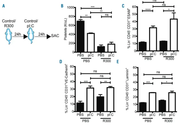
inflam-Figure 5. The IFN mimetic-, pI:C, mediated bone marrow (BM) endothelial cell (EC) stimulation is not affected by platelet abrogation. (A) Experimental design: mice were treated with the anti-platelet antibody R300 (2 mg/g) and either PBS or the IFN mimetic, pI:C, (5 mg/kg) for 24 h. (B) Platelet counts in the peripheral blood of wild-type (WT) mice following treatment as outlined in (A). (C-E) FACS analysis of the expression of (C) ESAM (D) VE-Cadherin and (E) Laminin on ECs (Lin- CD45 -CD31+) from WT mice treated as outlined in (A). Data are representative of 3 or more independent experiments, and are presented as mean±Standard Error of Mean
(SEM) (n≥3). Statistical analysis was performed using unpaired Student t-test (two-tailed). ns: not significant, *P<0.05, **P<0.01, ***P<0.001, ****P<0.0001.
A B C
mation,29,30 and megakaryocytes, which give rise to
platelets, regulate BM HSC quiescence.31 To test
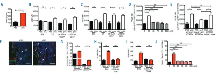
pI:C-mediated EC activation in the absence of platelets, mice were treated with a platelet depletion antibody, anti-GPIbα(CD42b) antibody (R300), prior to pI:C administra-tion (Figure 5A). Platelet levels were reduced upon R300 treatment (Figure 5B); however, EC activation markers were not altered following platelet depletion (Figure 5C-E). This suggested that platelet activation was not central to IFNα-induced BM EC stimulation. To investigate whether VEGF was regulated by acute pI:C treatment, we carried out a BM ELISA and intracellular staining for VEGF in BM cell types following pI:C treatment. After 24 h there was a significant increase in secreted VEGF in the BM supernatant of pI:C treated mice (Figure 6A). Intracellular VEGF did not increase in BM ECs (Figure 6B and C). However, there was a significant increase in intracellular VEGF in hematopoietic cells, including progenitors and HSCs (Lin–ckithi CD150+ CD48–CD34–), both after pI:C
(Figure 6B) and IFNα (Figure 6C) treatment. Consistent with the transient increase in activation of BM ECs (Figure 2G and H), the increase in intracellular VEGF levels in HSCs upon pI:C treatment was also transient. VEGF pro-duction peaked after 24 h and returned to homeostatic lev-els 72 h after treatment (Figure 6D). In addition, VEGF pro-duction was increased in WT HSCs in recipient IFNAR–/–
mice (WT BM into an IFNAR–/– niche) and in IFNAR–/–
HSCs in WT recipient mice (IFNAR–/–BM into a WT niche)
following pI:C treatment (Figure 6E). These data suggested that production of VEGF production in HSCs was stimu-lated both directly and indirectly by pI:C treatment. To assess whether VEGF signaling was consequently active in the BM,6the expression of the VEGF receptor, VEGFR2,
was analyzed in pI:C treated mice as an indicator of VEGF signaling. VEGFR2 expression was increased in BM
sec-tions (Figure 6F). In addition, VEGFR2 was up-regulated on the surface of ECs, but not on HSCs, in response to both pI:C and IFNα (Figure 6G-I). This suggested that VEGF signaling was active in ECs but differs between ECs and HSCs at this time point. As with VEGF production (Figure 6D), the increase in VEGFR2 expression on BM ECs was transient, peaking 24 h after pI:C treatment (Figure 6J). This time point correlated with the peak of increased expression of activation markers on BM ECs (Figure 2G and H). Taken together, these data indicate that pI:C and IFNαtreatment leads to an increase in VEGF pro-duction and signaling in the BM. In addition, they suggest that, upon pI:C treatment of mice, BM ECs responded to VEGF, which is produced by other BM cell types including HSCs in response to pI:C. Thus, VEGF may be a mediating
factor in the activation of BM ECs by
IFNα-stimulated hematopoietic cells.
IFN
α
-mediated stimulation of ECs
in vivo
is facilitated
by VEGF
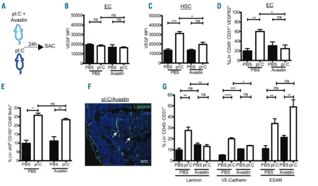
To test whether VEGF signaling was involved in BM EC activation, mice were co-treated with pI:C and the VEGF binding antibody, Avastin (Figure 7A). Avastin treatment did not affect the expression of VEGF or VEGFR2 in com-parison to PBS-treated mice (Figure 7B-D). While the expression level of VEGF in ECs was unchanged (Figure 7B), pI:C-induced VEGF expression in HSCs (LK SLAM CD34–)
was significantly reduced by co-treatment with Avastin (Figure 7C). In addition, the pI:C-induced expression of VEGFR2 on BM ECs was reduced upon Avastin co-treat-ment (Figure 7D). In contrast, Avastin treatco-treat-ment did not affect pI:C-mediated proliferation of HSCs (Figure 7E). This suggests that co-treatment with Avastin leads to reduced pI:C-mediated VEGF signaling in the BM. To assess the effect of diminished VEGF signaling on pI:C-mediated EC
Figure 6. Bone marrow (BM) vascular endothelial growth factor (VEGF) is modulated by the interferon mimetic, pI:C. (A) ELISA of BM VEGF from wild-type (WT) mice
treated with PBS or the IFN mimetic, pI:C, (5 mg/kg for 24 h). (B and C) FACS analysis of intra-cellular staining of VEGF in indicated BM cell types after treatment with either PBS, (B) pI:C (5 mg/kg for 24 h), or (C) IFNα(5x10⁶U/kg for 24 h). Data are presented as fold change in mean fluorescence intensity (MFI). (D) FACS
analy-sis of intra-cellular staining of VEGF in hematopoietic stem cells (HSCs) (Lin–ckithiCD150+CD48–CD34–) after treatment with either PBS (0 h) or pI:C (5 mg/kg, 0-120
h). Data are presented as fold change in MFI. (E) FACS analysis of intra-cellular staining of VEGF in HSCs (Lin–ckithiCD150+CD48–CD34–) from chimeric mice, as
described in Figure 4A, following treatment with either PBS or pI:C (5 mg/kg for 24 h). Data are presented as fold change in MFI. (F) Representative bone sections of VEGFR2 expression (Red) and Laminin (Green) after treatment with either PBS or pI:C (5 mg/kg for 24 h). (G) FACS analysis of VEGFR2 expression on (F) Lamininhigh/lowECs (Lin–CD45–CD31+, Lamininhigh/low) treated with PBS or pI:C (5 mg/kg for 24 h). (H and I) FACS analysis of VEGFR2 expression on ECs (Lin-
CD45-CD31+) and HSCs (Lin–ckithiCD150+CD48–CD34–) from mice treated with either (H) PBS or pI:C (mg/kg for 24 h) or (I) PBS or IFNα(5x10⁶U/kg for 24 h). (J) FACS
analysis of the expression of VEGFR2 on ECs from WT mice treated with either PBS or pI:C (5 mg/kg for up to 120 h). Data are representative of 3 or more inde-pendent experiments (A, B, D-G) and 2 or more (C and H) indeinde-pendent experiments, and are presented as mean±Standard Error of Mean (SEM) (n≥3). Statistical analysis was performed using unpaired Studentt-test (two-tailed). ns: not significant, *P<0.05, **P<0.01, ***P<0.001.
A B C D E
activation, the expression of EC activation markers follow-ing Avastin treatment was analyzed. While the increased expression of ESAM was not affected, the pI:C-induced expression of both VE-Cadherin and Laminin was signifi-cantly diminished upon co-treatment with Avastin (Figure 7F and G). This indicated that VEGF was involved, in part, in pI:C-mediated BM EC activation. Taken together, these data demonstrate that VEGF signaling is important for the stimulation of BM ECs following pI:C treatment.
Discussion
Bone marrow ECs are a primary defense against infec-tion, so understanding the effect of inflammation on the BM vasculature is critical. We demonstrate for the first time a rapid, transient activation of the BM vasculature in response to acute inflammatory signaling. We find that there is a direct and indirect effect of IFNαsignaling in the BM on ECs, mediated by an activated hematopoietic
sys-Figure 7. The IFN mimetic-, pI:C, mediated stimulation of bone marrow (BM) endothelial cells (ECs) is mediated by vascular endothelial growth factor (VEGF) sig-naling. (A) Experimental design: mice were treated with Avastin (2.5 mg/kg) and either PBS or the interferon mimetic, pI:C, (5 mg/kg) for 24 h. (B and C) FACS analysis of intra-cellular staining of VEGF in (B) ECs (Lin-CD45-CD31+) and (C) HSCs (Lin-ckithiCD150+CD48-CD34-) from wild-type (WT) mice treated as described in (A). Data are presented as fold change in mean fluorescence intensity (MFI). (D) FACS analysis of VEGFR2 expression on ECs (Lin-CD45- CD31+) from WT mice treated
as described in (A). (E) FACS analysis of percentage of BrdU positive HSCs (Lin- ckithiCD150+CD48-) from WT mice treated as described in (A) and BrdU (18 mg/kg, 16 h). (F) Representative bone marrow section (Laminin in green, DAPI in blue) from WT mice treated as described in (A). (G) FACS analysis of the expression of Laminin, VE-Cadherin and ESAM on ECs (Lin-CD45-CD31+) from WT mice treated as described in (A). Data are representative of 3 or more (A-D, E-G) and 2 or more
(E) independent experiments and are presented as mean±Standard Error of Mean (SEM) (n≥3). Statistical analysis was performed using unpaired Student t-test (two-tailed). ns (not significant), *P<0.05, **P<0.01, ***P<0.001, ****P<0.0001.
Figure 8. Acute IFNα-stimulation of BM ECs is mediated
by VEGF signaling in both hematopoietic and
non-hematopoietic cells. Model depicting BM vasculature
remodeling following stimulation of BM hematopoietic or non-hematopoietic cells by acute interferon α (IFNα) treatment.
A B C D
tem. Our data suggest a role for VEGF signaling in the BM in IFNα-mediated BM EC activation. This rapid, transient effect may be an emergency response to inflammatory sig-naling, coming from the hematopoietic system and affect-ing BM EC niche cells. This response may in turn facilitate the maintenance of BM homeostasis. In this acute setting, the vasculature may be rapidly 'primed' or activated, likely in anticipation of greater insult. In contrast to treatment with an isolated cytokine, initial inflammatory signaling during a full infection is followed by production of other cytokines, and stimulation of additional signaling.32 It is,
therefore, likely that the response elicited by an isolated cytokine differs to that elicited by a full infection, particu-larly with regard to continuation of signaling and recovery from the initial stimulation.
We have found that acute pI:C exposure results in a tran-sient expansion of the vasculature in the BM after 24 h. The integrity of this expanded vasculature was compromised (Figure 1). Increased BM vascular permeability is in keeping with an increase in the transit of immune cells or leakage of plasma during an inflammatory response.33 Acute pI:C
treatment induces production of IFNαand mimics an acute inflammatory response.16 Acute IFNαtreatment in vitromay
reduce apoptosis of endothelial cell lines.27 Whether IFNα
has a similar effect on apoptosis of ECs in vivo is unknown. Reduced BM EC apoptosis, mediated by IFNα during inflammatory stress, may influence vessel integrity. Permeability of different types of BM vasculature is distinct.7 Therefore, the integrity of specific BM vessels
fol-lowing an acute inflammatory response is likely influenced by vessel permeability under homeostasis. This may be a mechanism to maintain BM homeostasis during the early stages of an inflammatory response.
The effects of IFNαon ECs and other hematopoietic cell types in vivo are in contrast to the in vitro situation26,28,34-36
where cells are isolated from their niche environment. In the BM niche, IFNαtreatment rapidly and efficiently stim-ulates HSC proliferation in vivo, whereas in vitro, HSCs do not undergo increased proliferation.16 We have shown BM
EC activation in response to acute IFNα exposure in vivo
(Figures 2 and 3), in contrast to the described effect of IFNα on ECs in vitro. In addition to the differential effect of IFNα
in vitro and in vivo, IFNαhas also been described as being both stimulatory and inhibitory with regard to VEGF signal-ing.37-39 We find that VEGF production was increased in the
BM in response to pI:C (Figure 6). BM EC activation follow-ing acute pI:C treatment was dependent in part on VEGF signaling (Figure 7). pI:C mediated HSC proliferation was not affected by inhibition of VEGF, using Avastin treatment (Figure 7E). This corresponds with previous data showing that HSCs are directly activated by IFNα.16An IFNα
-medi-ated increase of VEGF is in contrast to previous studies, which suggest that VEGF is suppressed by long-term IFN treatment or in combination with chemotherapy.38-41
Together, these data highlight the contrast between chronic
versusacute IFN treatments, and between in vitro and in vivo
cytokine responses. As inflammatory stress is a complex signaling cascade, the in vivocytokine response in mice is, therefore, more reflective of the inflammatory response than the in vitrosituation.
In the BM niche, signaling between different cell types is imperative for maintenance of cellular homeostasis and a rapid response to inflammation. ECs and the vasculature itself have been ascribed many functions in the BM as regu-lators of HSCs.8,10,13,18,42-45Furthermore, there are distinct vessel
subtypes within the BM which differentially regulate hematopoiesis.7In addition, Notch signaling in BM ECs has
been shown to expand the HSC niche in vivo.8As Notch
sig-naling is involved in the inflammatory response of ECs,46
these data may further support a role for inflammatory sig-naling in BM niche remodeling. Furthermore, IFNαdoes not lead to mobilization of hematopoietic stem progenitor cells HSPCs that are not in the BM.16,35 However, the percentage
of HSCs found within 20 mm of arterioles in sternal BM is sig-nificantly reduced following treatment with pI:C in compar-ison to the control.18 These data suggest that the location of
HSCs in the BM is affected by pI:C. Relocating HSPCs can potentially affect many different BM cell types, and the BM vasculature, following treatment with pI:C. Whether pI:C-stimulated BM vasculature affects the location of HSPCs within the BM remains unclear. Many cytokines produced by hematopoietic cells, such as EPO and GCSF, have been shown to affect specific EC functions in isolation.47,48
However, signaling from the hematopoietic system to the ECs in the BM niche has not been examined in detail. To address this question, we have created BM chimeras in which only either hematopoietic cells or niche cells respond directly to IFNα. Using this system, we have demonstrated that inflammatory signaling from an activated hematopoietic system can affect the BM vasculature (Figure 4). Platelets, which can induce EC stimulation,30do not play a major role
in BM EC stimulation in this setting (Figure 5). As the inflam-matory response is complex, these data do not exclude the possibility that IFNα-mediated signaling from other cell types within the BM, in addition to hematopoietic cells, may be involved in this response. Further, these data cannot discrim-inate within the heterogeneous population of BM ECs. Whether there is crosstalk between pI:C-stimulated BM ECs and BM HSPCs within this context, remains to be elucidated. Importantly, we demonstrate that BM hematopoietic cells, including HSCs, are implicated in the pI:C-mediated BM EC stimulation, and thus in vasculature remodeling. This pro-vides evidence for crosstalk between BM HSPCs and ECs under inflammatory stress conditions (Figure 8).
Our findings demonstrate a novel response of the BM vasculature to primary inflammatory signaling. We have revealed potential crosstalk between the hematopoietic sys-tem and the BM vasculature under inflammatory stress. The transient activation of the BM vasculature represents a novel, emergency response of the BM stem cell niche to inflammation. Future studies will likely uncover other emergency situations in which HSCs influence the BM niche. Understanding this critical cellular relationship under stress conditions such as infection, but also chemotherapy, may reveal new mechanisms for the maintenance and recovery of BM homeostasis.
Acknowledgments
The authors would like to thank Drs M. Milsom, H. Augustin and A. Trumpp for helpful discussions, A. Atzberger and Dr S. Schmitt from the DKFZ Flow Cytometry Facility, M. Brom, Dr F. Bestvater and Dr D. Krunic from the DKFZ Imaging Core Facility, the DKFZ Animal Laboratory Services for their expertise and assistance, and L. Prendergast for critical reading of the manu-script.
Funding
This work was supported by FOR2033 NicHem and SFB873, both funded by the Deutsche Forschungsgemeinschaft, and the Dietmar Hopp Foundation.
References
1. Orozco AS, Zhou X, Filler SG. Mechanisms of the proinflammatory response of endothelial cells to Candida albicans infec-tion. Infect Immun. 2000;68(3):1134-1141. 2. Bazzoni G, Dejana E. Endothelial
cell-to-cell junctions: molecular organization and role in vascular homeostasis. Physiol Rev. 2004;84(3):869-901.
3. Pober JS. Endothelial activation: intracellu-lar signaling pathways. Arthritis Res. 2002; 4(Suppl 3):S109-16.
4. Granger DN, Rodrigues SF, Yildirim A, Senchenkova EY. Microvascular responses to cardiovascular risk factors. Microcirculation. 2010;17(3):192-205. 5. Muller WA. Getting leukocytes to the site
of inflammation. Vet Pathol. 2013;50(1):7-22.
6. Jones N, Iljin K, Dumont DJ, Alitalo K. Tie receptors: new modulators of angiogenic and lymphangiogenic responses. Nat Rev Mol Cell Biol. 2001;2(4):257-267. 7. Itkin T, Gur-Cohen S, Spencer JA, et al.
Distinct bone marrow blood vessels differ-entially regulate haematopoiesis. Nature. 2016;532(7599):323-328.
8. Kusumbe AP, Ramasamy SK, Itkin T, et al. Age-dependent modulation of vascular niches for haematopoietic stem cells. Nature. 2016;532(7599):380-384. 9. Cardier JE, Barbera-Guillem E.
Extramedullary hematopoiesis in the adult mouse liver is associated with specific hepatic sinusoidal endothelial cells. Hepatology. 1997;26(1):165-175.
10. Ding L, Saunders TL, Enikolopov G, Morrison SJ. Endothelial and perivascular cells maintain haematopoietic stem cells. Nature. 2012;481(7382):457-462. 11. Gattazzo F, Urciuolo A, Bonaldo P.
Extracellular matrix: a dynamic microenvi-ronment for stem cell niche. Biochim Biophys Acta. 2014;1840(8):2506-2519. 12. Li W, Johnson SA, Shelley WC, Yoder MC.
Hematopoietic stem cell repopulating abili-ty can be maintained in vitro by some pri-mary endothelial cells. Exp Hematol. 2004; 32(12):1226-1237.
13. Morrison SJ, Scadden DT. The bone mar-row niche for haematopoietic stem cells. Nature. 2014;505(7483):327-334. 14. Avecilla ST, Hattori K, Heissig B, et al.
Chemokine-mediated interaction of hematopoietic progenitors with the bone marrow vascular niche is required for thrombopoiesis. Nat Med. 2004;10(1):64-71. 15. Zhao YD, Huang X, Yi F, et al. Endothelial FoxM1 mediates bone marrow progenitor cell-induced vascular repair and resolution of inflammation following inflammatory lung injury. Stem Cells. 2014;32(7):1855-1864.
16. Essers MA, Offner S, Blanco-Bose WE, et al. IFNalpha activates dormant haematopoiet-ic stem cells in vivo. Nature. 2009;458(7240):904-908.
17. Muller U, Steinhoff U, Reis LF, et al. Functional role of type I and type II
interfer-ons in antiviral defense. Science. 1994; 264(5167):1918-1921.
18. Kunisaki Y, Bruns I, Scheiermann C, et al. Arteriolar niches maintain haematopoietic stem cell quiescence. Nature. 2013; 502(7473):637-643.
19. Radu M, Chernoff J. An in vivo assay to test blood vessel permeability. J Vis Exp. 2013;73):e50062.
20. Haas S, Hansson J, Klimmeck D, et al. Inflammation-Induced Emergency Megakaryopoiesis Driven by Hematopoietic Stem Cell-like Megakaryocyte Progenitors. Cell Stem Cell. 2015;17(4):422-434. 21. Kawamoto T. Use of a new adhesive film
for the preparation of multi-purpose fresh-frozen sections from hard tissues, whole-animals, insects and plants. Arch Histol Cytol. 2003;66(2):123-143.
22. Bentley K, Franco CA, Philippides A, et al. The role of differential VE-cadherin dynamics in cell rearrangement during angiogenesis. Nat Cell Biol. 2014;16(4):309-321.
23. Pietras EM, Lakshminarasimhan R, Techner JM, et al. Re-entry into quiescence protects hematopoietic stem cells from the killing effect of chronic exposure to type I interfer-ons. J Exp Med. 2014;211(2):245-262. 24. Wada H, Nagano H, Yamamoto H, et al.
Combination of interferon-alpha and 5-flu-orouracil inhibits endothelial cell growth directly and by regulation of angiogenic factors released by tumor cells. BMC Cancer. 2009;9:361.
25. Sgonc R, Fuerhapter C, Boeck G, Swerlick R, Fritsch P, Sepp N. Induction of apoptosis in human dermal microvascular endothelial cells and infantile hemangiomas by inter-feron-alpha. Int Arch Allergy Immunol. 1998;117(3):209-214.
26. Reynolds JA, Ray DW, Zeef LA, O'Neill T, Bruce IN, Alexander MY. The effect of type 1 IFN on human aortic endothelial cell function in vitro: relevance to systemic lupus erythematosus. J Interferon Cytokine Res. 2014;34(5):404-412.
27. Pammer J, Reinisch C, Birner P, Pogoda K, Sturzl M, Tschachler E. Interferon-alpha prevents apoptosis of endothelial cells after short-term exposure but induces replicative senescence after continuous stimulation. Lab Invest. 2006;86(10):997-1007. 28. Cheng X, Liu Y, Chu H, Kao HY.
Promyelocytic leukemia protein (PML) reg-ulates endothelial cell network formation and migration in response to tumor necro-sis factor alpha (TNFalpha) and interferon alpha (IFNalpha). J Biol Chem. 2012; 287(28):23356-23367.
29. Lee S, Chen TT, Barber CL, et al. Autocrine VEGF signaling is required for vascular homeostasis. Cell. 2007;130(4):691-703. 30. Stokes KY, Granger DN. Platelets: a critical
link between inflammation and microvas-cular dysfunction. J Physiol. 2012;590(Pt 5):1023-1034.
31. Bruns I, Lucas D, Pinho S, et al. Megakaryocytes regulate hematopoietic stem cell quiescence through CXCL4 secre-tion. Nat Med. 2014;20(11):1315-1320.
32. Mogensen TH. Pathogen recognition and inflammatory signaling in innate immune defenses. Clin Microbiol Rev. 2009; 22(2):240-273, Table of Contents. 33. Rajendran P, Rengarajan T, Thangavel J, et
al. The vascular endothelium and human diseases. Int J Biol Sci. 2013;9(10):1057-1069.
34. Borden EC, Sen GC, Uze G, et al. Interferons at age 50: past, current and future impact on biomedicine. Nat Rev Drug Discov. 2007;6(12):975-990. 35. Essers MA, Trumpp A. Targeting leukemic
stem cells by breaking their dormancy. Mol Oncol. 2010;4(5):443-450.
36. Rosa R, Monteleone F, Zambrano N, Bianco R. In vitro and in vivo models for analysis of resistance to anticancer molecular thera-pies. Curr Med Chem. 2014; 21(14):1595-1606.
37. Bauer EM, Zheng H, Lotze MT, Bauer PM. Recombinant human interferon alpha 2b prevents and reverses experimental pul-monary hypertension. PLoS One. 2014; 9(5):e96720.
38. Merkle M, Ribeiro A, Belling F, et al. Response of VEGF to activation of viral receptors and TNFalpha in human mesan-gial cells. Mol Cell Biochem. 2012;370(1-2):151-161.
39. von Marschall Z, Scholz A, Cramer T, et al. Effects of interferon alpha on vascular endothelial growth factor gene transcrip-tion and tumor angiogenesis. J Natl Cancer Inst. 2003;95(6):437-448.
40. Gambara G, Desideri M, Stoppacciaro A, et al. TLR3 engagement induces IRF-3-depen-dent apoptosis in androgen-sensitive prostate cancer cells and inhibits tumour growth in vivo. J Cell Mol Med. 2015; 19(2):327-339.
41. Raig ET, Jones NB, Varker KA, et al. VEGF secretion is inhibited by interferon-alpha in several melanoma cell lines. J Interferon Cytokine Res. 2008;28(9):553-561. 42. Boulais PE, Frenette PS. Making sense of
hematopoietic stem cell niches. Blood. 2015;125(17):2621-2629.
43. Ding L, Morrison SJ. Haematopoietic stem cells and early lymphoid progenitors occu-py distinct bone marrow niches. Nature. 2013;495(7440):231-235.
44. Kunisaki Y, Frenette PS. Influences of vas-cular niches on hematopoietic stem cell fate. Int J Hematol. 2014;99(6):699-705. 45. Lamagna C, Bergers G. The bone marrow
constitutes a reservoir of pericyte progeni-tors. J Leukoc Biol. 2006;80(4):677-681. 46. Quillard T, Charreau B. Impact of notch
signaling on inflammatory responses in car-diovascular disorders. Int J Mol Sci. 2013; 14(4):6863-6888.
47. Ribatti D, Vacca A, De Falco G, Ria R, Roncali L, Dammacco F. Role of hematopoietic growth factors in angiogen-esis. Acta Haematol. 2001;106(4):157-161. 48. Ribatti D, Vacca A, Roncali L, Dammacco F.
Hematopoiesis and angiogenesis: a link between two apparently independent processes. J Hematother Stem Cell Res. 2000;9(1):13-19.