© 2019 by the Serbian Biological Society How to cite this article: Tolić AZ, Rajić JJ, Đorđević MB, Đorđević MM, Dinić SS, 455 Grdović NM, Arambašić-Jovanović JD, Mihailović MV, Poznanović GĐ, Jurkowski TP, Vidaković MS, Uskoković AS. Enrichment of Cxcl12 promoter with TET2: A possible link between promoter demethylation and enhanced gene expression in the absence of PARP-1. Arch Biol Sci. 2019;71(3):455-62.
Enrichment of
Cxcl12
promoter with TET2: a possible link between promoter
demethylation and enhanced gene expression in the absence of PARP-1
Anja Z. Tolić1, Jovana J. Rajić1, Marija B. Đorđević1, Miloš M. Đorđević1, Svetlana S. Dinić1, Nevena M. Grdović1, Jelena D. Arambašić Jovanović1, Mirjana V. Mihailović1, Goran Đ. Poznanović1, Tomasz P. Jurkowski2,3, Melita S. Vidaković1 and Aleksandra S. Uskoković1,*
1Department of Molecular Biology, Institute for Biological Research “Siniša Stanković“ – National Institute of the Republic of
Serbia, University of Belgrade, Bulevar despota Stefana 142, 11060 Belgrade, Serbia
2Institute of Biochemistry, University of Stuttgart, Pfaffenwaldring 55, 70569 Stuttgart, Germany
3School of Biosciences, Cardiff University, Cardiff, Wales, Sir Martin Evans Building, Museum Avenue, Cardiff, CF10 3AX, UK
*Corresponding author: [email protected]
Received: April 4, 2019; Accepted: April 18, 2019; Published online: April 18, 2019
Abstract: Previously, we described the link between C-X-C motif chemokine 12 (Cxcl12) gene induction and DNA hypo-methylation in the absence of poly(ADP-ribose) polymerase 1 (PARP-1). We have now firmly established that dehypo-methylation is the primary cause of gene induction on the basis of Cxcl12 gene upregulation upon treatment with the demethylating agent 5-azacytidine (5-aza). Since the demethylation state of Cxcl12 is favored by PARP-1 absence, we investigated the pres-ence of ten-eleven translocation (TET) proteins on the Cxcl12 promoter in order to corroborate the relationship between the demethylation process and increased gene expression that occurs in the absence of PARP-1. Analysis was performed
on the promoter region within CpG islands of Cxcl12 from control mouse embryonic fibroblasts (NIH3T3) and PARP-1
knock-out mouse embryonic fibroblasts (PARP1-/-). The lack of PARP-1 increased the abundance of TET2 on the Cxcl12
promoter, suggesting that TET-mediated demethylation provoked by the absence of PARP-1 could account for the observed increased expression of this chemokine. Deciphering the regulation of DNA (de)methylation factors that control Cxcl12
expression may provide an additional therapeutic approach in pharmacological interventions where gene switching on or off based on targeted stimulation or inhibition is necessary.
Keywords: DNA demethylation; 5-aza; TET2; PARP-1; CXCL12
INTRODUCTION
DNA methylation is a covalent modification of genomic DNA that occurs predominantly at CG di-nucleotides (CpG) where DNA methyltransferase (DNMTs) enzymes mediate the transfer of a me-thyl group to the 5-position of cytosines, generating 5-methylcytosine (5mC), which is assumed to be a fifth DNA base [1-3]. Ten-eleven translocation 1-3 (TET1-3) proteins, members of the DNA hydroxylase family, affect the methyl group on 5mC by catalyzing the consecutive oxidation of 5mC into 5-hydroxym-ethylcytosine (5hmC), 5-formylcytosine (5fC) and 5-carboxylcytosine (5caC) [4,5]. Further, 5caC can be replaced by an unmethylated cytosine by
in the production of PAR polymers. Besides PARP-1 automodification where the enzyme PARylates itself, it can also PARylate covalently other target proteins, thereby modulating their activities and functions. PAR moieties can also bind non-covalently to target proteins that expand the number of PARP-1 inter-acting partners subjected to PARylation [9-11]. The link between PARP-1 and TET activity in DNA dem-ethylation is emerging as a newly discovered role of PARP-1 [11].
In our previous study we reported that PARP-1 promotes DNA methylation and the opposite, that lack of PARP-1 is connected with the demethyla-tion state of Cxcl12 gene; this also corresponds with changes in Cxcl12 expression, which tends to increase in the absence of PARP-1 [12]. CXCL12, also known as stromal cell-derived factor 1 (SDF-1), is a potent CXC chemokine produced by different cell types. It is involved in the migration of a variety of cells, including hematopoietic progenitor and stem cells, endothelial cells and most leukocytes [13]. CXCL12 is involved in physiological and pathological processes, such as development, cell survival, tissue repair and regenera-tion, as well as cancer [14,15]. The methylation status of the Cxcl12 gene plays an important role in tumor progression in human carcinogenesis. For example, epigenetically-controlled downregulation of the Cxcl12 by promoter hypermethylation has been detected in breast tumors [16] and in osteosarcomas [17], and is connected with metastatic potential and poor survival prognosis. Hence, the methylation status of Cxcl12 could be used as a target for therapeutic interventions. Since we previously found PARP-1 to be a po-tential upstream player in methylation events that modulated Cxcl12 expression, our aim was to examine whether a lack of PARP-1 would induce TET enzymes to demethylate Cxcl12.
MATERIALS AND METHODS Cell culture and treatment
The following cell lines were used: mouse embryonic fibroblasts, NIH3T3 (ATCC- CRL-1658), and PARP-1 knock-out (PARP1-/-) cells [18]. The cells were cul-tured in high glucose Dulbecco’s Modified Eagle’s
me-dium (DMEM) supplemented with 10% fetal bovine serum (FBS), L-glutamine and penicillin/streptomycin (all cell culture reagents were supplied by Biological Industries, Beit Haemek Ltd., Israel). NIH3T3 cells were treated with 15 µM 5-azacytidine (5-aza) solu-bilized in dimethyl sulfoxide (DMSO) for 72 h, with the treatment replaced every 24 h. In parallel with the 5-aza treatment, control NIH3T3 cells were treated with DMSO in the same manner.
RNA isolation, reverse transcription and real-time quantitative PCR (RT-qpcr)
Total RNA was isolated from NIH3T3 cells treated with 5-aza or DMSO and PARP1-/- cells using the GeneJET RNA Purification Kit (Thermo Fisher Scientific, USA). Reverse transcription was performed on 1 µg of DNase I-treated RNA by the RevertAid First Strand cDNA Synthesis Kit (Thermo Fisher Scientific, USA) with a mix of oligo(dT)18 and random hexamer primers. For RT-qPCR we used QuantStudio 3 Real-Time PCR system (Applied Biosystems, Carlsbad, CA, USA) and Maxima SYBR Green/ROX qPCR Master Mix (Thermo Fisher Scientific, USA). The thermal cycles were as fol-lows: initial denaturation at 95°C for 10 min and 40 cycles of two-step PCR at 95°C for 15 s and at 60°C for 60 s. Expression of Cxcl12 was estimated relative to GAPDH (as an internal control) by the delta Ct method (2dCt) with previously published primers [12] (Supple-mentary Table S1). The obtained data was log2 trans-formed for statistical testing and statistical significance was estimated using ANOVA with Dunnett’s post-hoc test used to compare NIH3T3 5-aza-treated and PARP-/- cells with control NIH3T3 cells mock-treated with DMSO. For graphs, mean values and error bars were transformed back to the linear scale.
DNA isolation, bisulfite conversion and methylation-specific PCR (MSP)
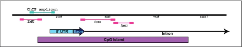
The EZ-DNA methylation kit (D5002; Zymo Research, Orange, CA, USA) was used for bisulfite conversion of genomic DNA, as per the provided in-structions. This bisulfite-converted DNA served as a template for MSP analysis of DNA methylation chang-es. The promoter (1MU), the exon-intron boundary (2MU) and part of the intron (3MU) of Cxcl12 were analyzed (Fig. 2) with three sets of MSP primers (Sup-plementary Table S1) that were previously published [12]. Each set was comprised of two pairs of primers targeted to the same location yet differentiating be-tween the methylation states, as one pair was com-plementary to the methylated (M) and the other to the unmethylated (U) bisulfite-converted target DNA sequence. Separate reactions were set up for each primer pair, ensuring that reactions for one sample with primers from the same set were always run in parallel, within the same PCR run. Each reaction had a volume of 10 µL and contained 2 µM primers, 60 ng of bisulfite-converted DNA and Maxima SYBR Green/ ROX qPCR Master Mix (Thermo Fisher Scientific, USA). The QuantStudio 3 Real-Time PCR system (Applied Biosystems, Carlsbad, CA, USA) was used for the runs with the following temperature cycling profile: initial denaturation at 95°C for 10 min and 40 cycles of two-step PCR at 95°C for 15 s and 58°C for 60 s. Finally, to assess the relative level of methylation, threshold cycle values (Ct) for M and U primer pairs from the same set were used to calculate the methyla-tion index [19] using the following equamethyla-tion:
2(Ct for U primer pair reaction)-(Ct for M primer pair reaction). The obtained data was log2 transformed for statis-tical testing and statisstatis-tical significance was estimated using ANOVA with Dunnett’s post-hoc test to com-pare the NIH3T3 5-aza-treated and PARP-/- cells with control NIH3T3 cells mock-treated with DMSO. For graphs, mean values and error bars were transformed back to the linear scale.
Chromatin immunoprecipitation (ChIP)
Chromatin immunoprecipitation was performed us-ing the Pierce™ Magnetic ChIP Kit (Thermo Fisher Scientific, USA) according to the protocol. In brief, NIH3T3 and PARP-/- chromatin were cross-linked with 1% formaldehyde (Zorka Pharma, Serbia) and
the process was stopped with a glycine solution after 10 min at room temperature. After extraction, the chro-matin was digested with 0.005 U/µL micrococcal nu-clease and sheared on ice by sonication (7 rounds of 20 s sonication and 30 s rest). Immunoprecipitation was performed using Protein A/G Magnetic Beads (USA) and the following antibodies: anti-RNA polymerase II (RNA pol II) (supplied with the ChIP kit), normal rab-bit IgG (supplied with the ChIP kit), 5 µg of anti-TET1 (Millipore Merck, USA) and 5 µg of anti-TET2 (Mil-lipore Merck, USA). The cross-linking was reversed at 65°C and the immunoprecipitated samples and sam-ples containing 10% of total input were treated with 40 µg proteinase at 65°C for 1.5 h. The abundance of the target sequence was assessed by quantitative PCR (qPCR) using QuantStudio 3 Real-Time PCR system (Applied Biosystems, Carlsbad, CA, USA) and Maxima SYBR Green/ROX qPCR Master Mix (Thermo Fisher Scientific, USA). Primers (Supplementary Table S1) were designed to amplify part of the promotor region directly adjacent to the transcription start site (TSS) of Cxcl12 (Fig. 2). To quantify the enrichment, first the quantity of ChIP and IgG samples was determined from a standard curve generated with qPCR data of 10-fold dilution series of the 10% total input samples. En-richment was calculated by normalizing to IgG sample quantity and fold enrichment was reported relative to the corresponding NIH3T3 samples (immunoprecipi-tated by the same antibody) from the same experiment. The experiment was repeated twice.
Co-immunoprecipitation (IP)
sulfo-nyl fluoride (PMSF) and a protease inhibitor cocktail. Proteins were eluted from the beads by boiling in a sample buffer (125 mM Tris-HCl, pH 6.8, 4% SDS, 20% glycerol, 100 mM dithiothreitol, 0.02% bromophenol blue), separated by 7% sodium dodecyl sulfate poly-acrylamide gel electrophoresis (SDS-PAGE) alongside 70 µg of proteins from the NIH3T3 lysate. The proteins were electrotransferred onto polyvinylidene difluoride (PVDF) membranes and analyzed by immunoblotting.
Immunoblot analysis
NIH3T3 and PARP-/- cells were lysed in ice-cold RIPA buffer (Sigma Aldrich, USA) supplemented with a protease inhibitor cocktail for 30 min at 4°C. Equal amounts of cell lysates were separated by 12% SDS-PAGE and electrotransferred onto PVDF mem-branes. Immunoblotting for lysates or IP samples was performed by overnight incubation at 4°C with anti-PARP-1 (H-250, Santa Cruz Biotechnology, Santa Cruz, CA, USA) or anti-TET2 primary antibodies (Millipore Merck, USA) and anti-GAPDH (FL-335, Santa Cruz Biotechnology, Santa Cruz, CA, USA) as loading con-trol for lysates. This was followed by incubation with horseradish peroxidase‐conjugated anti‐rabbit second-ary antibody at room temperature for 1 h. Staining was performed by the enhanced chemiluminescence detec-tion system (Santa Cruz Biotechnology, Santa Cruz, CA, USA) according to the manufacturer’s instructions.
RESULTS
Cxcl12 expression was increased in 5-aza-treated NIH3T3 and PARP-/- cells
Treatment of NIH3T3 cells with the demethylating agent 5-aza induced a higher level of Cxcl12 expres-sion as measured by real-time PCR. Treated NIH3T3 cells showed on average a 5.5-fold increase compared
to control NIH3T3 cells, while PARP-/- cells showed an even greater increase of 2x10⁵-fold compared to the control (Fig. 1). Both observed increases were statisti-cally significant (p5aza=0.02 and pPARP-/-=1.9x10-8).
Methylation changes of Cxcl12 in 5-aza-treated
NIH3T3 and PARP-/- cells
As previously established [12], murine Cxcl12 gene has a CpG island that spans a part of its promoter, first exon and part of the first intron (Fig. 2). The methyla-tion changes of Cxcl12 on these regions were exam-ined as follows: amplicons analyzed by MSP covered 8 CpGs in the promoter, 23 CpGs at the exon-intron boundary and 8 CpGs in the intron region. In accord-ance with the changes in expression, we observed that both the treatment of NIH3T3 cells with 5-aza and the absence of PARP-1 resulted in demethylation of Cxcl12. Cells treated with 5-aza exhibited a statistically significant decrease in methylation, on average
45.4-Fig. 1. Relative expression level of Cxcl12 in control NIH3T3 cells (mock-treated with DMSO), NIH3T3 cells treated with 15 µM 5-aza and PARP−/− cells (n = 5) measured by RT-qPCR. Statistical significance is reported for the comparison of NIH3T3 5-aza-treated and PARP-/- cells to the control NIH3T3 cells. Results are presented as the mean±standard error of the mean, *p≤0.05, ***p ≤ 0.001, n‐number of independent experiments.
fold in the promoter region (p=1x10⁻⁶) and 4.3-fold at the exon-intron boundary (p=3.9x10⁻⁴), while the intron region was not significantly affected (Fig. 3). PARP-/- cells showed a significantly higher level of demethylation (Fig. 3). Compared to NIH3T3 cells, methylation in PARP-/- cells was decreased 1032.4-fold in the promoter (p=3.6x10⁻⁸), 1002.4-fold in the exon-intron boundary (p=3.1x10⁻⁸) and even 285.3-fold in the intron region (p=5.2x10⁻⁸).
The level of TET2 at the Cxcl12 promoter is
higher in the absence of PARP-1
ChIP analysis showed that TET proteins interact with the Cxcl12 promoter. Control ChIP experiments per-formed with an anti- RNA pol II antibody confirmed that RNA pol II was more abundantly present (on aver-age 5.7-fold more) on the Cxcl12 promoter of PARP-/- than of NIH3T3 cells (Fig. 4). This result was in agree-ment with the Cxcl12 expression profile in these cells. The results for TET1 were deemed inconclusive as no consistent change was observed (data not shown). We therefore focused on TET2, as it was consistently pre-sent in higher abundance (on average 1.32-fold) on the analyzed region of the Cxcl12 promoter in PARP-/- when compared to NIH3T3 cells (Fig. 4).
TET2 interacts with PARP-1
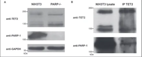
Western-blot analysis confirmed the absence of PARP-1 in PARP-/- cells and showed higher levels of TET2 isoforms (223 kDa and 130 kDa) in PARP-/- as compared to NIH3T3 cells (Fig. 5A). Results of IP analysis showed that TET2 was successfully immu-noprecipitated from NIH3T3 cell lysates and that it co-precipitated PARP-1, confirming their interaction in cellulo (Fig. 5B).
DISCUSSION
In the past decade, much attention has been focused on the role of epigenetic alterations in cancer progres-sion [20]. The increased metastatic potential in some types of cancers, such as colon, mammary and breast carcinomas, was shown to be associated with epige-netic silencing of Cxcl12 [16,21,22]. Furthermore, considering the ability of CXCL12 to stimulate the growth and survival of pancreatic β-cells, the
tran-Fig. 3. Methylation levels of different parts of Cxcl12 of control NIH3T3 cells (mock-treated with DMSO), NIH3T3 cells treated with 15 µM 5-aza and PARP−/− cells measured by methylation‐ specific PCR (MSP) (n = 4). Under each graph, the amplicon sequence is represented with highlighted CG dinucleotides. Statis-tical significance is reported for the comparison of NIH3T3 5-aza-treated and PARP-/- cells to the control NIH3T3 cells. Results are presented as the mean±standard error of the mean, *p≤0.05, ***p≤0.001, n‐number of independent experiments.
Fig. 4. ChIP-qPCR analysis of RNA pol II and TET2 occupancy
at the Cxcl12 promoter in NIH3T3 and PARP-/- cell lines (n=2). Results are presented as the mean±standard error of the mean.
Fig. 5. A – Immunoblot analysis performed with TET2,
scriptional regulation of this chemokine could provide an approach for diabetes treatment [23]. Hence, thera-pies that target CXCL12 expression via de/methylation possess the potential for therapeutic interventions, and identification of factors that affect the methyla-tion status of this chemokine is of great importance.
In this study we verified that PARP-1 affected the methylation status of Cxcl12 whereby the absence of PARP-1 promoted TET2 involvement on the Cxcl12 promoter, linking TET-dependent demethylation and increased gene expression in the absence of PARP-1. Restored expression of Cxcl12 after treatment with 5-aza, one of the most frequently used demethylating agents that inhibits DNMTs [24,25], confirmed the epigenetic transcriptional silencing of Cxcl12. While treatment with 5-aza is presumably based on inactiva-tion of methylainactiva-tion due to rapid loss of DNMT activity, our previous results indicated that in PARP-/- cells the promotion of TET-dependent active demethylation occurs rather than DNMT-related suppressed methyla-tion [12]. We assumed that this scenario also occurs in the Cxcl12 gene; however, a direct link between TET-mediated DNA demethylation and the transcriptional output is difficult to establish because of the existence of global and locus-specific effects that are difficult to discern [26]. Demethylation of the Cxcl12 promoter region was expected as it was already established that the Cxcl12 gene is exposed to epigenetic regulation by methylation of cytosine in CpG dinucleotides located in the promoter sequence [27-29]. In the intron region there was no statistically significant decrease in meth-ylation in NIH3T3 cells treated with 5-aza, whereas a significant decrease was observed in PARP-/- cells. This points to the stronger potential for demethyla-tion of Cxcl12 in PARP-/- compared to NIH3T3 cells treated with 5-aza, which could have also contributed to the higher increase in Cxcl12 expression.
Although the absence of PARP-1 in the demethyl-ation event is clear, it remains to be elucidated whether the TETs are responsible for the loss of methylation. Our previous results suggested that PARP-1 could be involved in demethylation as significant increases in both TET1 and TET2 expression were detected in the absence of PARP-1 [12]; herein we showed that the lack of PARP-1 increased TET2 occupancy on the Cxcl12 promoter. Furthermore, according to the ob-tained result there was no clear increase in TET1 on the Cxcl12 promoter (result not shown); however, this
does not rule out the possibility that TET1 was also affected by the absence of PARP-1 for recruitment and demethylation events on the Cxcl12 promoter. This assumption needs further validation.
TET proteins preferentially bind to DNA con-taining unmodified cytosine [30-32]. While TET1 is recruited to genomic target sites through direct bind-ing of CXXC domains to CpG-rich oligonucleotides [3,33,34], TET2 lacks the CXXC DNA-binding do-main, thus it is more likely that its recruitment on a specific gene is mediated by interactions with other DNA-binding factors [35]. It was found that TET2 in-teracts with IDAX protein whose CXXC domain binds to unmethylated CpG dinucleotides in the promoter but in a sequence-nonspecific manner [31]. A very recent report described the mechanism for targeting TET2 to specific promoters [36]. It was proposed that transcriptional co-activator SMAD nuclear interacting protein 1 (SNIP1) bridges TET2 to bind to a sequence-specific DNA-binding factor c-MYC, thus facilitating the recruitment of TET2 to regulate c-MYC target genes by promoting DNA demethylation at the pro-moters [36]. In addition, it is well established that the posttranslational modification, monoubiquitylation, which is catalyzed by CRL4VprBP E3 ligase, promotes the DNA binding of TET2 by stabilizing the conforma-tion of TET2 complexed with DNA (where the mon-oubiquitylation site directly contacts the DNA) [32,37]. The activity of TETs can be modulated by metabo-lites, cofactors and several post-translational modi-fications [35]. Our previous results marked TETs as potentially important factors in Cxcl12 expression in the absence of PARP-1. This was supported by the detected tendency of Cxcl12 expression to increase in the presence of TET activator (vitamin C), and to decrease upon treatment with the TET inhibitor di-methyloxaloylglycine (DMOG) in PARP-/- cells [12]. The findings presented herein encourage us to assume that the lack of PARP-1 was responsible for the in-creased recruitment of TET2 to the Cxcl12 promoter, which is a prerequisite step that leads to augmented activity and demethylation events.
in-hibitory role on the catalytic activity of TETs, since our previous results showed a global increase in TET ex-pression/activity in the absence of PARP-1 [12]. Hence the catalytic activation of TET enzymes together with the increased recruitment of TET2 in the absence of PARP-1 could have contributed to the local demeth-ylation event on the Cxcl12 promoter, accounting for its induced expression. This is in collision with some literature data showing that inhibition of PARylation suppresses the production of 5hmC by TET proteins and consequently region-specific demethylation [39]. Nevertheless, the role of PARP-1 in demethylation and its impact on TET activity was recently reviewed and it was pointed out that data implicating PARP/TET interplay in the regulation of (de)methylation that can produce opposite effects, resulting in either increased or decreased 5mC-hydroxylase activity, is still emerging [11]. Consistent with this view is the observation that after inhibition of PARylation, aside from a number of hypermethylated genes, several genes were also found to be hypomethylated [40].
CONCLUSION
Enrichment of the Cxcl12 promoter with TET2, to-gether with the previously described overall increased expression and activity of TETs in the absence of PARP-1, highlight the connection of PARP-1 and gene expression through (de)methylation events, and sup-port the inhibitory role of PARP-1 in demethylation, which explains the increased expression of Cxcl12. Future research should focus on exploring the effect of PARP activity (not only its presence) on Cxcl12 expression and on epigenetic events as this could en-hance the efficacy of PARP inhibitors in their poten-tial clinical translation.
Funding: This work was supported by the Alexander von
Hum-boldt foundation, program for funding a Research Group Linkage (to MV and TJ - 2015/2016). Financial support was also provided by the Ministry of Education, Science and Technological Develop-ment of the Republic of Serbia (Project No. 173020).
Acknowledgments: The authors are very grateful to Dr. Valérie
Schreiber (Département Intégrité du Génome, UMR7175-LC1 CNRS, Ecole Supérieure de Biotechnologie de Strasbourg, Illkirch Cedex, France) for providing the PARP-1 knock-out cells.
Author contributions: AU and MV designed the study; AT, JR
and MS performed the experiments; MĐ, MM and JAJ analyzed
the data statistically; AT and AU wrote the manuscript; TJ, SD, NG, and GP critically reviewed the manuscript.
Conflict of interest disclosure: The authors declare no conflict of interest.
REFERENCES
1. Goll MG, Bestor TH. Eukaryotic cytosine methyltransferases. Annu Rev Biochem. 2005;74(1):481-514.
2. Bernstein BE, Meissner A, Lander ES. The mammalian epig-enome. Cell. 2007;128(4):669-81.
3. Williams K, Christensen J, Helin K. DNA methylation: TET proteins-guardians of CpG islands? EMBO Rep. 2012;13(1):28-35.
4. Tahiliani M, Koh KP, Shen Y, Pastor WA, Bandukwala H, Brudno Y, Agarwal S, Iyer LM, Liu DR, Aravind L, Rao A. Conversion of 5-methylcytosine to 5-hydroxymethylcyto-sine in mammalian DNA by MLL partner TET1. Science. 2009;324(5929):930-5.
5. Ito S, Shen L, Dai Q, Wu SC, Collins LB, Carolina N, Hill C, Swenberg J a, He C, Zhang Y. Tet proteins can convert 5-methylcytosine to 5-formylcytosine and 5-carboxylcyto-sine. Science. 2011;333(6047):1300-3.
6. He Y, Li B-Z, Li Z, Liu P, Wang Y, Tang Q, Ding J, Jia Y, Chen Z, Li L, Sun Y, Li X, Dai Q, Song C-X, Zhang K, He C, Xu G-L. Tet-mediated formation of 5-carboxylcytosine and its excision by TDG in mammalian DNA. Science. 2011;333(6047):1303-7. 7. Tan L, Shi YG. Tet family proteins and
5-hydroxymeth-ylcytosine in development and disease. Development. 2012;139(11):1895-902.
8. Kong L, Tan L, Lv R, Shi Z, Xiong L, Wu F, Rabidou K, Smith M, He C, Zhang L, Qian Y, Ma D, Lan F, Shi Y, Shi YG. A primary role of TET proteins in establishment and mainte-nance of De Novo bivalency at CpG islands. Nucleic Acids Res. 2016;44(18):8682-92.
9. Caiafa P, Guastafierro T, Zampieri M. Epigenetics: poly(ADP-ribosyl)ation of PARP-1 regulates genomic methylation pat-terns. FASEB J. 2009;23(3):672-8.
10. Krishnakumar R, Kraus WL. The PARP side of the nucleus: molecular actions, physiological outcomes, and clinical tar-gets. Mol Cell. 2010;39(1):8-24.
11. Ciccarone F, Zampieri M, Caiafa P. PARP1 orchestrates epi-genetic events setting up chromatin domains. Semin Cell Dev Biol. 2017;63:123-34.
12. Tolić A, Grdović N, Dinić S, Rajić J, Đorđević M, Sinadinović M, Arambašić Jovanović J, Mihailović M, Poznanović G, Uskoković A, Vidaković M. Absence of PARP-1 affects Cxcl12 expression by increasing DNA demethylation. J Cell Mol Med. 2019;00:1-9.
13. Janssens R, Struyf S, Proost P. The unique structural and functional features of CXCL12. Cell Mol Immunol. 2018;15(4):299-311.
15. Liu Z, Habener JF. Stromal cell-derived factor-1 promotes survival of pancreatic beta cells by the stabilisation of beta-catenin and activation of transcription factor 7-like 2 (TCF7L2). Diabetologia. 2009;52(8):1589-98.
16. Ramos EAS, Camargo AA, Braun K, Slowik R, Cavalli IJ, Ribeiro EMSF, Pedrosa F de O, de Souza EM, Costa FF, Klas-sen G. Simultaneous CXCL12 and ESR1 CpG island hyper-methylation correlates with poor prognosis in sporadic breast cancer. BMC Cancer. 2010;10(1):23.
17. Li B, Wang Z, Wu H, Xue M, Lin P, Wang S, Lin N, Huang X, Pan W, Liu M, Yan X, Qu H, Sun L, Li H, Wu Y, Teng W, Wang Z, Zhou X, Chen H, Poznansky MC, Ye Z. Epigenetic regulation of CXCL12 plays a critical role in mediating tumor progression and the immune response in osteosarcoma. Can-cer Res. 2018;78(14):3938-53.
18. de Murcia JM, Niedergang C, Trucco C, Ricoul M, Dutril-laux B, Mark M, Oliver FJ, Masson M, Dierich A, LeMeur M, Walztinger C, Chambon P, de Murcia G. Requirement of poly(ADP-ribose) polymerase in recovery from DNA damage in mice and in cells. Proc Natl Acad Sci U S A. 1997;94(14):7303-7.
19. Akirav EM, Lebastchi J, Galvan EM, Henegariu O, Akirav M, Ablamunits V, Lizardi PM, Herold KC. Detection of cell death in diabetes using differentially methylated circulating DNA. Proc Natl Acad Sci. 2011;108(47):19018-23.
20. Jones PA, Baylin SB. The fundamental role of epigenetic events in cancer. Nat Rev Genet. 2002;3(6):415-28.
21. Wendt MK, Cooper AN, Dwinell MB. Epigenetic silencing of CXCL12 increases the metastatic potential of mammary carcinoma cells. Oncogene. 2008;27(10):1461-71.
22. Zhou W, Jiang Z, Liu N, Xu F, Wen P, Liu Y, Zhong W, Song X, Chang X, Zhang X, Wei G, Yu J. Down-regulation of CXCL12 mRNA expression by promoter hypermethylation and its association with metastatic progression in human breast carcinomas. J Cancer Res Clin Oncol. 2009;135(1):91-102. 23. Vidaković M, Grdović N, Dinić S, Mihailović M, Uskoković
A, Arambašić Jovanović J. The Importance of the CXCL12/ CXCR4 Axis in Therapeutic Approaches to Diabetes Mellitus Attenuation. Front Immunol. 2015;6:403.
24. Christman JK, Mendelsohn N, Herzog D, Schneiderman N. Effect of 5-azacytidine on differentiation and DNA methyla-tion in human promyelocytic leukemia cells (HL-60). Cancer Res. 1983;43(2):763-9.
25. Christman JK. 5-Azacytidine and 5-aza-2ʹ-deoxycytidine as inhibitors of DNA methylation: mechanistic studies and their implications for cancer therapy. Oncogene. 2002;21(35):5483-95. 26. Verma N, Pan H, Doré LC, Shukla A, Li Q V, Pelham-Webb B, Teijeiro V, González F, Krivtsov A, Chang C-J, Papapetrou EP, He C, Elemento O, Huangfu D. TET proteins safeguard bivalent promoters from de novo methylation in human embryonic stem cells. Nat Genet. 2018;50(1):83-95. 27. Mori T, Kim J, Yamano T, Takeuchi H, Huang S, Umetani N,
Koyanagi K, Hoon DSB. Epigenetic Up-regulation of C-C Che-mokine Receptor 7 and C-X-C CheChe-mokine Receptor 4 Expres-sion in Melanoma Cells. Cancer Res. 2005;65(5):1800-7. 28. Wendt MK, Johanesen PA, Kang-Decker N, Binion DG, Shah
V, Dwinell MB. Silencing of epithelial CXCL12 expression by DNA hypermethylation promotes colonic carcinoma metas-tasis. Oncogene. 2006;25(36):4986-97.
29. Kubarek Ł, Jagodzinski PP. Epigenetic up-regulation of CXCR4 and CXCL12 expression by 17 β-estradiol and tamox-ifen is associated with formation of DNA methyltransferase 3B4 splice variant in Ishikawa endometrial adenocarcinoma cells. FEBS Lett. 2007;581(7):1441-8.
30. Xu Y, Xu C, Kato A, Tempel W, Abreu JG, Bian C, Hu Y, Hu D, Zhao B, Cerovina T, Diao J, Wu F, He HH, Cui Q, Clark E, Ma C, Barbara A, Veenstra GJC, Xu G, Kaiser UB, Liu XS, Sugrue SP, He X, Min J, Kato Y, Shi YG. Tet3 CXXC domain and dioxygenase activity cooperatively regulate key genes for Xen-opus eye and neural development. Cell. 2012;151(6):1200-13. 31. Ko M, An J, Bandukwala HS, Chavez L, Aijö T, Pastor WA, Segal MF, Li H, Koh KP, Lähdesmäki H, Hogan PG, Aravind L, Rao A. Modulation of TET2 expression and 5-methylcyto-sine oxidation by the CXXC domain protein IDAX. Nature. 2013;497(7447):122-6.
32. Nakagawa T, Lv L, Nakagawa M, Yu Y, Yu C, D’Alessio AC, Nakayama K, Fan H-Y, Chen X, Xiong Y. CRL4(VprBP) E3 ligase promotes monoubiquitylation and chromatin binding of TET dioxygenases. Mol Cell. 2015;57(2):247-60.
33. Zhang H, Zhang X, Clark E, Mulcahey M, Huang S, Shi YG. TET1 is a DNA-binding protein that modulates DNA meth-ylation and gene transcription via hydroxmeth-ylation of 5-meth-ylcytosine. Cell Res. 2010;20(12):1390-3.
34. Xu Y, Wu F, Tan L, Kong L, Xiong L, Deng J, Barbera AJ, Zheng L, Zhang H, Huang S, Min J, Nicholson T, Chen T, Xu G, Shi Y, Zhang K, Shi YG. Genome-wide regulation of 5hmC, 5mC, and gene expression by Tet1 hydroxylase in mouse embryonic stem cells. Mol Cell. 2011;42(4):451-64. 35. Rasmussen KD, Helin K. Role of TET enzymes in DNA
methylation , development , and cancer. Genes Dev. 2016;30(7):733-50.
36. Chen L-L, Lin H-P, Zhou W-J, He C-X, Zhang Z-Y, Cheng Z-L, Song J-B, Liu P, Chen X-Y, Xia Y-K, Chen X-F, Sun R-Q, Zhang J-Y, Sun Y-P, Song L, Liu B-J, Du R-K, Ding C, Lan F, Huang S-L, Zhou F, Liu S, Xiong Y, Ye D, Guan K-L. SNIP1 recruits TET2 to regulate c-MYC target genes and cellular DNA damage response. Cell Rep. 2018;25(6):1485-1500.e4. 37. Hu L, Li Z, Cheng J, Rao Q, Gong W, Liu M, Shi YG, Zhu
J, Wang P, Xu Y. Crystal structure of TET2-DNA com-plex: insight into TET-mediated 5mC oxidation. Cell. 2013;155(7):1545-55.
38. Müller U, Bauer C, Siegl M, Rottach A, Leonhardt H. TET-mediated oxidation of methylcytosine causes TDG or NEIL glycosylase dependent gene reactivation. Nucleic Acids Res. 2014;42(13):8592-604.
39. Fujiki K, Shinoda A, Kano F, Sato R, Shirahige K, Murata M. PPARγ-induced PARylation promotes local DNA demethyl-ation by production of 5-hydroxymethylcytosine. Nat Com-mun. 2013;4:2262.
40. Nalabothula N, Al-jumaily T, Eteleeb AM, Flight RM, Xiaorong S, Moseley H, Rouchka EC, Fondufe-Mittendorf YN. Genome-wide profiling of PARP1 reveals an interplay with gene regulatory regions and DNA methylation. PLoS One. 2015;10(8):e0135410.
Supplementary Data
Supplementary Table S1.