JPRHC
Research Article
JPRHC Volume 2 Issue 4
293-301
PREPARATION AND EVALUATION OF NIOSOMES OF BRIMONIDINE TARTRATE AS OCULAR DRUG DELIVERY SYSTEM
P. PRABHU*, MARINA KOLAND, K. VIJAYNARAYAN, NM. HARISH, D. GANESH, RN CHARYULU, D. SATYNARAYANA.
For author affiliations, see end of text
This paper is available online at www.jprhc.in
ABSTRACT
Nioosomes of brimonidine tartrate were prepared by film hydration method. The prepared vesicles were evaluated for photomicroscopic characteristics, entrapment efficiency, in vitro, ex- in vitro drug release, in vivo intra ocular pressure lowering activity. Methods employed for the preparation of vesicles were found to be simple and reproducible, produced vesicles of acceptable shape and size with unimodal frequency distribution pattern. The in vitro, ex-in vitro
drug release studies showed that there was a slow and prolonged release of drug which followed zero order
kinetics. The intra ocular pressure lowering activity of prepared formulations were determined and compared with pure drug solution. It was found that intra ocular pressure lowering action was sustained for longer period of time. Stability study data revealed that the formulations were found to be stable when stored at refrigerator temperature (2 °C to 8 °C) and at 25 °C with no change in shape and drug content. Results of the study indicated that it is possible to develop a safe and physiological effective topical niosomal formulation which is patient compliance.
KEYWORDS: Niosome, film hydration,
bromonidine tartrate, intra ocular pressure.
INTRODUCTION
The main objective of drug delivery system to the eye is to improve existing ocular dosage forms and exploit newer drug delivery system for improving the therapeutic efficiency. Topical application of eye drops is the most common method of administering drugs to the eye in the treatment of ocular diseases. Topical and localized applications are still an acceptable and preferred route, such dosage forms are no longer sufficient to overcome the various ocular diseases like glaucoma due to poor bioavailability, due to the efficient mechanism protecting the eye from harmful materials and agents. This includes reflex, blinking, lachrymation, tear turnover, and drainage of tear results in the rapid removal of the drug from eye surface. Similarly frequent instillation of concentrated
JPRHC
Research Article
JPRHC Volume 2 Issue 4
293-301
several advantages over liposomes such as higher chemical stability, intrinsic skin penetration enhancing properties and lower costs. However, there may be problems of physical instability of niosomes during
the storage, which includes vesicles aggregation, fusion, leaking or hydrolysis of encapsulated drugs. This may affect the shelf life of the niosomes2
,3.
Brimonidine tartrate is α2-adrenergic agonist indicated
in open angle glaucoma, which is common form of glaucoma. Glaucoma is group of diseases of optic nerve involving loss of retinal ganglion cells. Increased intra ocular pressure (IOP) is significant risk factor for the development of glaucoma. At present the eye drops (0.2%) of the said drug is available in the market all over the world. However, the drug has to be instilled into the eye 3-4 times a day4, 5. To avoid such
frequent administration of the drug, in the present study an attempt was made to develop a niosomal drug delivery system of brimonidine tartrate for ocular administration and investigated its intraocular pressure lowering activity.
MATERIALS AND METHODS
Materials
Brimonidine tartrate was a gift sample from FDC Ltd, Aurangabad, India. Cholesterol, Span 60 were obtained from CDH laboratories Ltd, New Delhi. Diethyl ether, chloroform, methanol, potassium dihydrogen phosphate, disodium hydrogen phosphate were obtained from E-Merck India Ltd, Mumbai.
Methods
Preparation of niosomes:
In the present study three niosomal formulations of brimonidine tartrate were prepared by film hydration method6 as described by Bangham et.al. (1965). All the lipid components including surfactant, span 60, as per the formula were taken in round bottom flask and dissolved into sufficient quantity (10 ml) of organic solvent (chloroform). Organic solvent was evaporated under reduced pressure, at a
temperature about 60 °C, till the lipid film was formed. Dried lipid film obtained was hydrated with aqueous phase of phosphate buffer pH 7.4 (10 ml) containing drug. The flask was shaken for 1 h to get niosomal formulation. Niosomal formulations prepared were coded as NF1, NF2 and NF3. Once a stable suspension was produced, subjected to ultra probe sonication by transferring the colloidal suspension on to a glass vial. The probe tip of the ultra sonicator was just dipped into the suspension (care should be taken such that the probe tip does not touch the bottom of the glass vial during sonication). Sonication was done in 2 cycles. First the niosomal suspension was sonicated at 80% amplitude with a pulse of 0.5 cycles per second for a period of 3 min, followed by 3 min rest (excess heat may be generated during probe sonication, which may damage the lipids). After 3 min, second cycle was processed for 3 min at 80% amplitude with 0.5 sec pulse for another 3 min.
Photomicroscopic study of niosomes:
JPRHC
Research Article
JPRHC Volume 2 Issue 4
293-301
Drug entrapment efficiency determination:
Entrapment efficiency of brimonidine tartrate in the niosomes was determined as follows: After sonication, 1 ml of niosomal suspension (SUVs) was taken in a 1 ml micro-centrifuge tube. Centrifuged at 20,000 rpm for 1 h, at 4 ºC in a cold centrifuge to get a white pellet. This was settled at the bottom of the centrifuge tube. Supernatant was separated as it contains unentrapped drug which is highly soluble in PBS 7.4, using a micro-pipette. To the remaining pellet in the centrifuge tube 500 µl of 0.1 N NaOH (as drug is highly soluble in 0.1N NaOH) was added and vertexed thoroughly for 3 min. After vertexing a white
suspension was obtained and 1 ml of this suspension was taken in a micro-pipette and transferred to a test tube. To this 5 ml methanol was added which resulted in a clear solution, this was further vertexed in a vertex mixer for 2 min such that to ensure that the niosomes are lysed completely to release the drug7. This solution (1 ml) was further diluted with methanol and the absorbance was determined using a UV spectrophotometer (Jasco V-530). The entrapment efficiency (EE) was calculated using the following formula:
Entrapped drug (mg) Percentage entrapment (%EE) = --- × 100
Total drug added (mg)
In vitro drug release study:
In vitro drug release study of niosomal formulations was studied by membrane diffusion technique7. In vitro diffusion cell was made using cellophane membrane as a semipermeable membrane. The diffusion cell consists of a beaker, magnetic stirrer with temperature control and test tube with both ends open. One end of test tube was closed using treated cellophane membrane as semi permeable membrane and other end was open to introduce the niosomal formulation. The diffusion medium was freshly prepared phosphate buffer pH 7.48 solution (100 ml) equilibrated at 37± 0.5°C temperature. The niosomal formulation (5 ml) was placed inside the diffusion cell through open end of test tube on the cellophane membrane. The diffusion medium of freshly prepared phosphate buffer pH 7.4 solution (100 ml) was placed inside the beaker such way that the lower surface of cellophane membrane makes contact with the buffer. The temperature of buffer solution was maintained at 37± 0.5 °C and stirred with magnetic stirrer throughout the study period. Aliquots (5 ml) of the medium was withdrawn every hour and replaced with fresh diffusion medium of phosphate buffer pH 7.4, to maintain constant volume (sink condition). The samples were analyzed spectrophotometrically for concentration of brimonidine tartrate at 320 nm.
Ex- in vitro drug release study
Ex- in vitro drug release study of prepared niosomes was studied by membrane diffusion technique. In this study in vitro diffusion cell was made using porcine cornea as semipermeable membrane. All the procedures followed were similar to that explained under in vitro drug release study, except the cellophane membrane was replaced by fresh porcine cornea7.
In vivo intra ocular pressure lowering activity:
JPRHC
Research Article
JPRHC Volume 2 Issue 4
293-301
1: Drug formulations were administered 30 min before the administration of dextrose solution. In sequence 2: Drug formulations and dextrose solution were administered together. In sequence 3: Drug formulations were administered 30 min after the administration of dextrose solution. The intraocular pressure (IOP) changes were recorded every 30 min till the pressure difference between the control eye and treated eye is zero. Formulation was instilled on to corneal surface of one eye and contra lateral eye was remaining as control. Intraocular pressure (IOP) was measured by tonometry method with the help of Schniotz tonometere and mean was taken at three times fixed interval. All IOP measurements were carried out by the same operator, using same tonometere. Each rabbit was given washout period of three days after every treatment. The ocular hypotensive activity was expressed as the average difference in IOP between the treated and control eye of the same rabbit, according to the equation ∆ IOP = IOP of Treated Eye – IOP of control Eye8,9.
Stability study:
For stability testing, the sonicated niosomal suspension of was stored away from light in sealed 2 ml micro centrifuge eppendroff tubes in refrigerator (4-8 ºC) and at room temperature (25 ºC) for 3 months. Sampling was done by withdrawing 100 µl of the supernatant using a micro-pipette at different time intervals of 2nd day, 4th day, 10th day, 20th day, 40th day, 45th day, 60th day, 80th day and 90th day respectively. Suitable dilutions were made with PBS 7.4 whenever sample was withdrawn and UV absorbance was determined. The entrapment efficiency was calculated. from the regression equation In the present work, stability study was carried out for selected formulation NF3, at room temperature and refrigerator (2 °C to 8 °C), for 3 months and evaluated for the drug content10.
Determination of drug release kinetics:
To know the mechanism of drug release from these formulations, the data were treated according to first-order (log cumulative percentage of drug remaining vs. time), Higuchi’s (cumulative percentage of drug released
vs. square root of time), and zero order (cumulative amount of drug released vs. time) pattern11
RESULTS AND DISCUSSION
Niosomes were prepared by thin film hydration method as per the method described by Bangham et al.,
JPRHC
Research Article
JPRHC Volume 2 Issue 4
293-301
Table 1: Characterization of niosomal formulations
Formulation
code
Formulation Ratio (drug:
cholesterol: span 60)
Average particle
size µ (micron)
Percentage drug
entrappement
efficiency
NF 1 1:1:1 8.03 32.33
NF 2 1:1:2 8.27 39.20
NF 3 1:1:3 9.37 43.40
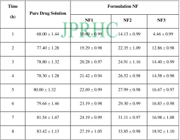
Table 2: Comparative In vitro dissolution profile of different formulations
Time
(h) Pure Drug Solution
Formulation NF
NF1 NF2 NF3
1 68.00 ± 1.44 10.69 ± 0.99 14.13 ± 0.99 4.46 ± 0.99
2 77.40 ± 1.28 19.29 ± 0.98 22.35 ± 1.09 12.86 ± 0.98
3 78.80 ± 1.32 20.28 ± 0.97 24.91 ± 1.16 14.40 ± 0.99
4 78.30 ± 1.28 21.42 ± 0.94 26.52 ± 0.98 14.58 ± 0.98
5 80.00 ± 1.32 22.69 ± 0.99 27.99 ± 0.98 16.67 ± 0.97
6 79.64 ± 1.46 23.19 ± 0.98 29.30 ± 0.99 16.83 ± 0.98
7 81.54 ± 1.67 24.19 ± 0.99 31.11 ± 0.97 16.98 ± 1.08
JPRHC
Research Article
JPRHC Volume 2 Issue 4
293-301
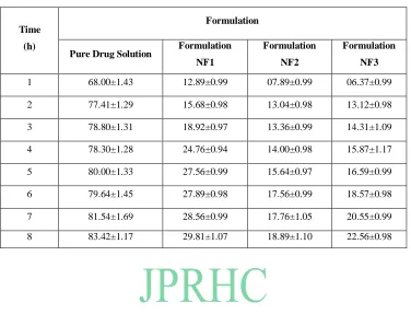
Table 3: Comparative Ex-In vitro dissolution profile of different formulations
Time
(h)
Formulation
Pure Drug Solution Formulation
NF1
Formulation
NF2
Formulation
NF3
1 68.00±1.43 12.89±0.99 07.89±0.99 06.37±0.99 2 77.41±1.29 15.68±0.98 13.04±0.98 13.12±0.98 3 78.80±1.31 18.92±0.97 13.36±0.99 14.31±1.09 4 78.30±1.28 24.76±0.94 14.00±0.98 15.87±1.17 5 80.00±1.33 27.56±0.99 15.64±0.97 16.59±0.99 6 79.64±1.45 27.89±0.98 17.56±0.99 18.57±0.98 7 81.54±1.69 28.56±0.99 17.76±1.05 20.55±0.99 8 83.42±1.17 29.81±1.07 18.89±1.10 22.56±0.98
Fig 1: Photomicrograph of niosomes
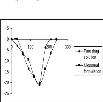
Fig. 2. Change in IOP. Sequence-1. Drug
formulations was administered 30 min before the administration of dextrose solution.
-20
-15
-10
-5
0
0
100
200
300
Time (min)
IO
P
D
iff
e
r
e
n
c
e
Pure drug
solution
JPRHC
Research Article
JPRHC Volume 2 Issue 4
293-301
Fig. 3. Change in IOP. Fig.4. Change in IOP.
The comparative in vitro drug release profile summarized in Table-3, for pure drug solution and for each formulation. It was observed that pure drug solution released approximately 78% of drug within 2 h, while niomal formulations NF1, NF2 and NF3 showed 18.92 %, 22.50 % and 29% drug release respectively in 8 h. The result of in vitro drug release profile of formulations showed that niosomal formulations provides the prolonged release of drug when compared to pure drug solution. Similarly, the comparative ex - in vitro drug release profile was summarized in Table 3, for pure drug solution and for each formulation. It was observed that pure drug solution released major amount of drug within 1 h, while the niosomal formulations NF3 and NF2 showed 18.89 % and 22.56 % drug release respectively in 8 h. Hence, from comparative in vitro and ex-in vitro drug release data of brimonidine tartrate from liposomes and pure drug solution, it has been observed that the amount of drug release remained similar. Further the delayed drug release rate may be attributed largely to the drug transport by diffusion controlled mechanism resulting in prolonged drug release
profile. The in vitro and ex- in vitro drug release studies showed that, there was slow and prolonged release of drug from all the formulations and followed zero order kinetics. This indicated that the drug release was independent of concentration of drug entrapped.
To study the in vivo performance of prepared formulations, intraocular pressure lowering activity was determined. It was found that in sequence 1, where drug formulations were administered 30 min before the administration of dextrose solution (Fig 2), intraocular pressure lowering activity with liposomal formulation was sustained for longer period (3-4 h). However marketed product though showed activity within 30 min, but could not sustain for more than 60 min. It was found that the IOP difference produced between pure drug solution and niosomes is very significant. Niosomal formulations sustained the action for prolonged period (Fig 2) of time (240 min). Hence, the difference in IOP lowering activity with pure drug solution did not last long and sustainability of action was also not observed. In sequence 2, formulation and dextrose solution were administered together (Fig 3), and sustained action
-20 -15 -10 -5 0 5
0 100 200 300
Pure drug solution Niosomal formulation
-25 -20 -15 -10 -5 0 5
0 100 200 300
JPRHC
Research Article
JPRHC Volume 2 Issue 4
293-301
was observed compared to pure drug solution. Further, extent of IOP lowering activity was found to be better in comparison to marketed product. Whereas for marketed product, the effect was observed immediately. The duration of intraocular pressure lowering activity remained more or less similar to that of sequence 1. In sequence 3, dextrose solution was administered before the administration of niosomal formulation and marketed product (Fig 4) and intraocular pressure lowering activity with niosomal formulation was not found to be significant. However, better duration of action was visible to some extent with niosomes. It was also observed that at the end of 240 min, the effect of all the formulations was found to be nil. This may be due to the fact that the induced IOP by injecting 5% dextrose solution did not last long. Nevertheless, the experimental data justified the sustained action of liposomal formulation in comparison to marketed eye drops. This may be the reason why in sequence 3, the effect of niosomal formulation was not observed to the greater extent as in the case of sequence 1 and sequence 2. The drug formulations were administered 30 minute after the induction of glaucoma.
However, the better reduction in IOP with niosomes may probably due to the better partitioning of drug between vesicle and eye corneal surface. Further, it is believed that the release of drug from niosome will increase the local concentration at corneal surface, after the release from vesicle depending on passive diffusion of drug molecule across the corneal barrier. The longer contacts time of vesicles at corneal surface, leads to higher bioavailability of drug. Thus the niosome acts as drug carrier, which changes
rate and extent of absorption resulting in reduction of IOP for prolonged period of time.
Result of stability study was found to be satisfactory and acceptable. The niosomes stored at refrigerator (2 °C to 8 °C), and room temperature, found to be sufficiently stable with no change in shape and no significant difference in drug content.
CONCLUSION:
Niosomes of brimonidine tartrate allowed a significant vesicular carrier system for therapeutic effectiveness in terms of duration of action and decrease in dose frequency. The in vitro and ex- in vitro drug release studies showed that, there was slow and prolonged release of drug from all the formulation and followed zero order kinetics. The in vivo intraocular pressure lowering activity of niosome formulation was found to be significant and sustained for long period of time which encourages its physiological effectiveness. Thus niosomes offer a promising avenue to fulfill the need for an ophthalmic drug delivery system that not only has the convenience of a drop, but that can localize and maintain drug activity at its site of action for a longer period of time thus allowing for a sustained action; minimizing frequency of drug administration with patient compliance.
ACKNOWLEDGEMENTS:
The authors wish to acknowledge Nitte University, Mangalore (Karnataka) India, for providing the necessary facilities and financial support to carry out this project
JPRHC
Research Article
JPRHC Volume 2 Issue 4
293-301
REFERENCES
1. Amaranth Sharma, Uma Sharma. Liposome in drug delivery: progress and limitation. Int J Pharma. 154;1997:123-40.
2. Sandeep KS, Meenakshi Chauhan* and b Narayanapillay Anilkumar. Span-60 niosomal oral suspension of fluconazole: Formulation and in vitro evaluation. J Pharm Res Health Care. 1(2); 2009: 142-56 3. Indu Pal K, Alka G, Anil K, Deepika A.
Vesicular systems in ocular drug delivery: An overview. Int J Pharma. 269(1); 200: 41-14.
4. Dong H Shin, Bernice K, Glover, Sooncha, Yong Y, Chaesik kim, Khoa D. Naguyen. Long term brimonidine therapy in glaucoma patients with apraclonidine allergy. Ame J Ophtha;.127(5); 1999:511-15.
5. Anuja Bhandari, George A, E Michale, Selim Orgul, Lin Wang. Effect of brimonidine on optic nerve blood flow in rabbits. Ame J Ophtha. 128(5-12);1999: 601-05.
6. Bangham AD, Standish MM, Watkens JC. Liposomes by film hydration technique. J C J Mol Biol. 13; 1965: 238.
7. Law SL, Shih CL. Characterization of calcitonin-containing liposomes formulations for intranasal delivery. J Microencapsul. 18; 2001:211-21.
8. Deepika Aggarwal, Alka Garg, Indupal K. Development of topical niosomal preparation of acetazolamide: preparation and evaluation. J Pharm Pharmacol. 56; 2002: 1509-17
9. Kaur IP, Singh M, Kanwar M. Formulation and evaluation of ophthalmic preparation of acetazolamide. Int J Pharm. 199; 2000:119-27.
10. Armengol X, Estelrich J. Physical stability of different liposome compositions obtained by extrusion method. J Microencapsul. 12; 1995:525-535.
11. Higuchi T. Mechanism of sustained action medication. Theoretical analysis of rate release of solid drugs dispersed in solid matrices. j pharm sci 52; 1963: 1145-49.
AUTHORS AFFILIATION AND ADDRESS FOR
COMMUNICATION:
Nitte Gulabi Shetty Memorial Institute Pharmaceutical Sciences, Mangalore.Karnataka 574160