IN VIVO TOXICITY OF NANOPARTICLES: MODALITIES AND
TREATMENT
Sufia Naseem
[a]*, Manzoor Ahmad Gatoo
[a]#, Ayaz Mahmood Dar
[b]§, and
Khusro Qasim
[c]Keywords: nanoparticles; surface charge; aggregation; lymphatic, circulatory system and respiratory system.
In the present scenario, the burgeoning field of nanotechnology is playing central role in various real world applications. Researches engrossing nanoparticles are evolving at a rapid pace owing to which engineered nanomaterials are increasingly becoming part of daily life in the form of cosmetics, food packaging, drug delivery, therapeutics, biosensors, etc. It is intrigued that the properties of nanoparticles which bestow them their unique physicochemical characteristics could also lead to adverse biological consequences such as increased uptake and interaction with the biological systems. Nanomaterials, due to their small size could enter the body through various semi open anatomical interfaces and can penetrate through cells and organelles and disrupt their normal function, which could lead to tissue inflammation, altered cellular redox balance or even cell death. Nanoparticles unlike larger particles can transverse through the circulatory/lymphatic to various vital organs of the body including nervous systems and brain. Nanomaterials could lead to various allied illnesses including bronchitis, asthma, lung and liver cancer, Parkinson’s disease, Alzheimer’s disease, Crohn’s disease, heart disease and colon cancer. Reckoning with the unprecedented applicability’s of nanomaterials in daily life, avenues for direct or indirect exposures of nanoparticles to human beings increases, which raises concern about their role in vivo toxicity. This necessitates intensive research to have knowledge of the various routes of nanoparticle exposure and their effects upon the human health. This review is an attempt to evaluate the various modes of exposure of nanoparticles in human beings, mechanism of toxicity, their fate inside body and adverse health effects.
Corresponding Authors
E-mail: [email protected]*, [email protected]#,
[a] Department of Biochemistry, Jawaharlal Nehru Medical
College, Aligarh Muslim University, Aligarh, India
[b] Department of Chemistry, Aligarh Muslim University,
Aligarh, India
[c] Department of Mechanical Engineering, Aligarh Muslim
University, Aligarh, India
Introduction
Nanotechnology has been defined as the design, characterization, production and application of structures, devices and systems by controlling shape and size at nanometre scale while nanoparticles are classified as particles having one or more dimensions in order of 100 nm or less.1 Research engrossing nanoparticles is currently an area of intense scientific interest due to their wide potential applications in diverse fields including biomedical, optical and electronics field. Semiconductors, metallic, magnetic and polymeric-nanosystems made possible the early diagnosis and new treatments for many diseases including multiple sclerosis, atherosclerosis and cancer.2 For instance, super paramagnetic iron oxide NPs (SPIONs) have been used for magnetic labelling, cell isolation, hyperthermia, imaging and controlled drug release.3 Nanomaterial based entities have potential applications in developing smart targeted drug delivery vehicles, biocompatible implants, sensor and diagnostic systems and besides biomedical applications, NPs are also used commercially in various products such as electronic components, scratch-free paint, sports equipment, cosmetics, food colour additives and surface coatings. Reckoning their potential applications, engineered nanomaterials with new chemical and physical
properties are being produced on daily basis; resulting in increased exposure to environment and human beings, yet there is little understanding of the unique toxicological properties of NPs and their long-term impact on human health and infact there is considerable gap between the available literature on the nanomaterials production and toxicity evaluations.
Lately, air pollution studies have generated indirect evidence for the role of combustion derived nanoparticles (CDNP) in driving adverse health effects in susceptible groups. Reasonably, owing to the nano size, NPs are capable of entering the human body by exploiting various semi open anatomical surfaces of human beings viz. skin, respiratory tract and gastrointestinal tract (GIT). It has been evidenced that through inhalation they gain entry into the respiratory tract (most common route of exposure) and through ingestion or nasopharyngeal route they could enter the GIT. They could also breach the intact skin tissues and beside these, they could translocate the systemic circulation and through the blood stream could reach various organs of the body such as liver, spleen, bone marrow and nervous system including brain. Moreover, implants and injections also serve minor role for their entry into the body.
Since anquity, human beings have been directly or indirectly exposed to NPs. Particle toxicology and the consequent adverse health effects of asbestos fibres and coal dust, serve as landmark references to the development of nanotoxicologic concepts.4 Nanomaterials owing to their diverse chemical, optical, magnetic and structural properties
may display different toxicological profiles, thus
titanium dioxide, alumina, zinc oxide, carbon black, and carbon nanotubes and “nano-C60”. Nanoparticle overload can instigate stress reactions that could lead to inflammation and weaken the body’s defence system. Non-degradable or slowly degrading nanoparticles not only accumulate in bodily organs but can also interact or interfere with biological processes inside the body.
Although size is the key factor in determining the potential toxicity of a particle, other properties including chemical composition, shape, surface structure, surface charge, aggregation, presence of functional group and solubility play their role in imparting toxicity. Chemical composition of nanoparticles is responsible for their reactivity while their surface charge is responsible for their electrostatic interactions. Surface area to volume ratio of these particles increases their interaction with the surrounding molecules. Of note, upon exposure to tissue and fluids, they could immediately adsorb onto their surface and affect their functionalities. Hydrophobicity and sometimes lipophilic groups of nanoparticles allows them to interact with proteins and membranes, respectively. Besides complementary nanostructure could cause inhibition of enzyme activity either competitive or noncompetitive while accumulation of an inert particle in the body could trigger tissue formation around the foreign entity leading to formation of a scar tissue.
Mechanism of nanoparticle toxicity in the body
Unlike larger particles, nanoparticles may be taken up by cell mitochondria and the cell nucleus and can cause DNA mutation and major structural damage to mitochondria, even resulting in cell death. Nanomaterials like silver-coated gold nanoparticles, fullerenes, block copolymer micelles and carbon nanotubes may be capable of localizing to mitochondria and inducing apoptosis, ROS formation, DNA damage, cell-cycle arrest, mutageneses which are possible intrigued as sources of in vivo toxicity.6
The toxicity of nanomaterials can occur through three different mechanisms in the body: i) dissolution process of nanomaterials in biological media, ii) catalyst properties of nanomaterials and iii) reduction and oxidation (Redox) evolution of the surfaces.7 It has been demonstrated that nanoparticles could penetrate into cells and by transcytosis could transverse through epithelial and endothelial cells into the lymphatic circulation to reach various sensitive parts of the body, such as bone marrow, brain, spleen, heart and nervous system including brain.
In experimental models, it has been observed that nanomaterial exposure ensues in oxidative stress, ROS generation, mitochondrial perturbation, inflammation, brain and peripheral nervous system injury, enzyme activity loss, atherogenesis, thrombosis, stroke, myocardial infarction, autoimmunity and DNA damage leading to mutagenesis and carcinogenesis. Of note, nanoparticles can reach the cell mitochondria and cell nuclei, which in turn cause DNA mutation and induce major structural damage and cell death 8 but most intracellular and in vivo toxicities from NPs arise
from the production of excess reactive oxygen species (ROS).9,6
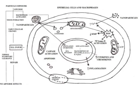
The Figure 1 depicts the interaction of nanoparticles with the cell and various mechanisms involved in nanotoxicity.
Figure 1. Nanoparticles interaction with the cell and various mechanisms involved in toxicity
Reactive oxygen species are both physiologically necessary and potentially destructive. Although moderate levels of ROS play specific roles in the modulation of several cellular events10 but increased ROS levels is indicative of oxidative stress and can damage cells by peroxidizing lipids, altering proteins, disrupting DNA, interfering with signalling functions and modulating gene transcription4,11,12 and finally ending up in cancer, renal disease, neurodegeneration, cardiovascular or pulmonary disease. Toxicity from ROS can be more pronounced in the central nervous system (CNS) due to the high content of unsaturated fatty acids, which are susceptible to peroxidation.12 ROS also play a role in the development of vasculopathies, including those that define atherosclerosis,
hypertension and restenosis after angioplasty.13
Accumulation of NPs in the liver and spleen leads to imbalance in ROS homeostasis and antioxidant defences, making these organs main targets of oxidative stress.
Nel et al.,14 described nanoparticle-induced oxidative stress affects cell signalling in three stages. A low level of oxidative stress enhances transcription of defence genes through transcription factor nrf2. A higher level of oxidative stress activates inflammation signalling through NFkB, and very high levels are connected with activation of apoptotic pathways and necrosis. Changing these signalling pathways in cells is associated with the carcinogenic effects of NPs. Peterson and Nelson15 reviewed the ROS toxicity of NPs towards the cell nucleus and DNA material and observed that it leads to double strand breaks, which are considered the most lethal type of oxidative DNA damage. Damage to mtDNA due to excess amount of ROS is reported to be associated with several clinical syndromes such as neurogenic muscle weakness, ataxia and retinitis pigmentosa, mitochondrial encephalomyopathy, lactic acidosis, stroke like episodes, retinitis pigmentosa, cardiac conduction defect and elevated cerebrospinal fluid protein.16
Nanoparticles: Routes of entry, translocation and
their clearance from the body
Nanostructures can enter the body via six principle routes viz. intra venous, dermal, subcutaneous, inhalation, intraperitoneal, oral and through inhalation and amongst these airborne inhalation of nanosized particles (NPs) i.e. entry through the respiratory tract is the most likely route of exposure to nanoparticles. Exposure via other routes has not been studied in detail and is less plausible unless it is by direct ingestion through food or drug delivery, dermal contact through application of oils and skin creams, or as a contaminant in water through a nanoparticle-treated membrane system. Absorption can occur where the nanostructures first interact with biological components and then they can distribute to various organs in the body and may remain the same structurally, be modified, or metabolized and can reside in the cells for an unknown amount of time before leaving to move to other organs or to be excreted.17 Figure 2 shows possible routes of entry of nanoparticles in the body and their adverse health effects.
Figure 2. Possible routes of entry of nanoparticles in the body and their adverse health effects.
Respiratory uptake of nanoparticles
Inhalation of airborne nanoparticles through the respiratory tract is the most common means of entry in humans as mentioned above. Of note, lungs represent the primary entry port for inhaled particles. It has been shown that the inhaled nanoparticles are efficiently deposited by diffusional mechanisms in all regions of the lungs and with decrease in the particle size, notably below 500 nm, the deposition increases due to increasing diffusion mobility. Hoet et al.18 summarized that most nanosized spherical solid materials easily enter the lungs. Particles of different sizes deposit differently in the airways, as well as the alveolar region so depict different effects in different parts of the lungs which is particularly important in children with developing lungs and in asthma and COPD patients. The smaller the particles, the higher the probability that the particle will hit the epithelium of a lung structure. Spherically shaped solid material with particle diameters less than 10 microns can reach the gas exchange surfaces.
Larger diameter particles, with size and diameters of 10 microns or more tend to be deposited further up in the respiratory tract as a result of gravitational settling, impaction, and interception.19 Many larger diameter fibres are deposited at “saddle points” in the branching respiratory tree. On the epithelium walls of the respiratory tract, particles contact first the mucous or serous lining fluid and its surfactant layer on top. Therefore, the fate of particle compounds soluble in this lining fluid of respiratory tract needs to be distinguished from slowly dissolving or even insoluble compounds.
alveolar macrophages, splenic macrophages and Kupfer cells, respectively. Translocation after inhalation of NP in the lung is not only towards the liver but also spleen, kidneys, brain and heart.25
Nervous system uptake of nanoparticles
Inhaled nanoparticles are shown to reach the nervous system via the olfactory nerves26,4 or by breaching the blood-brain-barrier.26,27 The nasal and tracheo-bronchial regions have many sensory nerve endings.4 More recent studies confirm the uptake of inhaled nanoparticles from olfactory mucosa via the olfactory nerves.4,26,28 Rat inhalation studies with 30 nm magnesium oxide29 and 20-30 nm carbon28 nanoparticles indicate that nanoparticles translocate to the olfactory bulb. 29
The passage of nanoparticle to the nervous system is also possible via the blood-brain-barrier. Regarding the passage of nanoparticles, the blood-brain-barrier permeability is dependent upon the charge of nanoparticles30 and allows a larger number of cationic nanoparticles to pass compared to neutral or anionic particles, due to the disruption of its
integrity30 Increased blood-brain-barrier permeability
observed in hypertension, brain inflammation26,respiratory tract inflammation27 allows nanoparticles access to the nervous system.
Lymphatic systems uptake of nanoparticles
Translocation of nanoparticles to lymph nodes is a topic of intense investigation today for drug delivery and tumor imaging.22 Several studies have shown that interstitially injected particles pass preferentially through the lymphatic system and not through the circulatory system, probably due to permeability differences22 and get located in the lymph nodes.22 The free nanoparticles reaching the lymph nodes are ingested by resident macrophages.31 Nanoparticles that are able to enter the circulatory system can also gain access to the interstitium and from there could be drained through the lymphatic system to the lymph nodes as free nanoparticles and/or inside macrophages. 22, 31
Circulatory system uptake of nanoparticles
Nanoparticles, unlike larger particles, are able to translocate across the respiratory epithelium after being deposited in the lungs.4,32 Once, they have crossed the respiratory epithelium, they may persist in the interstitium for years, or they may enter the lymphatic system and circulatory system.22 Inhalation or instillation studies in healthy animals have shown that metallic nanoparticles with size smaller than 30 nm pass rapidly into the circulatory system4,22,32 while non-metallic nanoparticles with size between 4 and 200 nm pass feebly or do not pass at all.33 In contrast, subjects suffering from respiratory and circulatory diseases have higher capillary permeability, allowing fast translocation of metallic or non-metallic nanoparticle into circulation.33 From the circulatory system, long-term translocation to organs such as the liver, heart, spleen, bladder, kidney, bone marrow is possible, depending on the duration of exposure.4 Evidence of rapid translocation of metal nanoparticles from lungs into the circulation and to organs has been provided by animal studies. These results have shown that nanoparticles with diameters of 30 nm
(Au)4, 22 nm (TiO2)32 could be located in pulmonary capillaries; whereas particles of 15 nm (Ag)25 and welding fumes34 could be located in blood, liver, kidney, spleen, brain, and heart. Animal studies on rats with inhalation of titanium dioxide nanoparticles (22 nm diameter) have shown that they could translocate to the heart as evidenced by their presence in the heart connective tissue (fibroblasts).32 Within 30 minutes post exposure, large quantities of intratracheally instilled gold nanoparticles (30 nm) has been found in platelets inside of pulmonary capillaries of rats.4
On the contrary, there is no conclusive evidence for fast translocation of carbon-based non metal nanomaterials in systemic circulation.
Nanoparticle uptake by red blood cells is entirely dictated by size, due to absence of phagocytic receptors27 while the nanoparticle charge or material type plays feeble roles. On the contrary, nanoparticle charge plays an essential role in their uptake by platelets; thereby influencing blood clot phenomena’s.35 Uncharged polystyrene particles do not have any effect on blood clots formation. Negatively charged nanoparticles significantly inhibit thrombi formation, while positively charge nanoparticles enhance platelet aggregation and thrombosis35 due to interaction of the positively charged nanoparticles with negatively charged platelets leading to reduction of their surface charge, making them more prone to aggregation. Until now, it was thought that blood clots can be formed due to three main causes: when the blood flow is obstructed or slowed down, when the vascular endothelial cells are damaged, or due to the blood chemistry. However, it seems possible, in the view of recent findings that nanoparticles may act as nucleating centres for blood clots.36 Microscopic and energy dispersive spectrometry (EDS) analysis of blood clots from patients with blood disorders revealed the presence of foreign nanoparticles.36 Most notably, patients with the same type of blood disorder show fibrous tissue clots embedding nanoparticle with different composition.
Organ uptake of nanoparticles
Gastro-intestinal tract uptake of nanoparticles
Endogenous sources of nanoparticles in the gastro-intestinal tract are derived from gastro-intestinal calcium and phosphate secretion.39 Exogenous sources are particles from food such as colorants – titanium oxide, pharmaceuticals, water, or cosmetics including toothpaste, lipstick, dental prosthesis debris38 and inhaled particles.25 The dietary consumption of nanoparticles in developed countries is estimated around 1012 particles/person per day38 and mainly consists of TiO2 and mixed silicates. These nanoparticles do not degrade in time and accumulate in macrophages. A portion of the particles cleared by the mucociliary escalator can be subsequently ingested into the gastro-intestinal tract. Also, a small fraction of inhaled nanoparticles was found to pass into the gastrointestinal tract.25 Particles that penetrate the mucus reach the enterocytes and are able to translocate further.18 Diseases, such as diabetes, may lead to higher absorption of nanoparticles in the gastrointestinal tract. 18 The extent of particles absorption in the gastro-intestinal tract is affected by size, surface chemistry and charge, length of administration, and dose.18 The absorption of particles in the gastro-intestinal tract decreases with increase in size of nanoparticles. For example study of polystyrene particles with size between 50 nm and 3 μm indicated that their uptake is 6.6%, 5.8%, 0.8% and 0% for nanoparticles of size 50 nm, 100nm, 1μm and 3 μm respectively.40 The kinetics of particles in the gastro-intestinal tract depends strongly on the charge of the particles. Positively charged latex particles are trapped in the negatively charged mucus while negatively charged latex nanoparticles diffused across the mucus layer and became available for interaction with epithelial cells.18
It is generally assumed that nanomaterials do not remain in gastro-intestinal tract for indefinite periods.18 Most of the studies of ingested nanoparticles have shown that they are eliminated rapidly; 98% in the faeces within 48 hours and most of the remainder via urine.4 However, other studies indicates that certain nanoparticles can translocate to blood, spleen, liver, bone marrow, lymph nodes, kidneys, lungs, and brain, and can also be found in the stomach and small intestine.40
Dermal uptake of nanoparticles
The skin is composed of three layers; epidermis, dermis and subcutaneous and the outer portion of the epidermis is called stratum corneum.18 As with many subjects involving nanoparticles, dermal penetration is still controversial.26 Several studies have shown that nanoparticles are able to
penetrate the stratum corneum.4,26,41,42 Nanoparticles
penetration through the skin typically occurs via the hair follicles41, flexed42 and broken skin.4 Intracellular nanoparticles penetration is also possible, as demonstrated by in vitro experiments.43 MWCNTs are internalized by human epidermal keratinocytes in cytoplasmic vacuoles and
induce the release of pro-inflammatory mediators.43
Spherical particles with diameter between 750 nm and 6 microns selectively penetrate the skin at hair follicles with a maximum penetration depth of more than 2400 microns (2.4 mm).43 Broken skin facilitates the entry of a wide range of larger particles (500 nm - 7 μm).4 Translocation of nanoparticles from skin into the lymphatic system occurs by soil particles found in lymph nodes as has been revealed in
patients with podoconiosis. Neuronal transport of small nanoparticles along sensory skin nerves may also be possible, in a similar way to the proven path for herpes virus.4 Penetration of the TiO2 nanoparticles (found in commercially available sunscreens) into the skin 44 is believed to depend on percentage of nanoparticles in the sunscreen. For example, the application of a sunscreen containing 8% nanoparticles (10-15nm) onto the skin of humans showed no penetration, while oil in water emulsions showed penetration.44
Dermal exposure is another important uptake source for NPs especially because of the increased interest in the use of TiO2, ZnO and other nanoparticles for protection against ultraviolet rays in various dermal creams, lotions, and cosmetics. Nanosized particles can enter through the unbroken skin during the flexing of the wrist. 41 It has been observed that the flexing of the skin can lead to uptake of micrometer-long fluorescent beads. TiO2 particles (5-20 nm) are able to penetrate into the skin cells and interfere with the immune system while anatase TiO2 nanoparticles (10 nm and 20 nm) induced oxidative DNA damage, lipid peroxidation, and micronuclei formation.
Nanoparticle uptake via injection
Injectable nanoparticles are been involved in drug delivery studies. The translocation of nanoparticles following injection depends on the site of injection;
intravenously injected nanoparticles quickly spread
throughout the circulatory system, with subsequent translocation to organs such as liver, spleen, bone marrow, lymph nodes4, small intestine, brain, lungs; whereas intradermal injection leads to lymph nodes uptake and intramuscular injection is followed by lymphatic and
neuronal system uptake.4 Nanoparticles injected
intravenously are retained longer in the body than ingested ones. For example, 90% of injected functionalized fullerenes
are retained after one week of exposure.4 Coating
nanoparticles with various types and concentrations of surfactants before injection significantly affects their distribution in the body.45 For example, coating with polyethylene glycol or other substances almost completely prevents hepatic and splenic localization.4,45 Similarly modification of nanoparticles surface with cationic compounds facilitates arterial uptake by up to 10 fold. A common side effect of injecting nanoparticles intravenously is hypersensitivity reaction.
Nanoparticle generation by implants
Adverse Effects of Nanoparticles and their
treatment
Recent researches has lead to changes in terminology of nanotechnological studies and brought about the realization that no particles are completely inert, and even low concentrations of particles could have negative health effects. It has been demonstrated the interaction of nanoparticles with biological systems can result in allergy47, fibrosis, organ failure, Inflammation, cytotoxicity48, ROS generation49 and DNA/tissue damage.50 Recent endeavours from the research fraternities has shown that nanoparticles inhalation can affect the immune system defence ability to combat infections51 and are able to modulate the intrinsic defensive function of macrophages, affecting their reactivity to infections. Several types of nanoparticles (such as ZrO2) enhance the expression of some viral receptors leading to
excessive inflammation51 while exposure to other
nanoparticles (SiO2, TiO2) leads to a decrease in the expression of some other viral and bacterial receptors, leading to lower resistance to some viruses or bacteria. Further, most human-made nanomaterials do not appear in the environment, so living organisms possibly do not embody an appropriate immune system to deal with these nanoscale products.
Adverse health effects of respiratory uptake of nanoparticles
The adverse health effects of nanoparticles uptake by respiratory system depend on the residence time in the respiratory tract27 as well as genetic susceptibility and health status.21 Smaller particles have a higher toxicological attributes than larger particles of the same composition and crystalline structure, and generates consistently higher inflammatory reaction in the lungs. Smaller nanoparticles are correlated with adverse reactions such as impaired macrophage clearance, inflammation, accumulation of particles, and epithelial cell proliferation, followed by
fibrosis, emphysema, and the appearance of tumors.20
Chronic (two year) high-dose inhalation exposures in rats with poorly soluble, low toxicity dusts can ultimately produce pulmonary fibrosis and lung tumours via an “overload mechanism” but same has not been reported in mice or hamsters, under similar chronic conditions. Treatments for nanoparticles inhalation include those that act to enhance mucociliary clearance, and those that reduce the effects of oxidation and inflammation. Anti-inflammatory medicine (sodium cromoglycate) was found to strongly reduce airway inflammation caused by diesel exhaust nanoparticles.52 Antioxidant vitamins (particularly vitamin C)53, rosmarinic acid54 and a high intake of fresh fruit and some vegetables have a protective effect against lung diseases.53
Adverse health effects of neuronal uptake of nanoparticles
Experimental evidence suggests that the initiation and
promotion of neurodegenerative diseases, such as
Alzheimer’s disease, Parkinson’s disease, Pick’s disease, are associated with oxidative stress and accumulation of high concentrations of metals (like copper, aluminum, zinc, but
especially iron) in brain regions associated with function loss and cell damage.55 However, it is not known if the
presence of metals in brain of subjects with
neurodegenerative diseases is due to nanoparticles themselves translocating to the brain or their soluble compounds.34 Recent studies on DNA damage in nasal and brain tissues of canines exposed to air pollutants shows evidences of chronic brain inflammation, neuronal dysfunction, and similar pathological findings with those of early stages of Alzheimer’s disease.27 Epidemiological studies show a clear association between inhalation of dust
containing manganese and neurological diseases in miners56
and welders.57 Some welders develop Parkinson’s disease much earlier in their life, usually in their mid forties, compared to the sixties in the general population.57
Antioxidants and metal chelators are treatment options for the adverse health effects caused by the neuronal uptake of nanoparticles. Functionalized fullerenes58 and nanoparticles made of compounds holding oxygen vacancies show great antioxidant properties.59 It appears that the antioxidant properties depend upon the structure of the particle but they are independent of its size within 6-1000 nm.
Adverse health effects of circulatory system uptake
Translocation of nanoparticles into the circulatory system was correlated with the appearance of thrombi (or blood clots)35,52 and cardiovascular malfunction.60 Thrombosis occurs during the first hour after exposure of nanoparticles. There is a clear dose-dependent response correlating the quantity of pollutant administered and the observed thrombus sizes.18,52 It is clear from clinical and experimental evidence that inhalation of nano and microparticles can cause cardiovascular effects.60 Although causal link between the particles in the lungs and cardiovascular effects is not entirely understood but it is believed that the pulmonary inflammation caused by the particles triggers a systemic release of cytokines, resulting in adverse cardiovascular effects. However, recent studies on animals25 and humans61 have shown that nanoparticles diffuse from the lungs into the systemic circulation, and then are transported to the organs, demonstrating that cardiovascular effects of instilled or inhaled nanoparticles can arise directly from the presence of nanoparticles within the organism.
Adverse health effects of liver and kidney uptake
Adverse health effects of gastro-intestinal tract uptake
In the intestinal tract there is a complex mix of compounds, enzymes, food, bacteria, etc., that can interact with ingested nanoparticles and reduce their toxicity.18 Nanoparticles of carbon, ceramic filosilicates, gypsum, sulphur, calcium, silicon, stainless steel, silver, and zirconium39 have been constantly found in colon tissue of subjects affected by cancer, Crohn’s disease, and ulcerative colitis, while in healthy subjects they could not be traced.38 Recently, it has been suggested that there is an association between high levels of dietary nanoparticles (100 nm-1μm) and Crohn's disease.63 Exogenous nanoparticles were found in macrophages accumulated in lymphoid tissue of the human gut, the lymphoid aggregates being the earliest sign of lesions in Crohn's disease.63 It is believed that genetic predisposition plays a role in development of Crohn’s disease increasing risk of some members of the population after intake of nanoparticles.11 Some evidence suggests that dietary nanoparticles may exacerbate inflammation in Crohn’s disease.64 The diseases associated with gastro-intestinal uptake of nanoparticles including Crohn’s disease and ulcerative colitis have no cure and often require surgical intervention.
Adverse health effects of dermal uptake
Many manufacturing processes pose an occupational health hazard by exposing workers to nanoparticles and small fibres, as suggested from the intracellular uptake of MWCNTs by human epidermal keratinocytes.43 This can explain beryllium sensitization in workers wearing inhalation protective equipment exposed to nanoparticulate beryllium.42 Also, this may be relevant for latex sensitivity and other materials that provoke dermatologic responses. Lymphatic system uptake of nanoparticles via the dermis is shown to cause podoconiosis65 and Kaposi’s sarcoma.66 Titanium dioxide commonly used as a physical sunscreen although reflects and scatters UVB and UVA light rays but can absorbs a substantial amount of UV radiation which in aqueous media leads to the production of reactive oxygen species which can cause substantial damage to DNA.67 Reports regarding the toxicity of titanium dioxide nanoparticles in the absence of UV radiation are contradictory. Nanoparticles were seen to have no inflammatory effect or genotoxicity in rats68 while several other studies reported that titanium dioxide caused chronic pulmonary inflammation in rats.69
It is known that silver has a beneficial antibacterial effect when used as a wound dressing, reducing inflammation and facilitating healing in the early phases70 but the same property that bestow their antimicrobial attributes may render them toxic to human cells. It has been reported that the concentrations of silver that are lethal for bacteria are also lethal for both keratinocytes and fibroblasts70 raising serious concerns on the applicabilities for human benefits.
Conclusion
With the rapid increase in the use of nanomaterials in everyday consumer products, manufacturing processes and medical products, it is obvious that people involved in
production as well as consumers could have exposure to these potentially toxic nanoparticles. It is argued that the unusual physiochemical properties of nanomaterials that lead advancements in the field of nanotechnology could also be responsible for their potential toxicity to the system. Nanoparticles show varied adverse health effects in respiratory, circulatory, nervous, gastrointestinal and dermal systems. There are various possible routes for their toxicities, however, interactions with the mitochondria and cell nucleus are being considered as main sources of in vivo toxicity. Although in vivo toxicities can occur through diverse mechanisms but the main molecular mechanism involved is induction of oxidative stress which cause damages to biological components through oxidation of lipids, proteins and DNA. Increasing exploration of nanotechnology reveals newer and newer particles with many unique properties; however, there exist considerable wide gap between the available data on the nanomaterials production and in vivo toxicity evaluations which could possibly subjugate further fabrication of NPs owing to increasing health concern. This review indicates that only few specific nanoparticles have been investigated in a limited number of test systems and extrapolation of this data to other materials is not possible. Therefore, despite the existing research database on nanoparticles, no uniformly applicable reports about human toxicity can be given at this time. In addition, limited ecotoxicological data for nanomaterials precludes a systematic assessment of the impact of nanoparticles on ecosystems. The large number of variables influencing toxicity means that it is difficult to generalise about health risks associated with exposure to nanoparticles and each new nanoparticle must be assessed individually and all material properties must be taken into account. For nanomaterials to enjoy status of repute in the application arena, it is crucial to have thorough understanding of their in vivo toxicities and our attempt to shed light on some of their toxicological aspects is a step forward in this regard.
References
1Welsher, K., Yang H., Nat. Nanotechnol., 2014, 9, 198-203 2Chauhan , A., Zubair , S., Tufail, S., Sherwani , A., Sajid , M.,
Raman , S. C., Azam, A., Owais, M., Int. J. Nanomedicine,
2011, 6, 2305-2319
3Hazra, S., Ghosh, N. N., J. Nanosci. Nanotechnol., 2014, 14,
1983-2000
4 Oberdorster, G., Oberdorster, E., Oberdorster, J. Environ. Health Perspect., 2005, 113, 823-839
5Bakand, S., Hayes, A., Dechsakulthorn, F., Inhal Toxicol., 2012, 24, 125-135
6Unfried, K., Albrecht, C., Klotz, L. O., Von Mikecz, A., Grether-
Beck, S., Schins, R. P. F., Nanotoxicology, 2007, 1, 52-71.
7Kumar, C., Nanomaterials-Toxicity, Health and Environmental Issues, Wiley-VCH, 2006
8Alarifi, S., Ali, D., Ahamed, M., Siddiqui, M. A., Al-Khedhairy,
A. A.,Int. J. Nanomedicine, 2013, 8, 189-199
9Sabella, S., Carney, R. P., Brunetti, V., Malvindi, M. A.,
Al-Juffali, N., Vecchio, G., Janes, S. M., Bakr, O. M., Cingolani, R., Stellacci, F., Pompa, P. P. Nanoscale,2014, 6, 7052-7061.
10Halliwell, B., Gutteridge, J. M. C., Free Radicals in Biology and Medicine, Oxford, University Press, Inc, New York, 4th edn,
11Jena, N. R., J. Biosci.,2012, 37, 503-17
12Ray, P. D., Huang, B. W., Tsuji,Y., Cell Signal.,2012, 24,
981-90
13Griendling, K. K., FitzGerald, G. A., Circulation, 2003, 108,
1912-1916
14Rallo, R., France, B., Liu, R., Nair, S., George, S., Damoiseaux,
R., Giralt, F., Nel, A., Bradley, K., Cohen, Y., Environ. Sci. Technol., 2011, 45, 1695-1702
15Petersen, E. J., Nelson, B. C., Anal. Bioanal. Chem., 2010, 398,
613-650
16Kirkinezos, G., Moraes, C. T., Seminars in Cell and Developmental Biology, 2001, 12, 449-457
17Arora, S., Rajwade, J. M., Paknikar, K. M., Toxicol. Appl. Pharmacol., 2012, 15, 151-65
18Hoet, P. H. M., Brüske-Hohlfeld, I., Salata, O. V., J. Nanobiotechnol., 2004, 2, 1-15
19Lippmann, M., Environ. Health Perspec., 1990, 88, 311-317 20Ferin, J., Toxicol. Lett.,2004, 72, 121-125
21Semmler, M., Seitz, J., Mayer, P., Heyder, J., Oberdörster, G.,
Kreyling, W. G., Inhalation Toxicol., 2004,16, 453-459
22Liu, J., Wong, H. L., Moselhy, J., Bowen, B., Wu, X. Y.,
Johnston, M. R., Lung Cancer, 2006, 51, 377-386
23Ng, A. W., Bidani, A., Heming, T. A., Lung, 2004,182, 297-317 24Brown, D. M., Donaldson, K., Stone, V., Respir. Res., 2014, 5,
29
25Takenaka, S., Karg, E., Roth, C., Schulz, H., Ziesenis, A.,
Heinzmann, U., Schramel, P., Heyder, J., Environ. Health Persp., 2001, 109, 547-551
26Borm, P. J. A., Robbins, D., Haubold, S., Kuhlbusch, T., Fissan,
H., Donaldson, K.,Schins, R. P. F., Stone, V., Kreyling, W., Lademann, J., Krutmann, J., Warheit, D., Oberdorster, E.,
Part. Fibre Toxicol., 2006, 3, 11
27Peters, A., Veronesi, B., Calderon-Garciduenas, L., Gehr, P.,
Chen, L. C., Geiser, M., Reed, W., Rothen-Rutishauer, B., Schurch, S., Schultz, H., Part. Fibre Toxicol., 2006, 3, 13
28Oberdörster, G., Sharp, Z., Atudorei, V., Elder, A., Gelein, R.,
Lunts, A., J. Toxicol. Environ. Health A, 2002, 65, 1531-1543.
29Elder, A., Gelein, R., Silva, V., Feikert, T., Opanashuk, L.,
Carter, J., Potter, R., Maynard, A., Ito, Y., Finkelstein, J., Oberdorster, G., Environ. Health Perspect., 2006, 114, 1172-1178
30Lockman, P. R., Koziara, J. M., Mumper, R. J., Allen, D. D., J. Drug Targ., 2004, 12, 635-641
31Shwe, T. T. W., Yamamoto, S., Kakeyama, M., Kobayashi, T.,
Fujimaki, H., Toxicol. Appl. Pharmacol., 2005, 209, 51-61
32Geiser, M., Rothen-Rutishauser, B., kapp, N., Schurch, S.,
Kreyling, W., Schultz, H., Semmler, M., Im Hof, V., Heyder, J., Gehr, P. Environ. Health Perspect., 2005, 113, 1555-1560
33Chen, J., Tan, M., Nemmar, A., Song, W., Dong, M., Zhang, G.,
Li, Y., Toxicol., 2006, 222, 195-201
34Donaldson, K., Tran, L., Jimenez, L. A., Duffin, R., Newby, D.
E., Mills, N., MacNee, W., Stone, V., Part. Fibre Toxicol.,
2005, 2, 10.
35Nemmar, A., Hoylaerts, M. F., Hoet, P. H. M., Dinsdale, D.,
Smith, T., Xu, H., Vermylen, J., Nemery, B., Am. J. Respir. Crit. Care Med., 2002, 166, 998-1004
36Gatti, A. M. Montanari, S. Monari, E. Gambarelli, A. Capitani, F.
Parisini, B., J. Mater. Sci. Mater. Med.,2004, 15, 469-472
37Gatti, M., Rivasi, F., Biomater.,2002, 23, 2381-2387
38Ballestri, M., Baraldi, A., Gatti, A. M., Furci, L., Bagni, A.,
Loria, P., Rapaa, M., Carulli, N., Albertazzi, A.,
Gastroenterology, 2001,121,1234-1238
39Gatti, A. M., Biomater., 2004, 25, 385- 392
40Jani, P., Halbert, G. W., Langridge, J., Florence, A. T., J. Pharm. Pharmacol., 1990, 42, 821-826
41Toll, R., Jacobi, U., Richter, H., Lademann, J., Schaefer, H.,
Blume-Peytavi, U., J. Invest. Dermatol., 2004, 123, 168-176.
42Tinkle, S. S., Antonini, J. M., Rich, B. A., Roberts, J. R., Salmen,
R., DePree, K., Adkins, E. J., Environ. Health Perspec.,
2003, 111, 1202-1208.
43Monteiro-Riviere, N. A., Nemanich, R. J., Inman, A. O., Wang,
Y. Y., Riviere, J. E., Toxicol. Lett., 2005, 155, 377-384.
44Tsuji, J. S., Maynard, A. D., Howard, P. C., James, J. T., Lam, C.
W., Warheit, D. B., Santamaria, A. B., Toxicol. Sci., 2006,
89, 42-50
45Araujo, L., Lobenberg, R., Kreuter, J., J. Drug Targ., 1999, 6,
373-385.
46Hasegawa, M., Yoshida, K., Wakabayashi, H., Sudo, A., Orthopedics, 2012, 36, 606-12
47Maynard, A. D., Nature,2006, 444, 267-269
48Nel, Xia, T., Madler, L., Li, N. Science,2006, 311, 622-627 49Manke, A., Wang, L., Rojanasakul, Y., BioMed Res. Inter.,2013,
doi: 10.1155/2013/942916
50Singh, N., Manshian, B., Jenkins, G. J. S., Griffiths, S. M.,
Williams, P. M., Maffeis, T. G. G., Wright, C. J., Doak, S. H.,
Biomaterials,2009, 30, 3891-3914
51Lucarelli, M., Gatti, A. M., Savarino, G., Quatronni, P.,
Martinelli, L., Monari, E., Boraschi, D., Eur. Cytokin. Net.,
2004, 15, 339-346
52Vermylen, J., Nemmar, A., Nemery, B., Hoylaerts, F., J. Thromb. Haemost., 2005, 3, 1955-1961
53Romieu, Int. J. Tuberc. Lung Diseases., 2005, 9, 362-374 54Risom, L., Moller, P., Loft, S., Mutat. Res., 2005, 592, 119-137 55Liu, G., Mena, P., Harris, P. R. L., Rolston, R. K., Perry, G.,
Smith, M. A., Neurosci. Lett., 2006, 406, 189-193
56Weiss, B., Neurotoxicology,2006, 27, 362-368, 2006
57Antonini, J. M., Santamaria, A. B., Jenkins, N. T., Albini, E.,
Lucchini, R., NeuroToxicol., 2006, 27, 304-310
58Bosi, S., da Ros, T., Spalluto, G., Prato, M., Eur. J. Med. Chem.,
2003, 38, 913-923
59Schubert, D., Dargusch, R., Raitano, J., Chan, S. W., Biochem. Biophys. Res. Commun.,2006, 342, 86-91
60Schulz, H., Hardewr, V., Ibald-Mulkli, A., Khandoga, A.,
Koenig, W., Krombach, F., Radykewicz, R., Stampfl, A., Thorand, B., Peters, A., J. Aerosol Med.,2005, 18, 1-22
61Nemmar, A., Hoet, P. H., Vanquickenborne, B., Dinsdale, D.,
Thomeer, M., Hoylaerts, M. F., Vanbilloen, H., Mortelmans, L., Nemery, B., Circulation, 2002, 105, 411-414,
62Lanone, S., Boczkowski, J., Curr. Mol. Med., 2006, 6, 651- 663 63Lomer, M. C. E., Thompson, R. P. H., Powell, J. J., Proc.
Nutrition Soc.,2002, 61, 123-130
64Lomer, M. C. E., Hutchinson, C., Volkert, S., Greenfield, S. M.,
Catterall, A., Thompson, R. P. H., Powell, J. J., British J. Nutrition, 2004, 92, 947-955
65Ali, M., Afzal, M., Bhattacharya, S. M., Ahmad, F. J., Dinda, A.
K., Expert Opin. Drug. Deliv., 2013, 10, 665-678
66Montella, M., Franceschi, S., Geddes da Filicaia, M., De Macro,
67Shi,H., Magaye,R., Castranova, V., Zhao, J., Part. Fibre Toxicol., 2013, doi:10.1186/1743-8977
68Rehn, Seiler, F., Rehn, S., Bruch, J., Maier, M., Toxicol. Appl. Pharmacol., 2003, 189, 84-95
69Oberdörster, G., Ferin, J., Gelein, R., Soderholm, S. C.,
Finkelstein, J., Environ. Health Perspect., 1992, 97, 193-199
70Poon, V. K., Burd, A., Burns, 2004, 30, 140-147