Multidisciplinary Management of Breast Cancer During Pregnancy
SHLOMITSTRULOVSHACHAR,a,b,fKRISTALYNGALLAGHER,b,cKANDACEMCGUIRE,b,cTIMOTHYM. ZAGAR,b,dAIMEEFASO,e HYMANB. MUSS,a,bRAESHALLSWEETING,gCAREYK. ANDERSa,b
a
Department of Medicine, Division of Hematology-Oncology,bLineberger Comprehensive Cancer Center,cDepartment of Surgery,
d
Department of Radiation Oncology, andeDepartment of Pharmacy, University of North Carolina, Chapel Hill, North Carolina, USA;fDivision of Oncology, Rambam Health Care Campus, Haifa, Israel;gDepartment of Surgery, Vanderbilt University, Nashville, Tennessee, USA
Disclosures of potential conflicts of interest may be found at the end of this article.
Key Words.Breast cancer • Pregnancy • Chemotherapy • Radiation • Surgery • Imaging • Outcomes
ABSTRACT
Background. Although breast cancer during pregnancy (BCDP) is rare (occurring with only 0.4% of all BC diagnoses in female patients aged 16–49 years), management decisions are challenging to both the patient and the multidisciplinary team.
Materials and Methods.Experts in breast cancer at the Uni-versity of North Carolina conducted a targeted literature search regarding the multidisciplinary treatment approaches to BCDP: medical, surgical, and radiation oncology. Supportive care, including antiemetic agents, and imaging approaches were also reviewed.
Results.Review of the literature revealed key points in the management of BCDP. Surgical management is similar to that in nonpregnant patients; pregnant patients may safely undergo breast-conserving surgery. Recommendations should be tai-lored to the individual according to the clinical stage, tumor biology, genetic status, gestational age, and personal
preferences. Anthracycline-based chemotherapy can be safely initiated only in the second and third trimesters. The rate of congenital abnormalities in children exposed to chemotherapy is similar to the national average (approximately 3%). Dosing of chemotherapy should be similar to that in the nonpregnant patient (i.e., actual body surface area). Antihuman epidermal growth factor receptor 2 therapy, radiation, and endocrine treatment are contraindicated in pregnancy and lactation. Care should include partnership with obstetricians. The literature regarding prognosis of BCDP is mixed.
Conclusion. To maximize benefit and minimize risk to the mother and fetus, an informed discussion with the patient and her medical team should result in an individualized treatment plan, taking into account the timing of the pregnancy and the stage and subtype of the breast cancer. Because BCDP is rare, it is essential to collect patient data in international registries.The Oncologist2017;22:324–334
Implications for Practice:Breast cancer during pregnancy is a major ethical and professional challenge for both the patient and the multidisciplinary treatment team. Although the oncologic care is based on that of the non-pregnant breast cancer patient, there are many challenges from regarding the medical, surgical and radiation oncology and obstetrical aspects of care that need to be considered to deliver the safest and best treatment plan to both the mother and developing fetus.
INTRODUCTION
While breast cancer diagnosis during pregnancy (BCDP) is rel-atively uncommon, accounting for only 0.4% of all breast cancer diagnoses ages 16–49 years [1], it represents a sub-stantial challenge to the patient and the multidisciplinary team making management decisions. Despite a desire to treat the pregnant patient with breast cancer as effectively as the nonpregnant patient with breast cancer, the selection and delivery of standard therapies must be modified to ensure safety to both the mother and the fetus. Pregnancy-associated breast cancer (PABC) has been defined in various ways throughout the literature, including but not limited to
breast cancer diagnosed during a pregnancy, during lactation, or up to 1 year (or more) after delivery. For the purposes of this review, we are most focused on the care of the pregnant patient with breast cancer (i.e., BCDP). However, we review literature that refers to the term PABC, which may also include patients diagnosed during lactation or soon after delivery of a child. The overarching goal of this comprehen-sive review is to summarize the oncologic approach of each subspecialty involved in the care of the pregnant patient with breast cancer, including medical, surgical, and radiation oncology perspectives. In addition, it is imperative to involve
maternal-fetal medicine early and throughout the care of the pregnant patient with breast cancer in partnership with the oncologic team. Herein, we provide evidence-based, practical guidance to the optimal approach and treatment of patients diagnosed with breast cancer during pregnancy.
MATERIALS ANDMETHODS
A targeted literature search in PubMed regarding BCDP (includ-ing the terms “pregnancy,” “breast cancer,” and “pregnancy-associated breast cancer,” as well as “breast cancer during pregnancy,” along with drug names [i.e., “trastuzumab” and “paclitaxel”] or other terms by topic [e.g., “survival outcomes”]) was performed by an expert in each respective discipline within our multidisciplinary team at the University of North Carolina at Chapel Hill: medical oncology, radiation oncology, surgical oncology, and pharmacology. The targeted literature search included epidemiology, diagnosis, treatment approach, and outcomes of mother and fetus. The type of evidence (retro-spective/prospective) is noted throughout the article and refer-enced appropriately.
INCIDENCE ANDEPIDEMIOLOGY
Compared with other malignancies, breast cancer is one of the most common cancer diagnoses during pregnancy [2], with an incidence of approximately 15 to 35 per 100,000 deliveries [3]. The incidence of cancer during pregnancy or lactation is increasing [1, 2]. This observation is partially due to increasing age at childbearing across the general population. The mean age of mothers has increased since 2000, especially age at first birth, which increased from 24.9 years in 2000 to 26.3 years in 2014 [4].
Investigators have evaluated the differential biology of PABC, including the incidence of breast cancer subtypes. Genin et al. conducted a retrospective study of 276 young women younger than age 43 years diagnosed with breast cancer, including 14.5% with PABC [5]. Those with PABC were signifi-cantly younger than those with non-PABC (NPABC) and were twice as likely to be diagnosed with a T3–T4 primary tumor. Moreover, PABCs were twice as likely to be human epidermal growth factor receptor 2 (HER2/neu) overexpressing and hor-mone receptor negative as were NPABC. A second study sup-ports the finding that PABC shares more aggressive features compared with NPABC [6]. Among a total of 175 parous and 114 nonparous women diagnosed with breast cancer, those within 2 years of last parity were more likely to have grade 3 tumors, positive lymph nodes, and triple-negative tumors. HER2/neu overexpression and p53 status were similar between groups.
Finally, and consistent with literature surrounding the biol-ogy of young women’s breast tumors [7, 8], Murphy et al. eval-uated the clinicopathologic features of PABC compared with NPABC and how these features affect outcome. As per prior reports, PABC was more likely than NPABC to be associated with tumors that were estrogen and/or progesterone receptor negative, larger in size, lymph node positive, and high grade. While overall survival (OS) was not significantly different between cases and controls (p5.078), hormone receptor sta-tus and nodal class were independent prognostic factors for survival in both groups [9]. Our understanding of the biology of BCDP continues to evolve and remains an area of active
research, with the goal of optimizing treatment options for patients diagnosed with BCDP.
MATERNALOUTCOMES
The literature on the prognosis of women diagnosed with PABC has been mixed (Table 1). It is important to recognize the out-comes of patients with BCDP compared with those who have non-BCDP when making global treatment recommendations for this group of patients. When adjusted for stage and subtype, survival has been reported to be the same for BCDP and non-BCDP, and routine termination of pregnancy after a diagnosis of breast cancer does not improve survival [10]. However, for women who present with advanced disease and poor progno-sis early in the first trimester, termination may be considered to avoid delay in treatment. Among patients who present with low-burden metastatic disease past the first trimester, empha-sis on single-agent medical therapy to control symptoms while allowing term delivery should be the focus.
Among patients who present with low-burden
meta-static disease past the first trimester, emphasis on
single-agent medical therapy to control symptoms
while allowing term delivery should be the focus.
Several studies report similar maternal outcomes for PABC and NPABC [11, 12]. For instance, in the prospective Cancer and Pregnancy Study, Cardonick et al. report on outcomes by stage for 130 women diagnosed with BCDP [10]. Evaluation of survival by stage for a primary diagnosis during pregnancy was similar to that in nonpregnant women matched for stage (mean follow-up, 3.1462.5 years) and was as follows: stage I, 100%; stage II, 86%; stage III, 86%; and stage IV, 0%.
Conversely, additional studies report worse survival for women diagnosed with PABC versus NPABC. Results from the California Cancer Registry (1991–1999) compared outcomes in 797 PABC cases (179 with BCDP) with outcomes in 4,177 NPABC controls. Rates of death were higher among those diag-nosed with PABC than in NPABC cases (39.2% vs. 33.4%;
include older series, during which delays in care and substan-dard chemotherapy were recommended for patients with BCDP. In addition, the population of patients studied ranged from those diagnosed with BCDP, breast cancer during lacta-tion, or breast cancer after delivery. Thus, on the basis of the heterogeneity of the patient populations and treatments pre-scribed, these results should be interpreted with caution.
CLINICALPRESENTATION
Not surprisingly, most PABC is diagnosed as a self-palpated breast mass. Most women diagnosed with BCDP are younger than age 40 years and are not undergoing routine screening mammography. Even for those age 40 years or older, screening mammography during pregnancy is generally not recom-mended because of the significantly reduced sensitivity of mammography in this setting; hormone changes during preg-nancy and lactation yield diffusely marked increases in breast density. Heterogeneous breast density (50% to 75% density) may obscure small masses, while extremely dense breasts (>75% density) outright reduce mammographic sensitivity.
During pregnancy, there is a proliferation of glandular tissue and differentiation of secretory units in preparation for lacta-tion. These physiologic processes result in tenderness and an increase in breast volume and density. Such changes compli-cate the clinical breast exam and can delay identification of sus-picious masses, resulting in more locally advanced disease at the time of diagnosis. Any breast or axillary mass that persists for longer than 2 weeks should be evaluated via diagnostic imaging and, if indicated, by biopsy. Eighty percent of breast masses identified during pregnancy are benign. The differential diagnosis of pregnancy-associated breast masses includes fibroadenoma; fibrocystic changes; galactocele; lactating
adenoma; lipoma; abscess; breast cancer; and, more rarely, sar-coma, leukemia, and lymphoma [16].
IMAGING ANDSTAGINGCONSIDERATIONS
Ultrasonography is the first line imaging modality for evaluation of a breast mass during pregnancy because it can differentiate between solid and cystic lesions and lacks ionizing radiation, which may be associated with birth defects. In several studies, ultrasonography had an extremely high sensitivity in the detec-tion of BCDP and detected both benign and malignant lesions. Robbins et al. reports a 100% sensitivity for ultrasonography compared with 78%–100% for mammography [17]. Ultrasonog-raphy is generally recommended for evaluating breast lesions in women younger than age 30 years because of increased breast density in this age group. However, in the dense breast of pregnancy, ultrasonography is appropriate at all ages. Ultrasonography-guided core biopsy can be performed for tis-sue diagnosis.
Once malignancy has been identified or is strongly sus-pected, mammography is also helpful to determine the extent of disease, visualize suspicious microcalcifications, and evaluate the contralateral breast, but with the limitations discussed in the “Clinical Presentation” section. There is minimal risk to the developing fetus with mammography because of modern shielding techniques. Ionizing radiation exposure to the fetus is less than 0.03mGy, several orders of magnitude less than the 50,000-mGy threshold above which teratogenic effects are a concern. Lead shielding reduces this risk an additional 50%, suggesting that mammography with proper shielding presents an exceptionally low risk to the developing fetus [18]. To put these doses of ionizing radiation into perspective, a fetus is exposed to an average of 1,000mGy of background radiation during normal development [19].
Table 1.Breast cancer-associated outcomes for women diagnosed with breast cancer during pregnancy
Study Sample size
Primary objective
and FU Results
Cardonick et al., 2010 [10]
n5130 BCDP
FU, 3.14 (62.5) yra OS Survival similar to that of nonpregnant, staged-matched women (stage I, 100%; stage II, 86%; stage III, 86%; and stage IV, 0%)
Rodriguez et al., 2008 [13]
n5797 PABC
(of these,n5179 BCDP)
n54,177 NPABC
OS Higher death rates for PABC (39.2%) vs. NPABC
(33.4%);p5.002
Ali et al., 2012 [14] n540 PABC
(of these,n517 BCDP)
n540 NPABC
Median FU, 100/103 monthsa
OS DFS
Survival worse for PABC (4.9 yr) vs. NPABC (6 yr), p5.002
DFS worse for PABC (2.7 yr) vs NPABC (5.1 yr), p5.01
Azim et al., 2012 [15]
n53,628 PABC
n537,100 NPABC
Very mixed cohorts: includes up to 2 yr after pregnancy
Meta-analysis OS
DFS
Survival worse for PABC vs. NPABC (HR, 1.44) DFS worse for PABC vs. NPABC (HR, 1.6)
Litton et al., 2013 [12]
n575 BCDP
n5150 NPABC
FU, 4.2 yr
OS DFS
Survival similar to/better than that in
nonpregnant, staged and age-matched women
5-year OS: BCDP, 77%; NBCDP, 71% (p5.0461)
DFS at 5 yr: BCDP, 72%; NBCDP, 57% (p5.0115)
Amant et al., 2013 [11]
n5311 BCDP
n5865 NBCDP
FU, 61 months
OS DFS
Survival similar to that in nonpregnant patient HR for OS, 1.34 (p5.14)
HR for DFS, 1.19 (p5.51)
Abbreviations: BCDP, breast cancer during pregnancy; DFS, disease-free survival; FU, follow-up; HR, hazard ratio; NBCDP, nonbreast cancer during pregnancy; NPABC, non-pregnancy-associated breast cancer; OS, overall survival; PABC, pregnancy-associated breast cancer.
Magnetic resonance imaging (MRI) with contrast is not rec-ommended during pregnancy. Gadolinium-based contrast has been shown to cross the blood-placental barrier, and although prospective clinical data are lacking, this agent is considered a potential teratogen [20]. In breastfeeding women in the post-partum period, MRI with contrast may safely be performed; however, the images may be difficult to interpret given increased background enhancement from hypervascularity and lactational changes. There is evidence that negligible doses of gadolinium-based contrast agents are excreted into breast milk, but the risk for potential complications, including direct toxicity or allergic reactions, is low and has not been reported in the lit-erature. The American College of Radiology endorses the safety of breastfeeding after MRI after potential risks have been dis-cussed. If the mother remains concerned, it is recommended she refrain from breastfeeding for 12–24 hours after adminis-tration of gadolinium [21].
For advanced cancers, systemic staging studies are indi-cated. The approach to the pregnant versus nonpregnant patient, however, varies because of the risks of radiation and/ or contrast agents to the developing fetus. In the nonpregnant patient, staging studies generally include contrast-enhanced computed tomography (CT) and bone scanning or positron emission tomography/CT. In the pregnant patient, staging stud-ies should be performed only if they will change the treatment recommendations. If they are necessary, they should include chest radiography with abdominal shielding, liver ultrasonogra-phy, and/or noncontrast skeletal MRI. Radionucleotide bone scanning with hydration and an indwelling catheter may be used when MRI is not available [22]. Contrast-enhanced CT is generally not recommended during pregnancy.
SURGICALCONSIDERATIONS
Surgical recommendations for women with BCDP are similar to recommendations for women who are not pregnant. Recom-mendations are based on the clinical stage, tumor biology, genetic status, gestational age, and surgical desires of the woman.
Gestational age at diagnosis is an important element of sur-gical planning (Table 2). Because BCDP commonly presents with locally advanced disease, systemic therapy has the poten-tial advantage of treating both locoregional and distant disease. Neoadjuvant chemotherapy has an added benefit of potentially reducing the size of the cancer, making the patient a better can-didate for breast conservation. Because BCDP patients are diag-nosed at a young age, genetic testing is usually indicated. Administration of neoadjuvant chemotherapy allows time for results to be interpreted to guide surgical planning.
Historically, modified radical mastectomy was the standard of care for women diagnosed with BCDP. The National Surgical Adjuvant Breast and Bowel Project B-06 trial and other randomized trials demonstrated that breast-conserving surgery is as effective as mastectomy; however, there is a higher risk for ipsilateral breast tumor recurrence in patients undergoing lum-pectomy alone compared with those having lumlum-pectomy fol-lowed by adjuvant radiation [23]. As will be discussed in detail later in this article, pregnant women cannot safely undergo radiation. This previously precluded the use of breast conserva-tion surgery in BCDP. Because many women will undergo chemotherapy as part of their treatment plan, this regimen will
often extend until delivery, allowing for adjuvant radiation to be safely given postpartum. This paradigm shift has made it possible for BCDP patients to be candidates for breast conser-vation surgery without compromising their cancer treatment. Kuerer et al. reported on a series of 22 pregnant women with stage I or II breast cancer from a prospective database; DFS and OS did not differ when breast conservation surgery was com-pared with modified radical mastectomy [24].
Historically, axillary lymph node dissection was considered the standard of care during pregnancy because of concerns about the safety of lymphoscintigraphy with 99m-Tc sulfur col-loid. Several studies have documented the feasibility of lym-phoscintigraphy during pregnancy and measured the uterine dose of radiation from lymphoscintigraphy for sentinel lymph node biopsy. Uterine radiation dose was calculated at 1.67mGy, much lower than the average background radiation of 8.2mGy per day and well below the 50,000mGy threshold for fetal tera-togenesis [25–28]. Sentinel lymph node biopsy should be con-sidered standard of care for BCDP in the clinically negative axilla. While the safety of 99m-Tc sulfur colloid has been estab-lished, isosulfan blue dye should not be used because no ani-mal or human studies have documented its safety. The U.S. Food and Drug Administration (FDA) has classified isosulfan blue dye as a category C drug.
Anesthetic considerations for surgery during pregnancy include concern for the safety of two patients: the mother and the fetus. Alterations in maternal anatomy and physiology induced by pregnancy have clinical anesthetic implications and present potential hazards for the mother and fetus undergoing anesthesia. The fetus may be subjected to hazard by (a) the risk for intraoperative hypoxemia or asphyxia caused by reduced uterine blood flow, maternal hypotension, excessive maternal mechanical ventilation or maternal hypoxia, and depression of the fetal cardiovascular system or central nervous system from placental passage of anesthetic agents; (b) exposure to terato-genic drugs; and (c) the risk for preterm delivery as a
Table 2.Suggested surgical management of breast cancer during pregnancy based on trimester at presentation
Trimester Surgical management
First 1. Chemotherapy not appropriate
2. Consider awaiting the second trimester to initiate therapy depending on disease severity and week of gestation
3. Consider surgery cautiously with RSI and FM
Second 1. Consider neoadjuvant chemotherapy to
downstage disease and allow for further workup
2. Consider surgery (using RSI and FM) followed by adjuvant chemotherapy
Third 1. Chemotherapy not appropriate unless it can
be halted approximately 3–4 wk before EDD; can resume after delivery or proceed with surgery after delivery
2. Consider surgery cautiously (using RSI, FM, and proper positioning) followed by adjuvant therapy after delivery
3. Consider awaiting or hastening delivery and treating in the postpartum period depending on disease severity and week of gestation/ fetal maturity
consequence of the surgical procedure or drugs administered [29]. In most circumstances, the fetus is a passive recipient of anesthesia administered to the mother, suffers no blood loss, and undergoes passive changes rather than direct stress or hemodynamic alterations caused by surgery.
Surgery can be safely performed at any time during preg-nancy as long as certain issues are addressed. Anesthetic con-siderations include the safety of both the mother and the fetus. A population study has suggested that adverse fetal outcomes after surgery may be due to the underlying maternal disease rather than the direct effect of anesthesia [30]. Although there is scant evidence of teratogenicity of commonly used anes-thetic agents, surgery is not generally recommended until after the first trimester, when organogenesis is complete, in order to minimize potential risks to the developing fetus. Other consid-erations include maternal physiologic changes, such as hyper-coagulability, delayed gastric emptying, increased blood volume and cardiac output, decreased functional residual capacity of the lungs, and decreased serum cholinesterase activity. Because of pregnancy-associated gastroesophageal reflux, rapid sequence induction of anesthesia is preferred over standard induction [16]. It is important to have a multidiscipli-nary team involved, including the surgeon, anesthesiologist, and maternal-fetal medicine specialist. The safest time to per-form surgery during pregnancy is during the second trimester. During the third trimester, the greatest risk to the fetus is pre-mature labor and prepre-mature delivery, which can be initiated by the stress of surgery. If surgery is necessary during the third tri-mester, placing the patient in a 158left lateral tilt position is helpful to avoid aortocaval compression; intraoperative fetal monitoring is imperative. Obstetric and neonatologic care should be readily available, with access to a neonatal intensive care unit.
As discussed earlier, neoadjuvant chemotherapy may increase the feasibility of breast-conserving surgery and allow time for the patient to make an informed surgical decision based on genetic information. Initiating chemotherapy in the second trimester and finishing in the third can allow for planned delivery after fetal maturity while minimizing the risk for chemotherapy-induced neutropenia at the time of parturi-tion. Surgery may then be performed postpartum. Timing of treatment initiation must be weighed against both the benefit of delivery after fetal maturity and the risk for neutropenia.
Perioperative care of the pregnant patient should include continued close fetal monitoring. Narcotic pain medications are widely used during pregnancy. Dosage and duration should be closely monitored to minimize infant dependency. Consultation with anesthesia and obstetrics may be considered if extended or high-dose analgesia regimens are required for adequate pain control.
SYSTEMICTHERAPY
Chemotherapy
When chemotherapy is considered for women diagnosed with BCDP, both pregnancy-related changes in maternal physiology and stage of fetal development must be taken into account. For instance, pregnancy is associated with dramatic changes in blood volume, hepatic metabolism, and renal clearance, all of which can affect adequate delivery of chemotherapy [31].
Although dose-adjusting chemotherapy is not recommended during pregnancy, it is important to acknowledge that altered metabolism and clearance may affect not only drug delivery but also toxicity profiles. Perhaps one of the most important considerations in the selection and timing of systemic therapy is the effect of chemotherapy on fetal development. Following implantation approximately 2 weeks after conception, organo-genesis occurs over the course of the next 8 to 10 weeks, at which time major malformations and fetal loss are most likely to occur [32]. While studies of chemotherapy delivery during the first trimester are few, fetal malformations, including neural tube defects, cleft lip/palate, cardiac defects, and fetal loss, have all been reported [32–34]. Thus, chemotherapy is contra-indicated during the first trimester of pregnancy.
Although most studies evaluating the safety of
chem-otherapy beyond the first trimester have been
retro-spective, rates of fetal malformations have been low:
on average, 3%–5% across several studies, similar to
the rates in the population at large in the United
States and to those reported in a large German study
(6.9%).
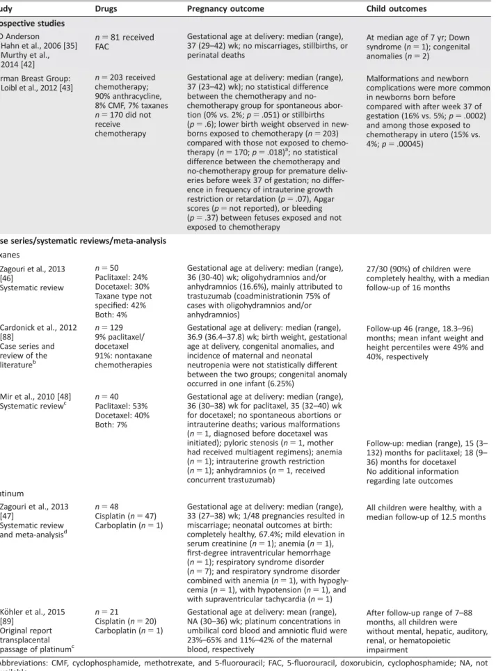
During the second and third trimesters, the use of cytotoxic chemotherapy is more widely accepted. Although most studies evaluating the safety of chemotherapy beyond the first trimes-ter have been retrospective, rates of fetal malformations have been low: on average, 3%–5% across several studies [10, 35–38], similar to the rates in the population at large in the United States [39] and to those reported in a large German study (6.9%) [40]. A multicenter, case-control study of 129 chil-dren who were exposed to maternal cancer with or without treatment revealed that cognitive, cardiac, or general develop-ment in early childhood was not impaired; however, prematur-ity was correlated with worse cognitive outcome independent of cancer treatment [41]. The fetal and maternal outcomes of two notable studies—Hahn and colleagues (updated by Murthy et al. [42]) and Loibl and colleagues (German report on the use and safety of chemotherapy during pregnancy in a prospective manner) [35, 42, 43]—are summarized in Table 3. Importantly, the German study results illustrate that although more compli-cations were reported for infants exposed to chemotherapy in utero, complications were more common among infants born prematurely, irrespective of exposure to chemotherapy [43].
Taking these data into account, we recommend that the careful decision regarding timing of chemotherapy during preg-nancy consider (a) standard clinicopathologic features (i.e., stage at diagnosis, lymph node status, grade, receptor status), (b) gestational age at breast cancer diagnosis, and (c) the likeli-hood of promoting a full-term delivery in an effort to maximize both maternal and fetal outcomes. On the basis of the available data, we would recommend initiation of systemic chemother-apy after completion of the first trimester, in the absence of a compelling contraindication.
Table 3.Studies reporting on use and safety of chemotherapy during pregnancy
Study Drugs Pregnancy outcome Child outcomes
Prospective studies
MD Anderson
Hahn et al., 2006 [35] Murthy et al., 2014 [42]
n581 received
FAC
Gestational age at delivery: median (range), 37 (29–42) wk; no miscarriages, stillbirths, or perinatal deaths
At median age of 7 yr; Down syndrome (n51); congenital anomalies (n52)
German Breast Group: Loibl et al., 2012 [43]
n5203 received chemotherapy; 90% anthracycline, 8% CMF, 7% taxanes
n5170 did not receive chemotherapy
Gestational age at delivery: median (range), 37 (23–42) wk); no statistical difference between the chemotherapy and no-chemotherapy group for spontaneous abor-tion (0% vs. 2%;p5.051) or stillbirths
(p5.6); lower birth weight observed in
new-borns exposed to chemotherapy (n5203) compared with those not exposed to chemo-therapy (n5170;p5.018)a; no statistical difference between the chemotherapy and no-chemotherapy group for premature deliv-eries before week 37 of gestation; no differ-ence in frequency of intrauterine growth restriction or retardation (p5.07), Apgar scores (p5not reported), or bleeding
(p5.37) between fetuses exposed and not
exposed to chemotherapy
Malformations and newborn complications were more common in newborns born before
compared with after week 37 of gestation (16% vs. 5%;p5.0002) and among those exposed to chemotherapy in utero (15% vs. 4%;p5.00045)
Case series/systematic reviews/meta-analysis Taxanes
Zagouri et al., 2013 [46]
Systematic review
n550 Paclitaxel: 24% Docetaxel: 30% Taxane type not specified: 42% Both: 4%
Gestational age at delivery: median (range), 36 (30-40) wk; oligohydramnios and/or anhydramnios (16.6%), mainly attributed to trastuzumab (coadministrationin 75% of cases with oligohydramnios and/or anhydramnios)
27/30 (90%) of children were completely healthy, with a median follow-up of 16 months
Cardonick et al., 2012 [88]
Case series and review of the literatureb
n5129 9% paclitaxel/ docetaxel 91%: nontaxane chemotherapies
Gestational age at delivery: median (range), 36.9 (36.4–37.8) wk; birth weight, gestational age at delivery, congenital anomalies, and incidence of maternal and neonatal neutropenia were not statistically different between the two groups; congenital anomaly occurred in one infant (6.25%)
Follow-up 46 (range, 18.3–96) months; mean infant weight and height percentiles were 49% and 40%, respectively
Mir et al., 2010 [48] Systematic reviewc
n540 Paclitaxel: 53% Docetaxel: 40% Both: 7%
Gestational age at delivery: median (range), 36 (30–38) wk for paclitaxel, 35 (32–40) wk for docetaxel; no spontaneous abortions or intrauterine deaths; various malformations
(n51, diagnosed before docetaxel was
initiated); pyloric stenosis (n51, mother had received multiagent regimens); anemia
(n51); intrauterine growth restriction
(n51); anhydramnios (n51, received
concurrent trastuzumab)
Follow-up: median (range), 15 (3– 132) months for paclitaxel; 18 (9– 36) months for docetaxel No additional information regarding late outcomes
Platinum
Zagouri et al., 2013 [47]
Systematic review and meta-analysisd
n548
Cisplatin (n547) Carboplatin (n51)
Gestational age at delivery: median (range), 33 (27–38) wk; 1/48 pregnancies resulted in miscarriage; neonatal outcomes at birth: completely healthy, 67.4%; mild elevation in serum creatinine (n51); anemia (n51), first-degree intraventricular hemorrhage
(n51); respiratory syndrome disorder
(n57); and respiratory syndrome disorder
combined with anemia (n51), with hypogly-cemia (n51), with hypotension (n51), and with supraventricular tachycardia (n51)
All children were healthy, with a median follow-up of 12.5 months
K€ohler et al., 2015 [89]
Original report transplacental passage of platinumc
n521
Cisplatin (n520) Carboplatin (n51)
Gestational age at delivery: mean (range), NA (30–36) wk; platinum concentrations in umbilical cord blood and amniotic fluid were 23%–65% and 11%–42% of the maternal blood, respectively
After follow-up range of 7–88 months, all children were without mental, hepatic, auditory, renal, or hematopoietic
impairment
Abbreviations: CMF, cyclophosphamide, methotrexate, and 5-fluorouracil; FAC, 5-fluorouracil, doxorubicin, cyclophosphamide; NA, not available.
a
After adjustment for gestational age. bBreast and ovarian cancer.
maternal and fetal outcomes has been with anthracycline-containing regimens. Perhaps the largest study of a single regi-men was of 5-fluourouracil, doxorubicin, and cyclophospha-mide) [35]. Data with other regimens and other classes of chemotherapy agents, including taxanes and platinum agents, are more limited [44–47] (Table 3). Specifically, fetal outcomes, although generally favorable after taxane and/or platinum ther-apy, are based on a relatively small number of carefully selected patients with relatively limited follow-up; thus, bias may be an issue. We believe that taxanes and platinum agents should be used cautiously to treat breast cancer during pregnancy, and only if standard anthracycline-based therapy, as the favored choice, is not feasible. Another concerning aspect of taxanes during pregnancy is the higher activity of cytochrome P-3A4 during the third trimester; thus, taxane clearance is higher, with possible limitations on expected efficacy [48]. This difficult deci-sion relies on weighing the risks and benefits to the patient and fetus.
In addition to type of chemotherapy, schedule of chemo-therapy must also be considered. In the nonpregnant patient, dose-dense chemotherapy has improved outcomes compared with treatments every 3 weeks [49]. However, the use of growth factors (i.e., granulocyte colony-stimulating factor) has only recently been investigated in pregnancy [50, 51]. As noted earlier, chemotherapy dosing in the pregnant patient should be the same as in the nonpregnant patient and should be based on actual body surface area calculation before every chemo-therapy cycle [52].
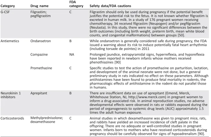
Data regarding the safety of supportive drugs during preg-nancy is relatively limited, and these medications should be used during pregnancy only if the potential benefit justifies the potential risk to the fetus (Table 4). The 5HT3 antagonists ondansetron, palonosetron, and granisetron are classified as pregnancy risk factor B. Few [53] or no reports have addressed granisetron and palonosetron in human pregnancy, respec-tively, but animal studies indicate that the risk of these agents during pregnancy is low [54, 55]. Regarding ondansetron, sev-eral studies support that this 5HT3 antagonist is not associated with an increased risk for birth defects [56–58]. However, the FDA has issued a warning regarding an increased risk for cardiac arrhythmia (i.e., QT prolongation and torsades) in the general population; therefore, maternal safety should be considered [59–61]. No data in humans are available for neurokinin1 inhibi-tors (i.e., aprepitant and fosaprepitant, pregnancy class B). Lim-ited animal data suggest the risk during pregnancy may be low. It is recommended to consider the dehydration and potential toxicity risks and use accordingly [62].
Animal and human studies with corticosteroids are conflict-ing but seem to suggest some increased risk for fetal risk, partic-ularly in the first trimester. The benefits of corticosteroids are generally thought to outweigh the risks [63]. Prednisolone and hydrocortisone are preferred over dexamethasone because they are highly metabolized in the placenta and have limited penetra-tion into the fetal compartment [64]. Antihistamines, H1 and H2 antagonists used to prevent allergic reactions with chemother-apy, are generally considered safe for use during pregnancy.
Diphenhydramine, a first-generation H1 antagonist and pregnancy risk factor B, has been associated with low fetal risk in human and animal studies [65]. It is the preferred parenteral H1 antagonist [66]. The H2 antagonists ranitidine, famotidine,
and cimetidine (pregnancy risk factor B) have low potential for birth defects, as animal and human studies demonstrate [67]. Ranitidine is considered the drug of choice over famotidine and cimetidine. Avoiding proton-pump inhibitors is recommended because they might have a muscle-relaxant effect [52]. As dis-cussed earlier, the use of granulocyte colony-stimulating factor during pregnancy has recently been investigated, and data are very limited. In a study of 176 pregnant women receiving chemotherapy, 34 received filgrastim (Neupogen, Amgen, Thousand Oaks, CA, http://www.amgen.com/) and/or peg-filgrastim (Neulasta, Amgen). Study found no significant differ-ences in the birth outcomes (including birth weight, preterm birth, newborn mean white blood counts, and congenital mal-formations) between groups [50].
Biologic Therapies
Because of adverse effects to the fetus, the use of trastuzumab, a monoclonal antibody that targets HER2/neu, is contraindi-cated during all trimesters of pregnancy. The FDA has catego-rized trastuzumab as a Category D drug because of updated safety information released in December 2010. Exposure to trastuzumab during pregnancy has been associated with oligo-hydramnios and oligooligo-hydramnios syndrome manifesting as pul-monary hypoplasia, skeletal abnormalities, renal insufficiency, and neonatal death. These findings have been noted I reports and postmarketing experience with trastuzumab [68–70]. In the unfortunate case of trastuzumab exposure during preg-nancy, treatment should be discontinued and the fetus should be closely monitored, with particular attention to amniotic fluid volumes. Pertuzumab, a monoclonal antibody that blocks the dimerization of HER2 and HER3, was approved by the FDA in the neoadjuvant setting in 2013 based on higher pathologic complete response rates for patients receiving pertuzumab in addition to trastuzumab [71]. Pertuzumab has not been studied in pregnant women and, similarly to trastuzumab, is contraindi-cated during pregnancy [72]. In the setting of early-stage, HER2-positive breast cancer diagnosed during pregnancy, tras-tuzumab and/or pertras-tuzumab can be administered after deliv-ery and at completion of lactation if a patient elects to breastfeed. There are no data regarding secretion of trastuzu-mab (or pertuzutrastuzu-mab) in human milk or effects on the breastfed infant or milk production with trastuzumab in humans [73]; thus, it is not recommended.
Lapatinib, a small molecule tyrosine kinase inhibitor of both the epithelial growth factor receptor (HER1) and HER2/neu, is also indicated in the treatment of advanced trastuzumab-refractory, HER2/neu-positive breast cancer [74]. Experience with lapatinib during pregnancy is limited, and thus lapatinib can-not be recommended during pregnancy. Collectively, anti-HER2 therapies are not recommended during pregnancy or breastfeed-ing [75].
Endocrine Therapy
associated with fetal malformations (Goldenhar syndrome, manifested by oculoauriculovertebral dysplasia, as well as ambiguous genitalia), vaginal bleeding, and miscarriage [34, 76–78]. Given these reports, initiation of tamoxifen is rec-ommended after delivery. Moreover, tamoxifen significantly delays postpartum milk production, and there are limited safety data regarding excretion of tamoxifen in human milk. Thus, we do not recommend tamoxifen during lactation [79]. Of note, the decision to delay endocrine treatment in order to allow lactation should be based on individual risk and include a balanced discussion of risk versus benefit.
Radiation Safety Issues
Pregnancy is considered one of the few absolute contraindica-tions to the use of radiotherapy (RT) because of the potential teratogenic and even lethal effects on the developing fetus. Most data regarding the effect of RT on pregnancy are based on animal models, although evidence in humans was also derived from the study of the atomic bombings of Hiroshima and Nagasaki, Japan [80]. The effects of RT on the developing fetus can be classified as lethal effects, malformations, and growth disturbances without malformations [81]. The two most important factors in discussing the effects of RT on the fetus are the actual dose received and the occurrence of the dose relative to the stage of pregnancy.
When one examines data from the atomic bombings in Japan, the most common effects seen in children whose
mothers received incidental RT are microcephaly and mental retardation [82–85]. Interestingly, RT-induced microcephaly was seen in children who were irradiated from 0 to 15 weeks’ gestation, whereas mental retardation was not seen from 0 to 8 weeks [84, 85]. The highest incidence of RT-induced mental retardation peaked at 8 to 15 weeks’ gestation, where the risk was postulated to be 40% for every 1 Gy (unit of radiation) received by the fetus; typical doses used for breast cancer are on the order of 60 Gy. The incidence drops by nearly four-fold if a fetus is irradiated from weeks 16 to 25.
Because RT is a known carcinogen, there is also concern for induction of malignancy in fetuses that are exposed. Although an issue of considerable debate, the exposure of a fetus in utero is postulated to increase the fetus’s risk for cancer [86]. The malignancy with the most convincing link to RT is leukemia. It is postulated that doses as low as 10 milli-Gy increase the risk, with an absolute increase in developing a cancer second-ary to RT of 6% per 1 Gy received.
RT’s acute effect on maternal lactation has not been stud-ied; however, there is some evidence that women who have received chest RT are capable of future lactation, although per-haps not as well as their non-irradiated age-matched cohort [87]. From a pragmatic standpoint, we recommend against breastfeeding during RT treatment because the suckling effect from the infant can augment skin toxicity secondary to the RT, resulting in discomfort, skin breakdown, and possible infections (i.e., mastitis).
Table 4.Safety information for selected supportive care agents administered during treatment for pregnancy-associated breast cancer
Category Drug name
FDA
category Safety data/FDA cautions
G-CSF Filgrastim;
pegfilgrastim
C Filgrastim should only be used during pregnancy if the potential benefit
justifies the potential risk to the fetus. It is not known whether filgrastim is excreted in human milk. In a study of 176 pregnant women receiving chemotherapy, 34 received filgrastim (Neupogen) and/or pegfilgrastim (Neulasta). In this study, there were no significant differences between the birth outcomes (including birth weight, preterm birth, mean white blood counts, and congenital malformations) between groups [50].
Antiemetics Ondansetron B While ondansetron is generally considered safe during pregnancy, the FDA
issued a warning about its risk to induce potentially fatal heart arrhythmia (including torsade de pointes) in 2011
Compazine NA Prolonged jaundice, extrapyramidal signs, hyperreflexia, and hyporeflexia
have been reported in newborn infants whose mothers received phenothiazines [90]
Promethazine C Specific studies to test the action of promethazine on parturition, lactation,
and development of the animal neonate were not done, but a general preliminary study in rats indicated no effect on these parameters. Although antihistamines have been found to produce fetal mortality in rodents, the pharmacologic effects of antihistamine in the rodent do not parallel those in humans.
Neurokinin 1 inhibitors
Aprepitant B There are insufficient data on use of aprepitant (Emend, Merck,
Whitehouse Station, NJ, http://www.merck.com) in pregnant women to inform a drug-associated risk. In animal reproduction studies, no adverse developmental effects were observed in rats or rabbits exposed during the period of organogenesis to systemic drug levels (AUC) approximately 1.5 times the adult human exposure.
Corticosteroids Methylprednisolone;
dexamethasone C Animal studies in which dexamethasone was given to pregnant mice, rats,and rabbits have yielded an increased incidence of cleft palate in the offspring. There are no adequate or well-controlled studies in pregnant women. Infants born to mothers who have received corticosteroids during pregnancy should be carefully observed for signs of hypoadrenalism [90].
CONCLUSIONS ANDRECOMMENDATIONS
BCDP is a major ethical and professional challenge for both the patient and the multidisciplinary treatment team. While the foundation of oncologic care is based on that of the NBCDP patient, there are many unique issues from the medical, surgi-cal, and radiation oncology perspectives that must be consid-ered to ensure safety to both the mother and developing fetus. Many of these key points and important contraindications are summarized in Table 5. An informed discussion between the patient and her medical team that generates an individualized treatment plan, taking into account the timing of the pregnancy and the stage and subtype of the breast cancer, is essential to maximize benefit and minimize risk to the mother and fetus.
ACKNOWLEDGMENTS
We dedicate this article to the memory of our beloved col-league and trusted surgeon, Dr. Keith Amos.
This review was funded, in part, by National Cancer Institute, National Institutes of Health, grant K23157728 (C.K.A.). The con-tent is solely the responsibility of the authors and does not
necessarily represent the official views of the National Institutes of Health.
AUTHORCONTRIBUTIONS
Conception/Design: Shlomit Strulov Shachar, Carey K. Anders
Provision of study material or patients: Shlomit Strulov Shachar, Carey K. Anders
Collection and/or assembly of data: Shlomit Strulov Shachar, Kristalyn Gallagher, Aimee Faso, Carey K. Anders
Data analysis and interpretation: Shlomit Strulov Shachar, Kristalyn Gallagher, Aimee Faso, Hyman B. Muss, Raeshall Sweeting, Carey K. Anders
Manuscript writing: Shlomit Strulov Shachar, Kristalyn Gallagher, Aimee Faso, Hyman B. Muss, Raeshall Sweeting, Carey K. Anders
Final approval of manuscript: Shlomit Strulov Shachar, Kristalyn Gallagher, Aimee Faso, Hyman B. Muss, Carey K. Anders
DISCLOSURES
Carey K. Anders: Novartis, Sanofi, toBBB, GERON, angiochem, Merrimack, PUMA, Lily, Kadmon (C/A); Novartis, Sanofi, toBBB, GERON, angiochem, Merrimack, PUMA, Lily, Kadmon (RF). The other authors indicated no financial relationships.
(C/A) Consulting/advisory relationship; (RF) Research funding; (E) Employment; (ET) Expert testimony; (H) Honoraria received; (OI) Ownership interests; (IP) Intellectual property rights/ inventor/patent holder; (SAB) Scientific advisory board
REFERENCES
1.Stensheim H, Moller B, van Dijk T et al. Cause-specific survival for women diagnosed with cancer during pregnancy or lactation: a registry-based cohort study. J Clin Oncol 2009;27:45–51.
2.Lee YY, Roberts CL, Dobbins T et al. Incidence and outcomes of pregnancy-associated cancer in Australia, 1994–2008: A population-based linkage study. BJOG 2012;119:1572–1582.
3.Smith LH, Danielsen B, Allen ME et al. Cancer associated with obstetric delivery: Results of linkage with the California cancer registry. Am J Obstet Gynecol 2003;189:1128–1135.
4.Mathews TJ, Hamilton BE. Mean age of mothers is on the rise: United States, 2000–2014. NCHS Data Brief 2016:1–8.
5.Genin AS, Lesieur B, Gligorov J et al. Pregnancy-associated breast cancers: Do they differ from other breast cancers in young women? Breast 2012;21: 550–555.
6.Pilewskie M, Gorodinsky P, Fought A et al. Asso-ciation between recency of last pregnancy and bio-logic subtype of breast cancer. Ann Surg Oncol 2012; 19:1167–1173.
7.Anders CK, Fan C, Parker JS et al. Breast carcino-mas arising at a young age: Unique biology or a sur-rogate for aggressive intrinsic subtypes? J Clin Oncol 2011;29:e18–e20.
8.Anders CK, Hsu DS, Broadwater G et al. Young age at diagnosis correlates with worse prognosis and defines a subset of breast cancers with shared
patterns of gene expression. J Clin Oncol 2008;26: 3324–3330.
9.Murphy CG, Mallam D, Stein S et al. Current or recent pregnancy is associated with adverse pathologic features but not impaired survival in early breast cancer. Cancer 2012;118:3254– 3259.
10.Cardonick E, Dougherty R, Grana G et al. Breast cancer during pregnancy: Maternal and fetal out-comes. Cancer J 2010;16:76–82.
11.Amant F, von Minckwitz G, Han SN et al. Prog-nosis of women with primary breast cancer diag-nosed during pregnancy: Results from an international collaborative study. J Clin Oncol 2013; 31:2532–2539.
Table 5.Key points and contraindications for breast cancer treatment during pregnancy
Key points
1. Ultrasonography is the first-line imaging modality. If concerning mass identified, bilateral mammography with appropriate
shielding is recommended.
2. Surgery can be safely performed at any time during pregnancy, but second trimester is preferred. Lumpectomy and
mastectomy are both reasonable surgical approaches.
3. The recommended method of lymphoscintigraphy is with 99m-Tc sulfur colloid alone.
4. Chemotherapy should not be administered in the first trimester of pregnancy; anthracycline-based chemotherapy can be
safely initiated in the second and third trimesters of pregnancy.
5. Chemotherapy should be stopped approximately 3–4 wk before delivery to avoid hematologic nadir during delivery that may
result in infectious or bleeding complications.
6. Dosing of chemotherapy in pregnant patient should be similar to that in nonpregnant patient (i.e., based on actual body
surface area). Contraindications
1. Gadolinium-based contrast for MRI is not recommended.
2. Isosulfan blue dye is contraindicated for lymphoscintigraphy as dual tracer for sentinel lymph node biopsy.
3. Chemotherapy is contraindicated in first trimester of pregnancy and during lactation.
4. Endocrine treatment is contraindicated during pregnancy and lactation.
5. Anti-HER2 therapy is contraindicated in pregnancy and lactation.
6. Radiation therapy is contraindicated during pregnancy and cautioned during lactation.
12.Litton JK, Warneke CL, Hahn KM et al. Case con-trol study of women treated with chemotherapy for breast cancer during pregnancy as compared with nonpregnant patients with breast cancer. Oncologist 2013;18:369–376.
13.Rodriguez AO, Chew H, Cress R et al. Evidence of poorer survival in pregnancy-associated breast cancer. Obstet Gynecol 2008;112:71–78.
14.Ali SA, Gupta S, Sehgal R et al. Survival out-comes in pregnancy associated breast cancer: A retrospective case control study. Breast J 2012;18: 139–144.
15.Azim HA Jr, Santoro L, Russell-Edu W et al. Prognosis of pregnancy-associated breast cancer: A meta-analysis of 30 studies. Cancer Treat Rev 2012; 38:834–842.
16.Woo JC, Yu T, Hurd TC. Breast cancer in preg-nancy: A literature review. Arch Surg 2003;138:91– 98; discussion 99.
17.Robbins J, Jeffries D, Roubidoux M et al. Accu-racy of diagnostic mammography and breast ultra-sound during pregnancy and lactation. AJR Am J Roentgenol 2011;196:716–722.
18.Vashi R, Hooley R, Butler R et al. Breast imaging of the pregnant and lactating patient: Physiologic changes and common benign entities. AJR Am J Roentgenol 2013;200:329–336.
19.Psyrri A, Burtness B. Pregnancy-associated breast cancer. Cancer J 2005;11:83–95.
20.Nguyen CP, Goodman LH. Fetal risk in diagnos-tic radiology. Semin Ultrasound CT MR 2012;33: 4–10.
21.American College of Radiology. ACR manual on contrast media. 2015. Available at: https://www. acr.org//media/ACR/Documents/PDF/QualitySafety/ Resources/Contrast-Manual/2016_Contrast_Media. pdf?la=en. Accessed January 10, 2017.
22.Amant F, Van Calsteren K, Halaska MJ et al. Long-term cognitive and cardiac outcomes after pre-natal exposure to chemotherapy in children aged 18 months or older: An observational study. Lancet Oncol 2012;13:256–264.
23.Fisher B, Anderson S, Bryant J et al. Twenty-year follow-up of a randomized trial comparing total mastectomy, lumpectomy, and lumpectomy plus irradiation for the treatment of invasive breast can-cer. N Engl J Med 2002;347:1233–1241.
24.Kuerer HM, Cunningham JD, Bleiweiss IJ et al. Conservative surgery for breast carcinoma associ-ated with pregnancy. Breast J 1998;4:171–176.
25.Mondi MM, Cuenca RE, Ollila DW et al. Senti-nel lymph node biopsy during pregnancy: Initial clini-cal experience. Ann Surg Oncol 2007;14:218–221.
26.Spanheimer PM, Graham MM, Sugg SL et al. Measurement of uterine radiation exposure from lymphoscintigraphy indicates safety of sentinel lymph node biopsy during pregnancy. Ann Surg Oncol 2009;16:1143–1147.
27.Morita ET, Chang J, Leong SP. Principles and controversies in lymphoscintigraphy with emphasis on breast cancer. Surg Clin North Am 2000;80: 1721–1739.
28.Keleher A, Wendt R 3rd, Delpassand E et al. The safety of lymphatic mapping in pregnant breast can-cer patients using Tc-99m sulfur colloid. Breast J 2004;10:492–495.
29.Rosen MA. Management of anesthesia for the pregnant surgical patient. Anesthesiology 1999;91: 1159–1163.
30.Mazze RI, Kallen B. Reproductive outcome after anesthesia and operation during pregnancy: A regis-try study of 5405 cases. Am J Obstet Gynecol 1989; 161:1178–1185.
31.Redmond GP. Physiological changes during pregnancy and their implications for pharmacologi-cal treatment. Clin Invest Med 1985;8:317–322.
32.Doll DC, Ringenberg QS, Yarbro JW. Antineo-plastic agents and pregnancy. Semin Oncol 1989;16: 337–346.
33.Paskulin GA, Gazzola Zen PR, de Camargo Pinto LL et al. Combined chemotherapy and teratogenicity. Birth Defects Res A Clin Mol Teratol 2005;73:634– 637.
34.Sukumvanich P. Review of current treatment options for pregnancy-associated breast cancer. Clin Obstet Gynecol 2011;54:164–172.
35.Hahn KM, Johnson PH, Gordon N et al. Treat-ment of pregnant breast cancer patients and out-comes of children exposed to chemotherapy in utero. Cancer 2006;107:1219–1226.
36.Peccatori FA, Azim HA Jr, Scarfone G et al. Weekly epirubicin in the treatment of gestational breast cancer (GBC). Breast Cancer Res Treat 2009; 115:591–594.
37.Azim HA Jr, Peccatori FA, Scarfone G et al. Anthracyclines for gestational breast cancer: Course and outcome of pregnancy. Ann Oncol 2008;19: 1511–1512.
38.Ring AE, Smith IE, Jones A et al. Chemotherapy for breast cancer during pregnancy: An 18-year experience from five London teaching hospitals. J Clin Oncol 2005;23:4192–4197.
39.Centers for Disease Control and Prevention National Center for Health and Statistics. Birth defects: data & statistics. April 2, 2016. Available at: http://www.cdc.gov/ncbddd/birthdefects/data.html. Accessed May 1, 2016.
40.Queisser-Luft A, Stolz G, Wiesel A et al. Malfor-mations in newborn: Results based on 30,940 infants and fetuses from the Mainz congenital birth defect monitoring system (1990–1998). Arch Gyne-col Obstet, 2002;266:163–167.
41.Amant F, Vandenbroucke T, Verheecke M et al. Pediatric outcome after maternal cancer diagnosed during pregnancy. N Engl J Med 2015;373:1824– 1834.
42.Murthy RK, Theriault RL, Barnett CM et al. Out-comes of children exposed in utero to chemotherapy for breast cancer. Breast Cancer Res 2014;16:500.
43.Loibl S, Han SN, von Minckwitz G et al. Treat-ment of breast cancer during pregnancy: An obser-vational study. Lancet Oncol 2012;13:887–896.
44.Berveiller P, Mir O, Taxanes during pregnancy: Probably safe, but still to be optimized. Oncology 2012;83:239–240.
45.Mir O, Berveiller P, Ropert S et al. Use of plati-num derivatives during pregnancy. Cancer 2008;113: 3069–3074.
46.Zagouri F, Sergentanis TN, Chrysikos D et al. Tax-anes for breast cancer during pregnancy: A system-atic review. Clin Breast Cancer 2013;13:16–23.
47.Zagouri F, Sergentanis TN, Chrysikos D et al. Platinum derivatives during pregnancy in cervical cancer: A systematic review and meta-analysis. Obstet Gynecol 2013;121:337–343.
48.Mir O, Berveiller P, Goffinet F et al. Taxanes for breast cancer during pregnancy: A systematic review. Ann Oncol 2010;21:425–426.
49.Bonilla L, Ben-Aharon I, Vidal L et al. Dose-dense chemotherapy in nonmetastatic breast can-cer: A systematic review and meta-analysis of randomized controlled trials. J Natl Cancer Inst 2010; 102:1845–1854.
50.Cardonick E, Irfan F, Torres N. The use of Neupogen (filgrastim) or Neulasta (pegfilgrastim) during pregnancy when chemotherapy is indi-cated for maternal cancer treatment. J Cancer Ther 2012;3:5.
51.Cardonick E, Gilmandyar D, Somer RA. Mater-nal and neonatal outcomes of dose-dense chemo-therapy for breast cancer in pregnancy. Obstet Gynecol 2012;120:1267–1272.
52.Loibl S, Schmidt A, Gentilini O et al. Breast can-cer diagnosed during pregnancy: Adapting recent advances in breast cancer care for pregnant patients. JAMA Oncol 2015;1:1145–1153.
53.Merimsky O, Le Chevalier T, Missenard G et al. Management of cancer in pregnancy: A case of Ewing’s sarcoma of the pelvis in the third trimester. Ann Oncol 1999;10:345–350.
54.Kytril (Granisetron HCl). Nutley, NRL, Inc., NJ: Roche, September 2009.
55.Aloxi (Palonosetron HCl injection) [package insert]. Bloomington MMP. Woodcliffe, NJ: Eisai, December 2015.
56.Pasternak B, Svanstrom H, Hviid A. Ondanse-tron in pregnancy and risk of adverse fetal out-comes. N Engl J Med 2013;368:814–823.
57.Einarson A, Maltepe C, Navioz Y et al. The safety of ondansetron for nausea and vomiting of preg-nancy: A prospective comparative study. BJOG 2004; 111:940–943.
58.Anderka M, Mitchell AA, Louik C et al. Medi-cations used to treat nausea and vomiting of pregnancy and the risk of selected birth defects. Birth Defects Res A Clin Mol Teratol 2012;94:22–30.
59.Ondansetron. Ondansetron Prescribing Infor-mation. Revised 11/2012. Available at: http:// www.accessdata.fda.gov/drugsatfda_docs/label/ 2012/020007s043lbl.pdf. Accessed May 1, 2016.
60.Koren G. Motherisk update. Is ondansetron safe for use during pregnancy? Can Fam Physician 2012;58:1092–1093.
61.US Food and Drug Administration. FDA drug safety communication: Abnormal heart rhythms may be associated with use of Zofran (ondanse-tron). Available at: http://www.fda.gov/Drugs/ DrugSafety/ucm271913.htm. Accessed May 1, 2016.
62.Emend (Aprepitant) [package insert]. White-house Station, NJ: Merck, December 2015. Accessed May 1, 2016.
63.Amant F, Halaska MJ, Fumagalli M et al. Gyne-cologic cancers in pregnancy: Guidelines of a second international consensus meeting. Int J Gynecol Can-cer 2014;24:394–403.
64.Blanford AT, Murphy BE. In vitro metabolism of prednisolone, dexamethasone, betamethasone, and cortisol by the human placenta. Am J Obstet Gynecol 1977;127:264–267.
65.Gilboa SM, Ailes EC, Rai RP et al. Antihistamines and birth defects: A systematic review of the litera-ture. Expert Opin Drug Saf 2014;13:1667–1698.
67.Briggs GE, Freeman RK. Drugs in Pregnancy and Lactation: A Reference Guide to Fetal and Neonatal Risk. 10th ed. Philadelphia, PA: Lippincott Williams & Wilkins, 2014.
68.Gottschalk I, Berg C, Harbeck N et al. Fetal renal insufficiency following trastuzumab treatment for breast cancer in pregnancy: Case report and review of the current literature. Breast Care (Basel) 2011;6: 475–478.
69.Sekar R, Stone PR, Trastuzumab use for meta-static breast cancer in pregnancy. Obstet Gynecol 2007;110:507–510.
70.Watson WJ. Herceptin (trastuzumab) ther-apy during pregnancy: Association with reversi-ble anhydramnios. Obstet Gynecol 2005;105: 642–643.
71.Gianni L, Pienkowski T, Im YH et al. Efficacy and safety of neoadjuvant pertuzumab and trastuzumab in women with locally advanced, inflammatory, or early HER2-positive breast cancer (NeoSphere): A randomised multicentre, open-label, phase 2 trial. Lancet Oncol 2012;13:25–32.
72.Pertuzumab. Pertuzumab Prescribing Infor-mation. March 7, 2016. Available at: http://www. gene.com/download/pdf/perjeta_prescribing.pdf. Accessed May 1, 2016.
73.Herceptin (trastuzumab). Herceptin Prescribing Information. Available at: http://www.gene.com/ download/pdf/herceptin_prescribing.pdf. Accessed May 1, 2016.
74.Geyer CE, Forster J, Lindquist D et al. Lapatinib plus capecitabine for HER2-positive advanced breast cancer. N Engl J Med, 2006;355:2733–2743.
75.Lambertini M, Peccatori FA, Azim HA Jr. Tar-geted agents for cancer treatment during pregnancy. Cancer Treat Rev 2015;41:301–309.
76.Cullins SL, Pridjian G, Sutherland CM. Golden-har’s syndrome associated with tamoxifen given to the mother during gestation. JAMA 1994;271:1905– 1906.
77.Isaacs RJ, Hunter W, Clark K, Tamoxifen as sys-temic treatment of advanced breast cancer during pregnancy—case report and literature review. Gyne-col OnGyne-col 2001;80:405–408.
78.Tewari K, Bonebrake RG, Asrat T et al. Ambigu-ous genitalia in infant exposed to tamoxifen in utero. Lancet 1997;350:183.
79.Nolvadex (tamoxifen citrate). Nolvadex Pre-scribing Information. Available at: http://www. accessdata.fda.gov/drugsatfda_docs/label/2005/ 17970s053lbl.pdf. Accessed May 1, 2016.
80.Committee on the Biological Effects of Ionizing Radiations. The Effects on Populations of Exposure to Low Levels of Ionizing Radiation. Washington, DC: National Academy of Sciences, 1980.
81.Hall EJ, Giaccia AJ. Radiobiology for the Radiol-ogist. 6th ed. New York, NY: Lippincott Williams & Wilkins, 2006.
82.Committee for the Compilation of Materials on Damage Caused by the Atomic Bomb in Hiroshima and Nagasaki, Hiroshima and Nagasaki. The Physical, Medical and Social Effects of the Atomic Bombings. New York, NY: Basic Books, 1981.
83.Otake M, Schull WJ. In utero exposure to A-bomb radiation and mental retardation: A reassess-ment. Br J Radiol 1984;57:409–414.
84.Otake M, Schull WJ. Radiation-related small head sizes among prenatally exposed A-bomb survivors. Int J Radiat Biol 1993;63:255– 270.
85.Otake M, Schull WJ, Lee S. Threshold for radiation-related severe mental retardation in pre-natally exposed A-bomb survivors: A re-analysis. Int J Radiat Biol 1996;70:755–763.
86.Doll R, Wakeford R. Risk of childhood cancer from fetal irradiation. Br J Radiol 1997;70:130– 139.
87.McCullough L, Ng A, Najita J et al. Breast-feeding in survivors of Hodgkin lymphoma treated with chest radiotherapy. Cancer 2010;116:4866– 4871.
88.Cardonick E, Bhat A, Gilmandyar D et al. Mater-nal and fetal outcomes of taxane chemotherapy in breast and ovarian cancer during pregnancy: Case series and review of the literature. Ann Oncol 2012; 23:3016–3023.
89.Kohler C, Oppelt P, Favero G et al. How much platinum passes the placental barrier? Analysis of platinum applications in 21 patients with cervical cancer during pregnancy. Am J Obstet Gynecol 2015; 213:206.e1–e5.
90.Compazine. Compazine Prescribing Informa-tion. Available at: http://www.accessdata.fda.gov/ drugsatfda_docs/label/2005/010571s096lbl.pdf. Accessed May 1, 2016.
91.Methylprednisolone. Methylprednisolone Pre-scribing Information. Available at: http://www. accessdata.fda.gov/drugsatfda_docs/label/2011/ 011856s103s104lbl.pdf. Accessed May 1, 2016