Common, low-frequency, and rare genetic
variants associated with lipoprotein
subclasses and triglyceride measures in
Finnish men from the METSIM study
James P. Davis1☯, Jeroen R. Huyghe2☯, Adam E. Locke2, Anne U. Jackson2, Xueling Sim2, Heather M. Stringham2, Tanya M. Teslovich2, Ryan P. Welch2, Christian Fuchsberger2,
Narisu Narisu3, Peter S. Chines3†, Antti J. Kangas4, Pasi Soininen4,5, Mika Ala-Korpela4,5,6,7,8, Johanna Kuusisto9, Francis S. Collins3, Markku Laakso9‡
*, Michael Boehnke2‡*, Karen L. Mohlke1‡*
1 Department of Genetics, University of North Carolina at Chapel Hill, Chapel Hill, NC, United States of America, 2 Department of Biostatistics and Center for Statistical Genetics, University of Michigan, Ann Arbor, MI, United States of America, 3 National Human Genome Research Institute, National Institutes of Health, Bethesda, MD, United States of America, 4 Computational Medicine, Faculty of Medicine, University of Oulu and Biocenter Oulu, Oulu, Finland, 5 NMR Metabolomics Laboratory, School of Pharmacy, University of Eastern Finland, Kuopio, Finland, 6 Population Health Science, Bristol Medical School, University of Bristol and Medical Research Council Integrative Epidemiology Unit at the University of Bristol, Bristol, United Kingdom, 7 Systems Epidemiology, Baker Heart and Diabetes Institute, Melbourne, Victoria, Australia, 8 Department of Epidemiology and Preventive Medicine, School of Public Health and Preventive Medicine, Faculty of Medicine, Nursing and Health Sciences, The Alfred Hospital, Monash University, Melbourne, Victoria, Australia, 9 Institute of Clinical Medicine, Internal Medicine, University of Eastern Finland and Kuopio University Hospital, Kuopio, Finland
☯These authors contributed equally to this work. † Deceased.
‡ ML, MB, and KLM also contributed equally to this work.
*[email protected](ML);[email protected](MB);[email protected](KLM)
Abstract
Lipid and lipoprotein subclasses are associated with metabolic and cardiovascular dis-eases, yet the genetic contributions to variability in subclass traits are not fully understood. We conducted single-variant and gene-based association tests between 15.1M variants from genome-wide and exome array and imputed genotypes and 72 lipid and lipoprotein traits in 8,372 Finns. After accounting for 885 variants at 157 previously identified lipid loci, we identified five novel signals near established loci at HIF3A, ADAMTS3, PLTP, LCAT, and LIPG. Four of the signals were identified with a low-frequency (0.005<minor allele fre-quency [MAF]<0.05) or rare (MAF<0.005) variant, including Arg123His in LCAT. Gene-based associations (P<10−10) support a role for coding variants in LIPC and LIPG with lipo-protein subclass traits. 30 established lipid-associated loci had a stronger association for a subclass trait than any conventional trait. These novel association signals provide further insight into the molecular basis of dyslipidemia and the etiology of metabolic disorders. a1111111111
a1111111111 a1111111111 a1111111111 a1111111111
OPEN ACCESS
Citation: Davis JP, Huyghe JR, Locke AE, Jackson
AU, Sim X, Stringham HM, et al. (2017) Common, low-frequency, and rare genetic variants associated with lipoprotein subclasses and triglyceride measures in Finnish men from the METSIM study. PLoS Genet 13(10): e1007079.https://doi.org/ 10.1371/journal.pgen.1007079
Editor: Ruth J. F. Loos, Icahn School of Medicine
at Mount Sinai, UNITED STATES
Received: March 23, 2017 Accepted: October 16, 2017 Published: October 30, 2017
Copyright: This is an open access article, free of all
copyright, and may be freely reproduced, distributed, transmitted, modified, built upon, or otherwise used by anyone for any lawful purpose. The work is made available under theCreative Commons CC0public domain dedication.
Data Availability Statement: All genome-wide
association results files are available at the University of Michigan website:http://csg.sph. umich.edu/boehnke/public/metsim-2017-lipoproteins/
Funding: This study was supported by Academy of
Author summary
Lipid and lipoproteins are heritable traits that differ in content and size and are correlated with coronary heart disease and mortality. To identify genetic variants associated with dif-ferent subclasses of lipoproteins, we conducted a genome-wide association study of 8,372 Finnish men. We curated a dataset of all genetic variants known to be associated with lipid or lipoprotein subclasses and used these data to conduct rigorous analyses to identify new associations in the same gene region or new ones. We identified five new signals at established lipid-associated loci revealing possible complex regulatory mechanisms under-lying the signals. Using the contribution of rare coding variants predicted to be protein truncating or missense, we uncovered novel associations for a set of variants atLIPCand
LIPGwith HDL subclasses. Investigating the genetic association of lipoprotein subclass traits may help lead to a better understanding of the etiology of cardio-metabolic diseases, and provide novel therapeutic targets.
Introduction
Genome-wide association studies (GWAS) have identified hundreds of common (MAF>
0.05) variants associated with conventional lipid and lipoprotein traits: high-density lipo-protein cholesterol (HDL-C), low-density lipolipo-protein cholesterol (LDL-C), total cholesterol (TC), and triglycerides (TG)[1–4]. While some low-frequency (0.005<MAF0.05) and rare variants (MAF0.005) have been associated with lipid and lipoprotein traits, additional loci remain to be identified[2,5,6]. High-throughput proton nuclear magnetic resonance (NMR)-based measurements of lipid and lipoprotein subclasses provide a more compre-hensive view of particle size and composition than conventional blood lipid profile mea-surements[7], and these expanded sets of traits have been associated with metabolic and cardiovascular diseases[8–10]. For example, HDL subclasses are differentially associated with incidence of coronary heart disease, and VLDL particle size is negatively associated with mortality[11,12].
Previous association studies for lipid traits have identified several genomic regions of<1 Mb that contain more than one association signal for which the lead variants are not in strong linkage disequilibrium (LD) (r2<0.8)[2,3,13]. Fine-mapping with higher density variants and conditional analyses can determine which signals are distinct (remain significant after condi-tional analysis) and which are independent, which we define here asr2<0.01. For example, Teslovichet al. used conditional analysis at 95 lipid loci to identify 26 loci that harbor at least two distinct association signals[3]. Association signals at the same locus can be population-specific or shared across populations, with potentially different effect sizes and/or lead variants [13]. Multiple association signals at a locus may indicate allelic heterogeneity in gene function or regulation or that more than one gene at the locus affects the trait[14]. Furthermore, identi-fying and accounting for additional independent association signals increases the variance in traits that can be explained by genetic loci[15,16].
In this study, we performed genome-wide single-variant and gene-based association analy-ses of 68 NMR lipid and lipoprotein subclass traits and four conventional traits (TC, TG, HDL-C, and LDL-C) in 8,372 non-diabetic Finnish men from the METabolic Syndrome In Men (METSIM) study[17]. To identify novel associations, we performed analyses with and without conditioning on lipid-associated variants at loci previously described in array- and sequence-based GWAs. We identified the most strongly associated lipid and lipoprotein sub-class traits at established loci for conventional lipid and lipoprotein traits. Since several (ML); and National Institutes of Health (https://
grants.nih.gov/) grants R01DK093757 (KLM), R01DK072193 (KLM), U01DK105561 (KLM), U01DK062370 (MB), T32 HL129982 (JPD) and National Human Genome Research Institute Division of Intramural Research (www.genome. gov/10000004/grant-information/) project number Z01HG000024 (FSC). The serum NMR
metabolomics platform has been supported by the Sigrid Juselius Foundation (sigridjuselius.fi) and the Strategic Research Funding from the University of Oulu (www.oulu.fi/university/node/35275). MAK works in a unit that is supported by the University of Bristol and UK Medical Research Council (www. mrc.ac.uk/) (MC_UU_1201/1). MAK has also been supported by Research Funding from the British Heart Foundation (www.bhf.org.uk/) and Wellcome Trust (wellcome.ac.uk/). The funders had no role in study design, data collection and analysis, decision to publish, or preparation of the manuscript.
Competing interests: I have read the journal’s
subclasses are associated with cardiovascular and metabolic diseases, identifying the variants that influence these traits is the first step to develop novel clinical treatments. These expanded association results have the potential to lead to advances in determining the etiological role of the variants and genes in cardiovascular and metabolic disease.
Results
Genome-wide association study
To identify genetic variants associated with the 72 lipid and lipoprotein traits, we analyzed 15.1M genotyped and imputed variants in 8,372 non-diabetic Finnish men (S1 Table,S1 Fig). Each trait was adjusted for age, age2, lipid-lowering medication use, and smoking status. Inverse normalized trait residuals were tested for association with each variant assuming additive allelic effects using a linear mixed model to account for relatedness among study par-ticipants[18]. Many of the traits are highly correlated with each other, with 104 trait-pair com-parisons having a pairwise Pearson correlation greater than 0.98 (S2 Fig). We used a genome-wide significance threshold ofP5×10−8, consistent with previous association studies of this scale and high trait correlation[19]. We note where associations meet a conservative experi-ment-wide Bonferonni-correctedP-value (P4.6×10−11). We identified 32,524 variant-trait associations (Pdiscovery<5×10⁻⁸) for the 72 lipid and lipoprotein traits (S3 Fig). 30,348 (93%) of
the 32,524 associations were for one of the 68 subclass traits and 2,176 (7%) for one of the four conventional lipid traits (TC, TG, HDL-C, LDL-C). More than half the associations were with the VLDL- (38%) or HDL-subclass (29%) traits. 3,784 unique variants comprise the total 32,524 trait-variant associations (S2 Table). 73% (2,780) of the 3,784 variants had a greater association with one of the 68 subclass traits, and 27% (1,004) were more highly associated with at least one of the four conventional traits (S2 Table). These variants cluster into 42 loci that were associated with at least one of the 72 traits (S1 Table). For example, at the well-characterizedAPOA5locus on chromosome 11, rs964184 was significantly associated (Pdiscovery<5×10⁻⁸) with 43 of the 72
lipid and lipoprotein traits. AtCETP, rs12446515 was significantly associated with 34 of the 72 traits. For such loci, the high correlation between the traits obscures identification of a causal trait underlying the signal.
Conditional analyses to identify novel loci and signals
To identify novel associations not reported previously for any conventional or subclass lipid or lipoprotein trait, we identified and curated a list of previously known associated variants to use in genome-wide conditional analyses (Methods). We identified 1,714 variants (S3 Table) that we clustered based on stringent LD (r2>0.95) into 885 representative variants (S4 Table). After genome-wide conditional analysis using these 885 variants, we defined novel association sig-nals using a significance threshold ofPconditional<5×10⁻⁸(S5 Table). Consistent with highly correlated traits, we observed that most of the associated variants were associated with multiple correlated traits. We considered variants located within 1 Mb of an established lipid or lipo-protein signal to be an additional signal in the region, and we define a locus as the region 1 Mb up- and downstream of a signal. We considered additional signals independent if the signal was not in LD (r2<0.01) with known lipid/lipoprotein signals, and remained significant
(Psingle<5×10⁻⁸) after single-variant conditional analyses. Associated variants with MAF<0.01
One novel common variant signal at HIF3A
Common variant rs73059724 (MAF = 0.09), associated with decreased (β= –0.14) concentra-tions of phospholipids in small VLDL, is located 3.5 kb upstream ofHIF3A(hypoxia inducible factor 3, alpha subunit) and 1.4 Mb fromAPOE(S4AandS5AFigs). Additionally, this signal is associated with decreased VLDL subclass traits (S6 Fig). This signal achieved significance after conditioning on known lipid GWAS variants (Pdiscovery= 3.8×10−
7
,Pconditional=1.4×10−
8
) (Table 1,S6 Table). When adjusted for total triglycerides, the strength of the association of rs73059724 with phospholipids in small VLDL was reduced (P = 3.6×10⁻²,S7 Table). This sig-nal is located in a gene-dense region on chromosome 19 that includes 10 previously reported lipoprotein-associated variants within 1 Mb of the index variant (S6 Table)[20]; none of these variants exhibited LD (r2>0.02) with rs73059724. Further analysis of theAPOElocus with additional samples may be necessary to elucidate the haplotype relationships between these signals. Twenty-nine proxy variants in LD (r2>0.7) with rs73059724 span a 25-kb region including the promoter and intron 1 ofHIF3A, and five of these variants overlap5 liver and adipose regulatory element (histone marks of transcriptional regulation and open chromatin) datasets (S8 Table). Hyper-methylation atHIF3Ais associated with increased adiposity and BMI in Asian infants and children[21,22]. HIF3A is a known negative regulator ofHIF1A
(hypoxia inducible factor 1, alpha subunit)[23], which has been shown to regulate the cellular uptake of cholesterol esters and VLDL by creating hypoxic conditions[24]. One or more of the associated variants may affectHIF3Atranscription or other genes in the region, leading to fewer phospholipids in small VLDL particles.
Two novel low-frequency variant signals at ALB and SYS1
We identified two new signals with low-frequency variants located nearALBandSYS1
(Table 1,S4BandS5BFigs). At theALBlocus, the low-frequency allele of rs187918276 (MAF = 0.017) located in intron 1 ofANKRD17was associated with increased (β= 0.60) con-centration of small LDL particles (Pdiscovery=6.3×10⁻²²,Pconditional=3.2×10⁻¹¹) and 26 addi-tional traits, including increased TC, LDL-C, esterified cholesterol, free cholesterol, and IDL/ LDL/VLDL subclasses (S6 Fig). When adjusted for total cholesterol, the strength of the associa-tion of rs187918276 with small LDL particles was reduced (P= 9.1×10⁻⁷,S7 Table). Variants in LD (r2>0.7, METSIM) with this variant span>1.2 Mb (S5B Fig,S8 Table), consistent with long haplotypes previously described in Finns[25]. The 885 variants used for the conditional analysis included established TC-associated signals at rs60873279 and rs182616603, located 337 kb and 1 Mb away; these variants exhibited low (r2<0.01) and moderate (r2= 0.39) pair-wise LD with rs187918276 (S6 Table). When conditioned on rs182616603, the association with rs187918276 was reduced but still highly significant (Psingle=5×10⁻¹⁵), suggesting the signals are distinct. An additional variant at this locus, rs115136538, was reported previously to be associated with albumin levels[5]. rs115136538 is located 710 kb away from and is not in LD with rs187918276 (r2<0.01 in METSIM), and the association of rs187918276 with small LDL particles was essentially unchanged when conditioned on rs115136538 (S6 Table). Taken together, theALBregion contains three distinct signals for lipid traits (rs60873279, rs1826 16603, and now rs187918276).
concentration of small LDL particles, or another of the candidate variants spanning 1.2 Mb may act on this or another nearby gene.
In an intergenic region downstream ofPIGT, we identified the low-frequency allele of lead variant rs184392658 (MAF = 0.008) associated with the increased (β= 0.45) concentration of large HDL particles (Pdiscovery=2.3×10⁻⁷,Pconditional=2.5×10⁻⁹,Table 1,S4C FigandS5C Fig). When adjusted for HDL-C, the association of rs184392658 with large HDL particles was reduced (P= 4.1×10⁻⁵,S7 Table). Two previously established lipid-associated variants are located within 1 Mb of rs184392658: rs1800961 nearHNF4Aand rs6065904 nearPLTP. rs184392658 was not in LD (r2<0.015) with either of these established variants, and condition-ing on the individual known variants did not substantially change the association signal (all
Psingle<3.7×10⁻⁶,S6 Table). Thus, rs184392658 represents a new distinct signal in this region.
Of six variants in high LD (r2>0.7) with lead variant rs184392658, only rs149985455 overlaps multiple epigenomic marks of transcription regulation from liver, blood, and adipose tissue datasets (S8 Table). This variant is located 2.2 kb upstream fromSYS1(Sys1 Golgi trafficking protein), which may have a role in lipid metabolism through an interaction with GTPases[28]. SYS1 targets ARFRP1 (ADP-ribosylation factor-related protein 1) and forms a complex in the Golgi membrane[29]; deletion ofArfrp1in mouse adipocytes led to lipodystrophy caused by failure in lipid droplet formation[30]. rs149985455 may mediate a regulatory effect onSYS1to increase the plasma concentration of large HDL particles, or another of the candidate variants spanning>500 kb may act on this or another nearby gene.
Two novel rare variant signals at LCAT and LIPG
We identified additional novel independent signals with rare variants nearLCATandLIPG
(Table 1). The rare allele (MAF = 0.005) of the missense variant rs199717050 (Arg123His) in exon 3 ofLCAT(lecithin-cholesterol acyltransferase) was associated with decreased (β= –0.72) HDL-C levels (Pdiscovery=5.9×10⁻¹⁰,Pconditional=2.5×10⁻¹²,Table 1,S4D FigandS5D Fig). This signal was not significantly associated with any of the HDL subclass traits or other traits from this study (S6 Fig). The association of rs199717050 with HDL-C was nominally reduced (P= 2.9×10⁻⁸) when adjusted for total cholesterol (S7 Table). Six variants at this locus, within 1 Mb of rs199717050, were reported previously to be associated with HDL-C[2,4] (S6 Table). However, these six variants all show low pairwise LD with rs199717050 (r2<0.01), and single-variant conditional analyses using any one of the six single-variants did not substantially change the association of rs199717050 with HDL-C (Psingle1.9×10−9,S6 Table). rs199717050 may be nearly specific to Finns; the Exome Aggregation Consortium (ExAC) database shows a total allele count of 16: fifteen in Finns and one in a non-European population. LCAT is responsible for cholesterol esterification for eventual transfer into the lipoprotein core, and facilitates the transport of cholesterol into the liver[31]. rs199717050 is predicted to be deleterious (SIFT, 0.02) or possibly damaging (PolyPhen, 0.55)[32], consistent with a plausible functional effect on LCAT to decrease levels of HDL-C.
association signals are located within 1 Mb of this variant, including rs74558535 (P= 2×10⁻¹⁰), rs10438978 (P= 7.7×10−36), rs77960347 (P= 3.6×10⁻¹¹), and rs2156552 (P= 2×10⁻¹²). The new signal is not in LD (r2<0.043) with the previously described variants and remained signifi-cant after single-variant conditional analyses (S6 Table,Fig 1C).LIPGencodes endothelial lipase (EL), which catalyzes HDL phospholipids and aids in the sequestration of HDL from cir-culation, and is expressed in several tissues and organs including the liver[34–36]. The associa-tion with phospholipids in medium-size HDL is consistent with the known phospholipase of EL[37]. Several variants inLIPGhave been shown to decrease endothelial lipase levels and increase HDL-C[38]. Based on the direction of effect in these previous studies, missense vari-ant (A172V) may decrease function of LIPG, leading to increased phospholipids in medium-size HDL and other HDL subclasses.
Gene-based tests of association
To test the association between lipid and lipoprotein subclasses and sets of coding variants within a gene, we performed gene-based tests of association using SKAT-O with four variant masks (Methods) based on the predicted function of the coding variants. Sets of variants in
LIPC(Pgene= 7.1×10⁻¹¹) andLIPG(Pgene=3.8×10⁻¹⁷) were associated with lipid and lipopro-tein subclasses using the gene-based method; these results remained significant after adjusting for nearby noncoding signals (LIPC P<1.3×10⁻¹⁰andLIPG P<1.2×10⁻¹⁷) (Fig 2,S9 Table).
AtLIPC, the set of five rare missense variants, R138C, A145T, R208H, R281Q, and R329H, showed the strongest association using the protein truncating variant (PTV)+missense mask with triglycerides in very large HDL (Fig 2A,Pgene= 7.1×10⁻¹¹). Of the five single-variant tests of association with triglycerides in very large HDL, A145T was individually the most significant
(Pdiscovery=5.3×10⁻⁸). Four of the variants (R138C, A145T, R208H, and R281Q) showed higher
trait levels (β= 0.72 to 1.8) and were predicted to be deleterious by Variant Effect Predictor (VEP), while R329H, observed in one individual, showed a modestly lower trait level (β= – 0.24) and was predicted to be benign[32]. While rare, A145T had 1.7-fold higher allele fre-quency in Finns (0.003%) than other populations[39]. Three of the variants, A145T, R138C, and R208H, were associated with increased HDL-C in a previous gene-based association study, consistent with our results[40]. Among the other variants, the relatively high trait values for R281Q suggest that it may also increase HDL-C. Based on previous data that decreasedLIPC
expression can result in increased large HDL levels[41], the rare alleles may lead to reduced LIPC function. Consistent with the gene-based test, deficiency in hepatic lipase activity resulted in increased concentration of triglycerides in plasma HDL[42].
AtLIPG, the PTV+missense mask showed five variants with the strongest association with phospholipids in medium-size HDL (Fig 2B,Pgene=3.8×10⁻¹⁷). Of the five single-variant tests, a rare missense (A172V) variant rs201922257 was the only one significantly associated (Pdiscovery= 8.6×10⁻⁹) with the subclass trait, and in three of four transcripts the amino acid substitution is predicted by VEP to be ‘deleterious’ and ‘probably damaging’ in most of the transcripts (Fig 2B). This variant is in LD (r2= 0.98) with the non-coding index variant rs538509310 for phospholipids
variant and a reference variant, rs538509310 or rs1943973, represented in red and blue, respectively. X-axis, genomic (GRCh37/hg19) position in Mb. Left y-axis, p- value of variant-trait association in–log10. Right y-axis, local estimates of
genomic recombination rate in cM/Mb, represented by blue lines. (A) Unconditional association with phospholipids in medium HDL. Black squares indicate the five coding variants (rs200435657, rs201922257, rs142545730, rs138438163, and rs77960347) used in the LIPG gene-based association tests. (B) Association with phospholipids in medium HDL after genome-wide conditional analysis of known lipid-associated variants (n = 885). (C) Association with phospholipids in medium HDL after conditioning on rs538509310. The association plots for four additional signals at HIF3A, ALB, SYS1, and LCAT are provided inS4 FigandS5 Fig.
in medium HDL (Table 1,Fig 1A). The other associated variants may also affect LIPG function despite less-significantP-values. A splice variant rs200435657 (MAF = 0.0035,Pdiscovery= 4.0×10⁻⁶) is located at the 3’ end of intron 1; this variant has only been observed once (1/ 121,029; 0.0008%) in non-Finnish ExAC samples. Based on position, this splice variant is pre-dicted to cause skipping of exon 2, which would lead to four aberrantly coded amino acids and a stop codon in exon 3. The remaining missense variants are predicted by VEP to be deleterious except for E391K. N396S and E391K have been reported previously to be associated with increased HDL-C levels[43–45]. However, our data suggest that all five variants increase phos-pholipids in medium HDL (β= 0.01 to 0.75) (Fig 2B). Together, the gene-based tests suggest that additional rare variants may influenceLIPGfunction and HDL-C subclass levels.
Lipid and lipoprotein associations at known lipid and coronary artery
disease loci
We next asked whether any of 157 previously known loci associated with one or more of the four conventional lipid and lipoprotein traits exhibited stronger evidence of association with
Fig 2. Gene-based tests of association with HDL subclass traits for LIPC and LIPG. The distribution of the inverse normalized residuals of the trait values for all individuals (histogram) compared to individuals carrying variants included in the gene-based tests of association (triangles) (A) at LIPC with triglycerides in very large HDL and (B) at LIPG with phospholipids in medium HDL. The histograms indicate counts of individuals per trait bin in the METSIM study, and the dashed gray line below the histograms indicates the mean trait level. The rows of black and red triangles represent individuals that are heterozygous and homozygous, respectively for each variant indicated, and the solid black lines indicate the mean trait level for variant carriers. Pdiscovery, p-value for the individual variant-trait association; Pgene, p-value for the gene-based test of association; Annotation, functional annotation of the
variants; Splice accept., splice acceptor variant. Figure created with VARV (https://github.com/shramdas/varv).
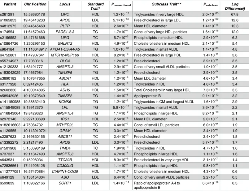
one of the lipid or lipoprotein subclass traits. Among the 157 loci associated (P<5×10⁻⁸) here with at least one subclass trait, 30 showed stronger association with a subclass trait than any conventional trait (Table 2,S7 Fig). For example, atPLTP(phospholipid transfer protein), rs4812975 was much more strongly associated with HDL diameter (Psubclass= 1.4×10⁻¹⁵) than with HDL-C (Pconventional= 2.6×10⁻³), consistent with PLTP mediating the net transfer of phos-pholipids between lipoproteins and uptake of phosphos-pholipids into the HDL-C core[46]. In addi-tion, atANGPTL3(angiopoietin-like 3),ANGPTL4(angiopoietin-like 4), andLPL(lipoprotein lipase), the variants were all more strongly associated with VLDL subclass traits than with the conventional traits (Table 2), consistent with studies showing that mouseAngptl3knockout and
Angptl4overexpression may act viaLplto decrease or increase VLDL, respectively[47,48]. At less well-characterized and gene-dense loci, lipid and lipoprotein subclass associations may help suggest target genes or biological roles. At the gene-denseMTCH2-NUP160locus, rs4752801 was>3 log units more strongly associated with decreased free cholesterol in large HDL levels (Psubclass= 1.4×10⁻⁹) than any conventional trait (HDL-C,Pconventional= 8.2×10⁻⁶,
Table 2). The pattern of association of rs4752801 with all 72 subclass traits (S7 Fig) is similar to the pattern of association and direction of effect for at least two other signals, rs737337 at
ANGPTL8and rs1129555 atGPAM.ANGPTL8andGPAMare both regulated directly or indi-rectly by LXR, encoded byNR1H3,[49,50] which is a positional candidate gene at this locus [49,50]. Thus, the global pattern of association supports a contribution ofNR1H3at the
MTCH2-NUP160locus and suggests that the lipid and lipoprotein subclass traits can be a use-ful tool to help determine which genes underlie association signals.
We performed a similar analysis of lipoprotein associations at coronary artery disease (CAD) loci (S10 Table). Variants at theAPOA5/APOA1locus were 5.2 log units more strongly associated with triglycerides in small VLDL than total triglycerides, andAPOE/APOC1was 2.3 log units more strongly associated with ratio of apoA-I/apoB than any conventional trait.
APOA5has been shown to affect VLDL concentrations and TG-rich particle metabolism, and the stronger association with the subclass trait is consistent with the known functions of these genes[51].
Discussion
In this study we conducted GWAS for 72 lipid and lipoprotein subclass traits in 8,372 Finn-ish men participating the METSIM study, and focused on identifying association signals that had not been identified previously with any lipid or lipoprotein trait. From the litera-ture of existing lipid and lipoprotein association studies, we identified 1,714 cholesterol, TG, lipid, and lipoprotein-associated variants. We trimmed this list based on LD (r2>0.95) to 885 variants to account for multiple known signals in a genome-wide conditional analy-sis. With this approach, we identified five novel signals at established lipid loci. We con-firmed that signals were independent by reciprocal conditional analyses.
This analysis focused on NMR measurements of 72 lipid and lipoprotein subclasses, includ-ing four conventionally measured lipid traits: TC, TG, HDL-C, and LDL-C. 892 of the associa-tion signals were located at or near loci previously associated with one or more of the four conventional traits. Lipid and lipoprotein subclass traits have been linked to metabolic and cardiovascular diseases, which underline their clinical importance[8–10]. We identified vari-ants at 30 loci that showed a more significant association with a subclass trait than one of the conventional lipid traits, consistent with previous observations[52].
gene, or more than one coding variant that affect the same gene’s function, provide stronger evidence for a gene’s role in determining trait variability. Multiple signals can be critical to understanding the relationship between genetic variants and gene function, quantitative traits, and disease[53]. Multiple association signals at established loci can also be used to detect molecular interactions between coding and regulatory variants on protein levels[54]. In addi-tion, multiple signals at the same locus may suggest that more than one nearby gene affects trait variation, and the association signals may represent different routes of transcriptional reg-ulation. Further study of the multiple association signals at a locus may more precisely define the functional genetic mechanisms.
Table 2. Comparison of METSIM association data between conventional lipid traits and subclass traits at established loci.
Variant Chr:Position Locus Standard
Trait*
Pconventional Subclass Trait** Psubclass Log Difference§
rs261291 15:58680178 LIPC HDL 1.2×10⁻¹⁷ Triglycerides in very large HDL 2.0×10−85 67.8 rs1065853 19:45413233 APOE LDL 5.1×10−50 Free cholesterol in large LDL 1.2×10−62 12.6
rs4812975 20:44545460 PLTP HDL 2.6×10⁻³ Mean HDL diameter 1.4×10⁻¹⁵ 12.3
rs174554 11:61579463 FADS1-2-3 TG 1.7×10⁻⁴ Conc. of very large HDL particles 1.6×10⁻¹⁶ 12.0 rs2156552 18:47181668 LIPG TC 5.7×10⁻³ Phospholipids in medium HDL 2.9×10⁻⁹ 6.3 rs10864726 1:230296153 GALNT2 HDL 4.9×10⁻⁷ Cholesterol esters in medium HDL 2.1×10⁻¹² 5.4
rs964184 11:116648917 APOA1-C3-A4-A5 TG 1.0×10⁻³⁰ Triglycerides in small VLDL 1.4×10⁻³⁵ 4.8
rs4752801 11:47907641 MTCH2-NUP160 HDL 8.2×10⁻⁶ Free cholesterol in large HDL 1.4×10⁻⁹ 3.8
rs55714927 17:7080316 DLG4 TG 1.2×10⁻⁵ Free cholesterol 3.9×10⁻⁹ 3.5
rs12130333 1:63191777 ANGPTL3 TC 2.9×10⁻⁴ Conc. of very small VLDL particles 1.0×10⁻⁷ 3.5
rs193042029 17:4667984 TM4SF5 TG 1.2×10⁻⁵ Free cholesterol 3.9×10⁻⁹ 3.5
rs3890182 9:107647655 ABCA1 HDL 1.2×10⁻⁴ Mean LDL diameter 4.6×10⁻⁸ 3.4
rs3177928 6:32412435 HLA HDL 1.2×10⁻⁴ Triglycerides in IDL 4.6×10⁻⁸ 3.4
rs2602836 4:100014805 ADH5 HDL 1.5×10⁻² Total Cholesterol in very large HDL 7.3×10⁻⁶ 3.3
rs58542926 19:19379549 TM6SF2 TG 1.4×10⁻⁸ Apolipoprotein B 9.1×10⁻¹² 3.2
rs141150988 19:38832410 KCNK6 TG 1.2×10⁻² Triglycerides in CM and largest VLDL 1.6×10⁻⁵ 2.9 rs115849089 8:19912370 LPL TG 5.8×10⁻¹³ Triglycerides in small VLDL 3.6×10⁻¹⁵ 2.2
rs116843064 19:8429323 ANGPTL4 TG 1.1×10⁻⁷ Phospholipids in large HDL 8.2×10⁻¹⁰ 2.1
rs2972146 2:227100698 IRS1 HDL 2.5×10⁻⁵ Mean HDL diameter 2.0×10⁻⁷ 2.1
rs182616603 4:75084732 MTHFD2L TC 4.4×10⁻⁷ Conc. of small LDL particles 6.0×10⁻⁹ 1.9
rs1129555 10:113910721 GPAM TG 3.0×10⁻⁴ Mean HDL diameter 3.4×10⁻⁶ 1.9
rs2287623 2:169830155 ABCB11 TC 2.4×10⁻³ Free cholesterol 3.4×10⁻⁵ 1.8
rs13392272 2:21217490 APOB LDL 3.0×10⁻⁸ Free cholesterol 5.7×10⁻¹⁰ 1.7
rs1501908 5:156398169 TIMD4 TC 1.9×10⁻³ Triglycerides in IDL 4.7×10⁻⁵ 1.6
rs737337 19:11347493 ANGPTL8 HDL 4.7×10⁻⁴ Phospholipids in large HDL 1.1×10⁻⁵ 1.6 rs643531 9:15296034 TTC39B HDL 8.3×10⁻⁶ Free cholesterol in very large HDL 3.1×10⁻⁷ 1.4
rs72836561 17:41926126 CD300LG HDL 1.1×10⁻⁴ Phospholipids in large HDL 9.8×10⁻⁶ 1.1
rs112777051 16:57470884 CIAPIN1-COQ9 HDL 1.7×10⁻⁴ Cholesterol esters in medium HDL 4.3×10⁻⁵ 0.6 rs649129 9:136154304 ABO LDL 6.4×10⁻⁷ Conc. of very small VLDL particles 2.2×10⁻⁷ 0.5 rs599839 1:109822166 SORT1 LDL 1.4×10⁻¹⁴ Ratio of apolipoprotein A-I to
apolipoprotein B
6.6×10⁻¹⁵ 0.3
Variants at established lipid trait loci for which the METSIM association (P<5×10⁻⁵) for a subclass trait was stronger than for any of four conventional lipid traits (HDL, LDL, TC, or TG). CM, chylomicrons. Chr, chromosome. IDL, intermediate-density lipoprotein.
§Log difference,−log10(Psubclass/Pconventional).
*Strongest associated conventional trait for the variant. **Strongest associated subclass trait for the variant.
The gene-based tests of association atLIPCandLIPGidentified new rare coding variants that may alter the function of these genes, and of lipid and lipoprotein subclass levels. While the missense variants identified here all have mean normalized lipoprotein trait values above the population mean, this type of analysis can help distinguish variants that lead to loss or decrease vs gain or increase of gene function[53]. As well, the rise of whole exome sequencing will likely uncover many more rare coding variants, including variants with unknown signifi-cance on gene function. While it is still unclear which variants included in the gene-based tests forLIPCandLIPGtruly affect gene function, the comparison of trait values between carriers of different variants may be used to help interpret the potential role of these variants in indi-vidual carriers.
In conclusion, this GWAS of 72 lipid and lipoprotein subclass traits in 8,372 Finnish partic-ipants in the METSIM study identified associations with 42 loci previously identified only with the conventional lipid and lipoprotein traits[2], five novel signals associated with lipoprotein subclasses, and eight rare, potentially functional, coding variants atLIPCandLIPG. Our use of a dense reference panel of>15M variants combined with the high-throughput NMR-mea-sured traits allowed us to conduct higher-resolution genetic analyses than reported previously. Functional analysis of the variants identified in this study is the next step to determine which variants and genes are affected, and replication of these lipid and lipoprotein subclass associa-tions in women and in other ancestry groups will be useful to better understand the genetic architecture of lipid and lipoprotein metabolism.
Materials and methods
Ethics statement
The METSIM study was performed in accordance with the Helsinki Declaration and was approved by the Research Ethics Committee, Hospital District of Northern Savo (number 171/ 2004). All study participants gave their written informed consent.
Study participants
Among the 10,197 participants in the METSIM study, we analyzed 8,372 non-diabetic individ-uals (mean age 57±7 SD years and BMI 26.8±3.8 kg/m2)[55].
Subclass trait measurements
We measured the 72 lipid and lipoprotein traits from blood serum samples by proton NMR, as previously described[56]. Briefly, lipid samples are extracted and measured by proton NMR, and the NMR-spectra and automated phasing are compared to plate, background, and serum controls. Regression modeling is used to quantify the spectral areas to produce the quantified molecular data. The samples included 60 lipoprotein subclasses, 6 cholesterol and triglyceride measures, 3 cholesterol diameter measures, and 3 apolipoprotein measurements (S1 Table). Definition of the subclass traits has been previously described[56,57]. We visualized the Pear-son correlation matrix between lipoprotein traits using a corrgram with the ellipse (https:// cran.r-project.org/web/packages/ellipse/) and lattice packages (http://lattice.r-forge.r-project. org/) within R (S2 Fig)[58].
Genotyping and imputation
and large deletions) based on whole-genome sequence of 2,657 Europeans consisting of Ger-man, Swedish, Finnish, and British participants; with the majority of the cohort comprised of Finns[59]. The resulting 15,144,991 variants were subjected to quality controls including sam-ple- and variant-level controls for detecting sample contamination, sex and relatedness confir-mation, and detection of sample outliers using principal-component analysis. To exclude samples with evidence of DNA contamination, we used BAFRegress v0.9 (http://genome.sph. umich.edu/wiki/BAFRegress). Based on principal component analysis, eighteen exome array sample duplicates, one individual each from seven monozygotic twin pairs, and twelve popula-tion outliers were removed from analysis. Due to sex chromosome inconsistencies, fourteen OmniExpress samples were removed. Samples with low genotype call rate (<95%) for either array were removed. Variants with low-mapping quality to build hg19, low genotype com-pleteness (<95% for OmniExpress and<98% for exome array), or multi-allelic variants were removed. The remaining high quality variants were phased using Shape-It v2[60].
Single-variant analysis
We tested for association using imputed dosages for all variants with summed minor allele count dosage>1 with each of the 72 lipid and lipoprotein traits assuming an additive model and accounting for cryptic relatedness using the EMMAX linear mixed model approach as implemented in EPACTS (http://genome.sph.umich.edu/wiki/epacts). Traits were adjusted for age, age2, smoking status, and lipid lowering medication. Residuals were inverse normalized. To assess the level of genomic inflation, we calculated the genomic control statistic (λGC) for
all of the trait-variant associations using R[58] (S1 Fig). Reported effect size regression coeffi-cients (betas) sizes are given in standard deviation units. The rare lead associated variants that were imputed and had MAF<0.01 were tested for genotype accuracy by using TaqMan assays (Thermo Fisher Scientific) or Sanger sequencing in 499 METSIM participants who carried one or more rare alleles at these variants. Variants that had>10% discordance between the imputed genotype and the sequenced genotype in the examined individuals were removed from the analysis. Variants with MAF<0.001 were excluded from analysis (S5 Table).
Compilation of existing lipid and lipoprotein trait associated variants
To identify variant association signals distinct or independent from those reported previously, we identified variants previously reported to be associated with any cholesterol, lipid, lipopro-tein, or triglyceride trait. We performed a literature review of GWAS and sequencing studies using PubMed (https://www.ncbi.nlm.nih.gov/pubmed/) and Google Scholar (https://scholar. google.com/), screened a GWAS Catalog (http://www.ebi.ac.uk/gwas/), and used SNIPPER (https://csg.sph.umich.edu/boehnke/snipper/) to query publicly accessible databases. The resulting curated list contained 1,714 variants, at>150 loci from 33 studies (S3 Table). The resulting curated list contained 1,714 variants, at>150 loci from 33 studies (S3 Table). We used this list to represent the known genome-wide lipid and lipoprotein-associated variants.
Conditional analyses
conditional analyses (S6 Table). Signals that remained significant (Psingle<5×10⁻⁸) after single-variant conditional analysis were considered independent. Signals that only achieved a signifi-cance threshold ofPsingle<5×10⁻⁶after single-variant conditional analysis were considered dis-tinct. The single-variant conditional analyses considered variants within 1 Mb of the signal, which accounts for<1% of the genome. Therefore, the significance thresholds for the distinct and independent additional signals are conservative. At each locus, we validated that signals were distinct/independent by reciprocal conditional analysis with the putative novel lead asso-ciated variant for the trait. Additionally, the association data for the novel signals was adjusted for each of the four conventional traits (HDL-C, LDL-C, TC, and TG), and the effect of the association is reported inS7 Table.
Comparison of subclass trait associations with conventional lipid and
lipoprotein trait associations
For each of the 885 lipid/lipoprotein-associated variants (S4 Table), we determined whether the variant showed stronger association with one of the 68 subclass traits compared to the four conventional lipid traits (TC, TG, LDL-C, HDL-C). Variants were included for comparison if the variant association with a subclass trait satisfiedP<5×10⁻⁵and if–log10pvalue for subclass
trait association was greater than any of the four conventional lipid traits.
Gene-based tests of association
To determine the contribution of rare coding variants, we used the Optimal Sequence Kernel Association Test (SKAT-O) with EMMAX, as implemented in EPACTS, to test for gene-based associations with the 72 lipoprotein traits[61]. Only coding variants directly genotyped on the OmniExpress or Exome array were included, resulting in 709,600 variants. Since SKAT-O requires no missing data, we imputed missing genotype data with the variant mean genotype. We annotated coding variants using VEP. These annotations were the basis for four masks that we implemented in the gene-based tests, as previously described[53]. Briefly, the four masks were: Protein-Truncating Variants (PTV): no MAF limit; variants are nonsense, frame-shift, or essential splice variants. PTV+missense: MAF<1%; all PTVs and missense variants. PTV+Nonsynonymous strict (NSstrict): no MAF limit; all PTVs and missense variants
pre-dicted as deleterious by five variant annotation algorithms: LRT, Mutation Taster, PolyPhen2--HumDiv, PolyPhen2-HumVar, and SIFT. PTV+NSstrict+NSbroad: MAF<1%; all variants in
PTV+NSstrictand variants predicted to be deleterious by any of the five algorithms above. Only
genes containing two or more variants in a given mask were tested. We conducted gene-based conditional analyses to determine whether a single variant or a net-effect of multiple variants could explain the observed association signal.
Variant annotation
Supporting information
S1 Fig. Distribution of the METSIM lambda genomic control (GC) for the 72 lipoproteins.
(PDF)
S2 Fig. Correlogram showing Pearson correlations between lipoprotein traits. Correlogram
showing Pearson correlations between lipoprotein traits. Rows and columns are ordered according to a complete linkage hierarchical clustering of traits based on the Pearson correla-tion matrix. Percent correlacorrela-tion is shown for each trait-trait comparison. The color scale is from red (negative correlation) to blue (positive correlation). The shapes are representations of the correlation with a circle representing ‘no correlation’, ellipse representing ‘moderate correlation’, and a diagonal line representing ‘high correlation’.
(PDF)
S3 Fig. Manhattan plots of traits. Manhattan plots for the five traits with signals identified
from this study. X-axis shows the chromosomes, and the y-axis is the–log10(Pvalue) for the
variant-trait association. The horizontal line is at the cutoffP= 5×10⁻⁸. (PDF)
S4 Fig. Plots of the five association signals after conditioning on the 885 known lipid sig-nals. Each circle represents a single variant. The color is based on LD (r2) between each variant and the reference variant (purple diamond); X-axis, genomic (GRCh37/hg19) position in Mb; Left y-axis, p- value of variant-trait association in–log10; Right y-axis, local estimates of
geno-mic recombination rate in cM/Mb, represented by blue vertical lines. (PDF)
S5 Fig. Unconditional plots of the five lipid and lipoprotein subclass-associated loci. Each
circle represents a single variant. The color is based on LD (r2) between each variant and the reference variant (purple diamond); X-axis, genomic (GRCh37/hg19) position in Mb; Left y-axis, p-value of variant-trait association in–log10; Right y-axis, local estimates of genomic
recombination rate in cM/Mb, represented by blue vertical lines. (PDF)
S6 Fig. Association of all 72 lipoprotein/lipid traits with the variants inTable 1. ThePvalue is shown in -log10and in the direction (+ or−) of the effect (Beta). The red line denotes the
sig-nificance cutoff of P5E-8. The red asterisk indicates the most significantly associated trait. CAD, coronary artery disease.
(PDF)
S7 Fig. Association of all 72 lipoprotein/lipid traits with the variants inTable 2. ThePvalue is shown in -log10and in the direction (+ or−) of the effect (Beta). The red line denotes the
sig-nificance cutoff of P5E-8. The red asterisk indicates the most significantly associated trait. CAD, coronary artery disease.
(PDF)
S1 Table. Characteristics of 8,372 METSIM study participants and 72 lipoprotein sub-classes and triglyceride measures.
(XLSX)
minor allele frequency; P-value, best unconditional p-value for any of the 72 traits; Beta, effect size of the alternate allele; Trait, trait with strongest association.
(XLSX)
S3 Table. Previously reported associations between 1,714 variants and one or more lipid or lipoprotein traits. Variants and association data in this table were collected from 33 published
GWAS, fine-mapping, and exome-sequencing studies. The reported trait, Beta, and P-value are taken from the study with the lowest p-value. N.R., data not reported in the study; Chr, chromosome; Position, hg19; Trait, strongest associated trait from study shown; EA, effect allele; NEA, non-effect allele; N, sample size of the study; Reference, the first study in the list is the study with the lowest p-value for the given trait.
(XLSX)
S4 Table. 885 variants used for the conditional analysis (seeMethods). Variants on this
table were trimmed from the 1,714 variants onS3 Tableby using an r2>0.95 cutoff. The reported trait, Beta, and P-value are taken from the study with the lowest p-value. N.R., data not reported in the study; Chr, chromosome; Position, hg19; Trait, strongest associated trait from study shown; EA, effect allele; NEA, non-effect allele; N, sample size of the study; Refer-ence, the first study in the list is the study with the lowest p-value for the given trait.
(XLSX)
S5 Table. Variants that remained significantly associated (P<5×10⁻⁸) with at least one of the 72 lipid or lipoprotein traits after conditioning on 885 known variants. Trait, subclass
trait (defined inS1 Table); Chr, chromosome; Genotype counts for homozygous reference allele/heterozygous/homozygous alternate allele; MAC, minor allele count; MAF, minor allele frequency; P-value, p-value for the variant after conditioning on 885 known lipid-associated variants (S4 Table); Beta, effect size of the alternate allele; R2, variance explained.
(XLSX)
S6 Table. Results of single-marker and reciprocal conditional analyses for the associated variants from this study. Lead variant, the most associated variant for the Lead Trait at a
given locus; Lead trait, the lipoprotein subclass or triglyceride measure with lowest p-value across the 72 traits; Locus, biologically relevant gene within 1 Mb of lead variant; Chr, chromo-some; MAF, minor allele frequency;Pdiscovery, unconditional p-value for the lead variant and trait;Pconditional, p-value for the lead variant after conditioning on 885 known lipid GWAS vari-ants (S4 Table); Conditional variant, known lipid or lipoprotein associated variants used for single-marker conditional analyses;Psingle, p-value for the Lead variant after conditioning on the Conditional variant;Ptrait, unconditional p-value of the Conditional variant for the Lead trait;Preciprocal, p-value of the Conditional variant after conditioning on the Lead variant; Conc, concentration; At chromosome 18, the variants identified in the gene-based test for
LIPGare in italics. § The effect of the unconditional p-value for the lead variant and trait after adjusting for each of the four conventional traits.
(XLSX)
S7 Table. Results of conventional trait conditional analyses for the associated variants from this study. Lead variant, the most associated variant for the Lead Trait at a given locus;
traits. (XLSX)
S8 Table. Lipoprotein subclass and triglyceride measure loci associated variants that over-lap epigenetic evidence of regulatory elements. Variants listed were in LD (r2>0.7) with the lead trait variant (in bold) from this study and overlapped a regulatory element in relevant tis-sue datasets. Abbreviations of the tistis-sues tested for overlapping of regulatory elements are: A, adipose; B, blood; L, liver. Chr, chromosome; Nearest coding TSS, distance from nearest GEN-CODEv12 basic annotation transcription start site. Negative distance indicates the variant is upstream of the TSS relative to the direction of transcription; N, total number of overlapping datasets across experiments and cell types; Open chromatin, variants overlapping FAIRE/ DNase hypersensitivity elements; ChIP-seq Peak, Variant overlaps ChIP-seq peaks. Transcrip-tion factor name:Tissue.
(XLSX)
S9 Table. Results of single-marker conditional analyses for the gene-based association data from this study. Gene, gene tested in gene-based association tests; Lead trait, the lipoprotein
subclass or triglyceride measure with lowest p-value across the 72 traits;Pgene, p-value for the gene and trait; Conditional variant, known lipid or lipoprotein associated variants used for sin-gle-marker conditional analyses; Chr:Position, the hg19 chromosome and base-pair position of the Conditional variant; P, p-value for the gene-based association after conditioning on the Conditional variant.
(XLSX)
S10 Table. Comparison of METSIM association data between conventional lipid traits and subclass traits at established coronary artery disease loci. Variants at established CAD loci
for which the METSIM association (P<5×10⁻⁵) for a subclass trait was stronger than for any of four conventional lipid traits (HDL, LDL, TC, or TG). Log difference, log10(Psubclass/P
conven-tional). Chr, chromosome.
(XLSX)
Acknowledgments
We thank the participants of the METSIM study, the Exome Aggregation Consortium, and the groups that provided exome variant data for comparison. The data used for the analyses described in this manuscript were obtained from: theGTEx Portalon 07/11/17. We dedicate this manuscript in memory of our friend and colleague Peter Chines.
Author Contributions
Conceptualization: James P. Davis, Jeroen R. Huyghe, Francis S. Collins, Markku Laakso,
Michael Boehnke, Karen L. Mohlke.
Data curation: James P. Davis, Jeroen R. Huyghe, Anne U. Jackson, Heather M. Stringham,
Antti J. Kangas, Pasi Soininen, Mika Ala-Korpela, Francis S. Collins, Markku Laakso.
Formal analysis: James P. Davis, Jeroen R. Huyghe, Anne U. Jackson, Francis S. Collins,
Markku Laakso, Michael Boehnke, Karen L. Mohlke.
Funding acquisition: James P. Davis, Mika Ala-Korpela, Francis S. Collins, Markku Laakso,
Investigation: James P. Davis, Jeroen R. Huyghe, Adam E. Locke, Anne U. Jackson, Xueling
Sim, Narisu Narisu, Peter S. Chines, Antti J. Kangas, Pasi Soininen, Mika Ala-Korpela, Johanna Kuusisto, Markku Laakso, Michael Boehnke, Karen L. Mohlke.
Methodology: James P. Davis, Jeroen R. Huyghe, Adam E. Locke, Anne U. Jackson, Xueling
Sim, Heather M. Stringham, Tanya M. Teslovich, Ryan P. Welch, Christian Fuchsberger, Narisu Narisu, Peter S. Chines, Mika Ala-Korpela, Francis S. Collins, Markku Laakso, Michael Boehnke, Karen L. Mohlke.
Project administration: Mika Ala-Korpela, Francis S. Collins, Markku Laakso, Michael
Boehnke, Karen L. Mohlke.
Resources: Anne U. Jackson, Heather M. Stringham, Ryan P. Welch, Peter S. Chines, Mika
Ala-Korpela, Francis S. Collins, Markku Laakso, Michael Boehnke.
Software: Anne U. Jackson, Heather M. Stringham, Ryan P. Welch, Peter S. Chines, Mika
Ala-Korpela.
Supervision: Mika Ala-Korpela, Francis S. Collins, Markku Laakso, Michael Boehnke, Karen
L. Mohlke.
Validation: Anne U. Jackson, Heather M. Stringham, Narisu Narisu.
Visualization: James P. Davis, Anne U. Jackson, Karen L. Mohlke.
Writing – original draft: James P. Davis, Anne U. Jackson, Michael Boehnke, Karen L.
Mohlke.
Writing – review & editing: James P. Davis, Jeroen R. Huyghe, Anne U. Jackson, Xueling
Sim, Heather M. Stringham, Narisu Narisu, Mika Ala-Korpela, Michael Boehnke, Karen L. Mohlke.
References
1. Willer CJ, Mohlke KL. Finding genes and variants for lipid levels after genome-wide association analy-sis. Curr Opin Lipidol. 2012; 23: 98–103.https://doi.org/10.1097/MOL.0b013e328350fad2PMID:
22418572
2. Willer CJ, Schmidt EM, Sengupta S, Peloso GM, Gustafsson S, Kanoni S, et al. Discovery and refine-ment of loci associated with lipid levels. Nat Genet. 2013; 45: 1274–1283.https://doi.org/10.1038/ng. 2797PMID:24097068
3. Teslovich TM, Musunuru K, Smith A V, Edmondson AC, Stylianou IM, Koseki M, et al. Biological, clinical and population relevance of 95 loci for blood lipids. Nature. 2010; 466: 707–713.https://doi.org/10. 1038/nature09270PMID:20686565
4. Kathiresan S, Willer CJ, Peloso GM, Demissie S, Musunuru K, Schadt EE, et al. Common variants at 30 loci contribute to polygenic dyslipidemia. Nat Genet. 2009; 41: 56–65.https://doi.org/10.1038/ng.291
PMID:19060906
5. Kettunen J, Tukiainen T, Sarin A-P, Ortega-Alonso A, Tikkanen E, Lyytika¨inen L-P, et al. Genome-wide association study identifies multiple loci influencing human serum metabolite levels. Nat Genet. 2012; 44: 269–276.https://doi.org/10.1038/ng.1073PMID:22286219
6. Kettunen J, Demirkan A, Wu¨rtz P, Draisma HHMM, Haller T, Rawal R, et al. Genome-wide study for cir-culating metabolites identifies 62 loci and reveals novel systemic effects of LPA. Nat Commun. 2016; 7: 11122.https://doi.org/10.1038/ncomms11122PMID:27005778
7. Soininen P, Kangas AJ, Wu¨rtz P, Tukiainen T, Tynkkynen T, Laatikainen R, et al. High-throughput serum NMR metabonomics for cost-effective holistic studies on systemic metabolism. Analyst. 2009; 134: 1781–1785.https://doi.org/10.1039/b910205aPMID:19684899
8. Kathiresan S, Otvos JD, Sullivan LM, Keyes MJ, Schaefer EJ, Wilson PWF, et al. Increased small low-density lipoprotein particle number: A prominent feature of the metabolic syndrome in the Framingham Heart Study. Circulation. 2006; 113: 20–29.https://doi.org/10.1161/CIRCULATIONAHA.105.567107
9. Petersen AK, Stark K, Musameh MD, Nelson CP, Ro¨misch-Margl W, Kremer W, et al. Genetic associa-tions with lipoprotein subfracassocia-tions provide information on their biological nature. Hum Mol Genet. 2012; 21: 1433–1443.https://doi.org/10.1093/hmg/ddr580PMID:22156577
10. Wu¨rtz P, Havulinna AS, Soininen P, Tynkkynen T, Prieto-Merino D, Tillin T, et al. Metabolite profiling and cardiovascular event risk: a prospective study of 3 population-based cohorts. Circulation. 2015; 131: 774–785.https://doi.org/10.1161/CIRCULATIONAHA.114.013116PMID:25573147
11. Akinkuolie AO, Paynter NP, Padmanabhan L, Mora S. High-density lipoprotein particle subclass hetero-geneity and incident coronary heart disease. Circ Cardiovasc Qual Outcomes. 2014; 7: 55–63.https:// doi.org/10.1161/CIRCOUTCOMES.113.000675PMID:24248942
12. Fischer K, Kettunen J, Wu¨rtz P, Haller T, Havulinna AS, Kangas AJ, et al. Biomarker profiling by nuclear magnetic resonance spectroscopy for the prediction of all-cause mortality: an observational study of 17,345 persons. PLoS Med. 2014; 11: e1001606.https://doi.org/10.1371/journal.pmed.1001606PMID:
24586121
13. Wu Y, Waite LL, Jackson AU, Sheu WHH, Buyske S, Absher D, et al. Trans-Ethnic Fine-Mapping of Lipid Loci Identifies Population-Specific Signals and Allelic Heterogeneity That Increases the Trait Vari-ance Explained. PLoS Genet. 2013; 9: e1003379.https://doi.org/10.1371/journal.pgen.1003379PMID:
23555291
14. Shungin D, Winkler TW, Croteau-Chonka DC, Ferreira T, Locke AE, Ma¨gi R, et al. New genetic loci link adipose and insulin biology to body fat distribution. Nature. 2015; 518: 187–196.https://doi.org/10. 1038/nature14132PMID:25673412
15. Wu Y, Waite LL, Jackson AU, Sheu WHH, Buyske S, Absher D, et al. Trans-Ethnic Fine-Mapping of Lipid Loci Identifies Population-Specific Signals and Allelic Heterogeneity That Increases the Trait Vari-ance Explained. PLoS Genet. 2013; 9: e1003379.https://doi.org/10.1371/journal.pgen.1003379PMID:
23555291
16. Sanna S, Li B, Mulas A, Sidore C, Kang HM, Jackson AU, et al. Fine mapping of five loci associated with low-density lipoprotein cholesterol detects variants that double the explained heritability. PLoS Genet. 2011; 7.https://doi.org/10.1371/journal.pgen.1002198PMID:21829380
17. Stanca´kova´ A, Javorsky´ M, Kuulasmaa T, Haffner SM, Kuusisto J, Laakso M. Changes in insulin sensi-tivity and insulin release in relation to glycemia and glucose tolerance in 6,414 Finnish men. Diabetes. 2009; 58: 1212–1221.https://doi.org/10.2337/db08-1607PMID:19223598
18. Kang HM, Sul JH, Service SK, Zaitlen NA, Kong SY, Freimer NB, et al. Variance component model to account for sample structure in genome-wide association studies. Nat Genet. 2010; 42: 348–354.
https://doi.org/10.1038/ng.548PMID:20208533
19. Surakka I, Horikoshi M, Magi R, Sarin A-P, Mahajan A, Lagou V, et al. The impact of low-frequency and rare variants on lipid levels. Nat Genet. 2015; 47: 589–597.https://doi.org/10.1038/ng.3300PMID:
25961943
20. van Leeuwen EM, Karssen LC, Deelen J, Isaacs A, Medina-Gomez C, Mbarek H, et al. Genome of The Netherlands population-specific imputations identify an ABCA6 variant associated with cholesterol lev-els. Nat Commun. 2015; 6: 6065.https://doi.org/10.1038/ncomms7065PMID:25751400
21. Wang S, Song J, Yang Y, Zhang Y, Wang H, Ma J. HIF3A DNA Methylation Is Associated with Child-hood Obesity and ALT. PLoS One. 2015; 10: e0145944.https://doi.org/10.1371/journal.pone.0145944
PMID:26717317
22. Pan H, Lin X, Wu Y, Chen L, Teh AL, Soh SE, et al. HIF3A association with adiposity: the story begins before birth. Epigenomics. 2015; 7: 937–950.https://doi.org/10.2217/epi.15.45PMID:26011824
23. Forristal CE, Wright KL, Hanley NA, Oreffo ROC, Houghton FD. Hypoxia inducible factors regulate plur-ipotency and proliferation in human embryonic stem cells cultured at reduced oxygen tensions. Repro-duction. 2010; 139: 85–97.https://doi.org/10.1530/REP-09-0300PMID:19755485
24. Shen G, Zhao Y, Chen M, Zhang F-L, Liu X-L, Wang Y, et al. Hypoxia-inducible factor-1 (HIF-1) pro-motes LDL and VLDL uptake through inducing VLDLR under hypoxia. Biochem J. 2012; 441: 675–683.
https://doi.org/10.1042/BJ20111377PMID:21970364
25. Mohlke KL, Lange EM, Valle TT, Ghosh S, Magnuson VL, Silander K, et al. Linkage disequilibrium between microsatellite markers extends beyond 1 cM on chromosome 20 in Finns. Genome Res. 2001; 11: 1221–1226.https://doi.org/10.1101/gr.173201PMID:11435404
26. Roopenian DC, Low BE, Christianson GJ, Proetzel G, Sproule TJ, Wiles M V. Albumin-deficient mouse models for studying metabolism of human albumin and pharmacokinetics of albumin-based drugs. MAbs. 2015; 7: 344–351.https://doi.org/10.1080/19420862.2015.1008345PMID:25654695
28. Hsu J, Smith JD. Genetic-genomic replication to identify candidate mouse atherosclerosis modifier genes. J Am Heart Assoc. 2013; 2: e005421.https://doi.org/10.1161/JAHA.112.005421PMID:
23525445
29. Behnia R, Panic B, Whyte JRC, Munro S. Targeting of the Arf-like GTPase Arl3p to the Golgi requires N-terminal acetylation and the membrane protein Sys1p. Nat Cell Biol. 2004; 6: 405–413.https://doi. org/10.1038/ncb1120PMID:15077113
30. Hommel A, Hesse D, Vo¨lker W, Jaschke A, Moser M, Engel T, et al. The ARF-like GTPase ARFRP1 is essential for lipid droplet growth and is involved in the regulation of lipolysis. Mol Cell Biol. 2010; 30: 1231–1242.https://doi.org/10.1128/MCB.01269-09PMID:20038528
31. Maeda E, Naka Y, Matozaki T, Sakuma M. Lecithin-cholesterol with a missense acyltransferase (LCAT) deficiency mutation in exon 6 of the LCAT gene enzymatic amvlification of aenomic DNA SeauencinP of amolified sir & e-stranded DNA. Biochem Biophys Res Commun. 1991; 178: 460–466. PMID:
1859405
32. McLaren W, Pritchard B, Rios D, Chen Y, Flicek P, Cunningham F. Deriving the consequences of geno-mic variants with the Ensembl API and SNP Effect Predictor. Bioinformatics. 2010; 26: 2069–2070.
https://doi.org/10.1093/bioinformatics/btq330PMID:20562413
33. Kathiresan S, Melander O, Guiducci C, Surti A, Burtt NP, Rieder MJ, et al. Six new loci associated with blood low-density lipoprotein cholesterol, high-density lipoprotein cholesterol or triglycerides in humans. Nat Genet. 2008; 40: 189–197.https://doi.org/10.1038/ng.75PMID:18193044
34. Ishida T, Choi S, Kundu RK, Hirata K-I, Rubin EM, Cooper AD, et al. Endothelial lipase is a major deter-minant of HDL level. J Clin Invest. 2003; 111: 347–355.https://doi.org/10.1172/JCI16306PMID:
12569160
35. Ma K, Cilingiroglu M, Otvos JD, Ballantyne CM, Marian AJ, Chan L. Endothelial lipase is a major genetic determinant for high-density lipoprotein concentration, structure, and metabolism. Proc Natl Acad Sci U S A. 2003; 100: 2748–2753.https://doi.org/10.1073/pnas.0438039100PMID:12601178
36. Jin W, Millar JS, Broedl U, Glick JM, Rader DJ. Inhibition of endothelial lipase causes increased HDL cholesterol levels in vivo. J Clin Invest. 2003; 111: 357–362.https://doi.org/10.1172/JCI16146PMID:
12569161
37. Jaye M, Lynch KJ, Krawiec J, Marchadier D, Maugeais C, Doan K, et al. A novel endothelial-derived lipase that modulates HDL metabolism. Nat Genet. 1999; 21: 424–428.https://doi.org/10.1038/7766
PMID:10192396
38. Edmondson AC, Brown RJ, Kathiresan S, Cupples LA, Demissie S, Manning AK, et al. Loss-of-function variants in endothelial lipase are a cause of elevated HDL cholesterol in humans. J Clin Invest. 2009; 119: 1042–1050.https://doi.org/10.1172/JCI37176PMID:19287092
39. Exome Aggregation Consortium (ExAC). (Cambridge, MA, Aug 2015) [Internet].
40. Service SK, Teslovich TM, Fuchsberger C, Ramensky V, Yajnik P, Koboldt DC, et al. Re-sequencing expands our understanding of the phenotypic impact of variants at GWAS loci. PLoS Genet. 2014; 10: e1004147.https://doi.org/10.1371/journal.pgen.1004147PMID:24497850
41. Zambon A, Deeb SS, Hokanson JE, Brown BG, Brunzell JD. Common variants in the promoter of the hepatic lipase gene are associated with lower levels of hepatic lipase activity, buoyant LDL, and higher HDL2 cholesterol. Arterioscler Thromb Vasc Biol. 1998; 18: 1723–1729.https://doi.org/10.1161/01. ATV.18.11.1723PMID:9812910
42. Carlson LA, Holmquist L, Nilsson-Ehle P. Deficiency of hepatic lipase activity in post-heparin plasma in familial hyper-alpha-triglyceridemia. Acta Med Scand. 1986; 219: 435–447. PMID:3739751
43. Helgadottir A, Gretarsdottir S, Thorleifsson G, Hjartarson E, Sigurdsson A, Magnusdottir A, et al. Vari-ants with large effects on blood lipids and the role of cholesterol and triglycerides in coronary disease. Nat Genet. 2016; 48: 634–639.https://doi.org/10.1038/ng.3561PMID:27135400
44. Peloso GM, Auer PL, Bis JC, Voorman A, Morrison AC, Stitziel NO, et al. Association of low-frequency and rare coding-sequence variants with blood lipids and coronary heart disease in 56,000 whites and blacks. Am J Hum Genet. 2014; 94: 223–232.https://doi.org/10.1016/j.ajhg.2014.01.009PMID:
24507774
45. Singaraja RR, Sivapalaratnam S, Hovingh K, Dub?? MP, Castro-Perez JJ, Collins HL, et al. The impact of partial and complete loss-of-function mutations in endothelial lipase on high-density lipoprotein levels and functionality in humans. Circ Cardiovasc Genet. 2013; 6: 54–62.https://doi.org/10.1161/
CIRCGENETICS.111.962613PMID:23243195
46. Yazdanyar A, Jiang X-C. Liver phospholipid transfer protein (PLTP) expression with a PLTP-null back-ground promotes very low-density lipoprotein production in mice. Hepatology. 2012; 56: 576–584.
47. Wang Y, McNutt MC, Banfi S, Levin MG, Holland WL, Gusarova V, et al. Hepatic ANGPTL3 regulates adipose tissue energy homeostasis. Proc Natl Acad Sci. 2015; 112: 11630–11635.https://doi.org/10. 1073/pnas.1515374112PMID:26305978
48. Dijk W, Kersten S. Regulation of lipoprotein lipase by Angptl4. Trends Endocrinol Metab. Elsevier Ltd; 2014; 25: 146–155.https://doi.org/10.1016/j.tem.2013.12.005PMID:24397894
49. Wendel AA, Lewin TM, Coleman RA. Glycerol-3-phosphate acyltransferases: rate limiting enzymes of triacylglycerol biosynthesis. Biochim Biophys Acta. 2009; 1791: 501–506.https://doi.org/10.1016/j. bbalip.2008.10.010PMID:19038363
50. Lee J, Hong SW, Park SE, Rhee EJ, Park CY, Oh KW, et al. AMP-activated protein kinase suppresses the expression of LXR/SREBP-1 signaling-induced ANGPTL8 in HepG2 cells. Mol Cell Endocrinol. Elsevier Ireland Ltd; 2015; 414: 148–155.https://doi.org/10.1016/j.mce.2015.07.031PMID:26254015
51. Nilsson SK, Heeren J, Olivecrona G, Merkel M. Apolipoprotein A-V; a potent triglyceride reducer. Ath-erosclerosis. 2011; 219: 15–21.https://doi.org/10.1016/j.atherosclerosis.2011.07.019PMID:21831376
52. Wu¨rtz P, Kangas AJ, Soininen P, Lehtima¨ki T, Ka¨ho¨nen M, Viikari JS, et al. Lipoprotein subclass profil-ing reveals pleiotropy in the genetic variants of lipid risk factors for coronary heart disease: A note on mendelian randomization studies. J Am Coll Cardiol. 2013; 62: 1906–1908.https://doi.org/10.1016/j. jacc.2013.07.085PMID:24055740
53. Mahajan A, Sim X, Ng HJ, Manning A, Rivas M a, Highland HM, et al. Identification and Functional Characterization of G6PC2 Coding Variants Influencing Glycemic Traits Define an Effector Transcript at the G6PC2-ABCB11 Locus. PLoS Genet. 2015; 11: e1004876.https://doi.org/10.1371/journal.pgen. 1004876PMID:25625282
54. Lappalainen T, Montgomery SB, Nica AC, Dermitzakis ET. Epistatic selection between coding and reg-ulatory variation in human evolution and disease. Am J Hum Genet. The American Society of Human Genetics; 2011; 89: 459–463.https://doi.org/10.1016/j.ajhg.2011.08.004PMID:21907014
55. Laakso M, Kuusisto J, Stancakova A, Kuulasmaa T, Pajukanta P, Lusis AJ, et al. METabolic Syndrome In Men (METSIM) Study: a resource for studies of metabolic and cardiovascular diseases. J Lipid Res. 2017; 58: jlr.O072629.https://doi.org/10.1194/jlr.O072629PMID:28119442
56. Soininen P, Kangas AJ, Wu¨rtz P, Suna T, Ala-Korpela M. Quantitative serum nuclear magnetic reso-nance metabolomics in cardiovascular epidemiology and genetics. Circ Cardiovasc Genet. 2015; 8: 192–206.https://doi.org/10.1161/CIRCGENETICS.114.000216PMID:25691689
57. Wang J, Stanča´kova´ A, Soininen P, Kangas AJ, Paananen J, Kuusisto J, et al. Lipoprotein subclass profiles in individuals with varying degrees of glucose tolerance: a population-based study of 9399 Finn-ish men. J Intern Med. 2012; 272: 562–572.https://doi.org/10.1111/j.1365-2796.2012.02562.xPMID:
22650159
58. R Core Team. R: A Language and Environment for Statistical Computing. Vienna, Austria; 2015. 59. Fuchsberger C, Flannick J, Teslovich TM, Mahajan A, Agarwala V, Gaulton KJ, et al. The genetic
archi-tecture of type 2 diabetes. Nature. 2016; 536: 41–47.https://doi.org/10.1038/nature18642PMID:
27398621
60. Delaneau O, Zagury J-F, Marchini J. Improved whole-chromosome phasing for disease and population genetic studies. Nat Methods. 2012; 10: 5–6.https://doi.org/10.1038/nmeth.2307PMID:23269371
61. Lee S, Abecasis GR, Boehnke M, Lin X. Rare-variant association analysis: Study designs and statistical tests. Am J Hum Genet. 2014; 95: 5–23.https://doi.org/10.1016/j.ajhg.2014.06.009PMID:24995866
62. The ENCODE Project Consortium. An integrated encyclopedia of DNA elements in the human genome. Nature. 2012; 489: 57–74. nature11247 [pii]\n10.1038/nature11247https://doi.org/10.1038/
nature11247PMID:22955616