Kaposi’s Sarcoma-Associated Herpesvirus Latency Locus Compensates
for Interleukin-6 in Initial B Cell Activation
Sang-Hoon Sin,aSun Ah Kang,aYongbaek Kim,bAnthony Eason,aKelly Tan,aHyowon An,cDirk P. Dittmera
Department of Microbiology and Immunology, Program in Global Oncology, Lineberger Comprehensive Cancer Center, and Center for AIDS Research, The University of North Carolina at Chapel Hill, Chapel Hill, North Carolina, USAa; Laboratory of Veterinary Clinical Pathology, College of Veterinary Medicine, Seoul National University, Seoul, South Koreab; Department of Statistics & Operations Research, The University of North Carolina at Chapel Hill, Chapel Hill, North Carolina, USAc
Interleukin 6 (IL-6) is considered a proliferation and survival factor for B cells. To assess the role of IL-6 in Kaposi
sarcoma-asso-ciated herpesvirus (KSHV) latency, KSHV latency locus-transgenic mice (referred to as latency mice) lacking IL-6 were
evalu-ated. IL-6
ⴚ/ⴚlatency mice had the same phenotypes as the latency mice, i.e., increased frequency of marginal zone B cells,
hyper-plasia, and hyperglobulinemia, indicating that the KSHV latency locus, which includes all viral microRNAs (miRNAs), can
compensate for lack of IL-6 in premalignant B cell activation.
A
berrant interleukin 6 (IL-6) signaling is associated with
tu-morigenesis in preclinical and clinical models of lymphoma.
Mice overexpressing IL-6 develop IgG1 plasmacytoma (
1
,
2
),
while IL-6 knockout (IL-6
⫺/⫺) mice exhibit a lower incidence of
chemically induced liver cancer and resistance to
pristane-in-duced plasmacytoma (
3
,
4
). Anti-IL-6 (siltuximab) and anti-IL-6
receptor (tocilizumab) antibodies have clinical efficacy against
multicentric Castleman’s disease (MCD) (
5–8
). MCD is a
preneo-plastic hyperplasia of B cells, the plasmablastic variant of which is
associated with Kaposi sarcoma-associated herpesvirus (KSHV).
KSHV is also the etiologic agent of Kaposi sarcoma, primary
effu-sion lymphoma (PEL) (
9
), and an IL-6-associated disorder called
KSHV inflammatory cytokine syndrome (KICS) (
10
,
11
). PELs
produce IL-6 (
12
,
13
), and an anti-IL-6 antibody inhibited growth
of PELs both
in vitro
and
in vivo
(
14
,
15
); however, some PEL cell
lines, such as BCBL-1, do not express or depend on IL-6 (
15
,
16
).
KSHV encodes a viral IL-6 homolog which is expressed at various
levels in PEL (
17
,
18
). To understand the role of endogenous IL-6
in premalignant KSHV pathogenesis, we investigated KSHV
transgenic mice without IL-6.
KSHV latency-associated nuclear antigen (LANA)-transgenic
mice develop B cell hyperplasia, which is dependent on CD19 (
19
,
20
). C57BL/6J KSHV latency locus-transgenic mice (referred to as
latency mice), which in addition to LANA express all viral
mi-croRNAs (miRNAs), exhibit consistent expansion of the marginal
zone (MZ) and plasma cells (PCs), as well as
hypergammaglobu-linemia (
21
). These mice were crossed to isogenic IL-6
⫺/⫺knock-out mice (B6;129S2-
Il6
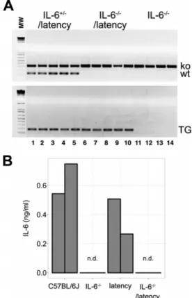
tm1Kopf/J). Genotyping was performed
ac-cording to the supplier’s protocol and as published elsewhere (
Fig.
1A
) (
20
). Splenocytes from 7- to 8-week-old mice were cultured
with 100 ng/ml lipopolysaccharides (LPS) for 48 h, and IL-6 levels
were measured by enzyme-linked immunosorbent assay (ELISA)
(eBioscience). In response, cells from C57BL/6J and the latency
mice secreted IL-6, while cells from IL-6
⫺/⫺and IL-6
⫺/⫺latency
mice did not (
Fig. 1B
).
IL-6 plays important roles in immunoglobulin (Ig) secretion
by sustaining long-lived PCs (reviewed in reference
22
). IgG
pro-duction is impaired in IL-6
⫺/⫺mice (
23–25
), whereas IgG
hyper-globulinemia is a consistent phenotype of the latency mice (
Fig.
2A
). To examine the genetic interaction between KSHV latency
genes and lack of IL-6, we examined serum Ig levels by ELISA as
Received23 November 2015Accepted1 December 2015
Accepted manuscript posted online9 December 2015
CitationSin S-H, Kang SA, Kim Y, Eason A, Tan K, An H, Dittmer DP. 2016. Kaposi’s sarcoma-associated herpesvirus latency locus compensates for interleukin-6 in initial B cell activation. J Virol 90:2150 –2154.doi:10.1128/JVI.02456-15.
Editor:R. M. Sandri-Goldin
Address correspondence to Sang-Hoon Sin, [email protected].
Copyright © 2016, American Society for Microbiology. All Rights Reserved.
FIG 1IL-6⫺/⫺ latency transgenic mice. (A) Agarose gel of PCR products
obtained with primers specific for IL-6⫺/⫺and a latency gene. (B) Level of IL-6
FIG 2Phenotypes of IL-6⫺/⫺latency mice. Box plots show the 1st and 3rd
quartiles, with the median indicated by the band. Whiskers extend to 1.5⫻the interquartile range. (A) Peripheral Ig levels were plotted from 9 C57BL/6J and 6 latency mice as determined by ELISA. This represents a meta-analysis. Some of the data points were previously reported (21). (B) Peripheral Ig levels were plotted from IL-6⫺/⫺and IL-6⫺/⫺latency mice as determined by ELISA (n⫽
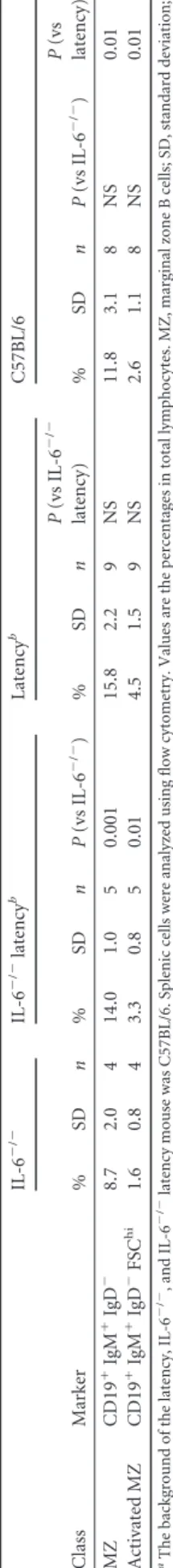
5). (C) Splenic marginal zone B cells (CD19⫹IgM⫹IgD⫺) and activated mar-ginal zone B cells (CD19⫹IgM⫹IgD⫺FSChi) (n⫽5). (D) Immature B cells (CD19⫹IgM⫹IgDlo) and plasma cells (CD19⫺B220⫺CD138⫹) in BM were
described previously (
21
). Total IgG1, IgG2a, IgG2b, and IgG3
levels were higher in IL-6
⫺/⫺latency than IL-6
⫺/⫺mice (
Fig. 2B
).
This demonstrates that KSHV latent genes (and miRNAs) in B
cells can compensate for the absence of IL-6 in B cell maturation.
To assess the effect of IL-6 on B cell development, cells were
isolated from the spleen or bone marrow (BM) of 7- to
11-week-old IL-6
⫺/⫺and IL-6
⫺/⫺latency mice and analyzed by flow
cy-tometry. IL-6
⫺/⫺latency mice displayed the same phenotypes as
the latency mice, specifically, increased frequencies of MZ cells
(CD19
⫹IgM
⫹IgD
⫺) and activated MZ B cells (CD19
⫹IgM
⫹FIG 3The degree ofex vivoproliferation of B cells was plotted in response to LPS (A) or anti-IgM antibody (B). The box plots (n⫽5) show the 1st and 3rd quartiles, with the median indicated by the band. Whiskers extend to 1.5⫻the interquartile range. Significance levels were determined by ANOVA. Immunostaining of spleen sections (5 IL-6⫺/⫺or IL⫺/⫺latency mice; 3 C57BL/6 or latency mice) with Ki-67 (C) and PNA (D). Representative images are shown. Magnification,⫻400.
TABLE 2Ex vivoproliferation of splenic B cells from IL-6⫺/⫺, IL-6⫺/⫺latency, latency, and C57BL/6 micea
Treatment
IL-6⫺/⫺ IL-6⫺/⫺latency Latencyb C57BL/6b
Slope Slope P(vs IL-6⫺/⫺) Slope
P(vs IL-6⫺/⫺
latency) Slope P(vs IL-6⫺/⫺)
P(vs IL-6⫺/⫺
latency)
LPS 1,474.0 1,348.9 NS 1,629.5 NS 910.4 NS NS
Anti-IgM 53.8 29.4 NS 229.3 0.001 158.1 0.04 0.007
aThe background of the latency, IL-6⫺/⫺, and IL-6⫺/⫺latency mice was C57BL/6. The degree ofex vivoproliferation of splenic B cells was analyzed by ANOVA. Five mice per
IgD
⫺FSC
hi) in spleen (
Fig. 2C
;
Table 1
). Frequencies of mature B
cells and plasma cells (PC) were not significantly different. This
held true for spleen (data not shown) and BM (
Fig. 2D
). This
suggests that the KSHV latency-associated hyperplasia of naive,
pre-GC B cells was not dependent on IL-6.
Ex vivo
hyperresponsiveness to B cell stimuli is a distinct
phe-notype of KSHV latency mice (
21
). To test the hypothesis that this
phenotype was dependent on IL-6, splenic B cells from 5- to
6-week-old IL-6
⫺/⫺and IL-6
⫺/⫺latency mice were purified by
negative selection and cultured with LPS or anti-IgM antibody.
Proliferation was measured using a Click-iT EdU assay
(Invitro-gen). The Toll-like receptor (TLR)-driven responsiveness to LPS
persisted in the absence of IL-6 (
Fig. 3A
;
Table 2
); however, the B
cell receptor (BCR)-driven responsiveness was damped in the
ab-sence of IL-6 (
Fig. 3B
;
Table 2
). This is consistent with a
mecha-nism whereby BCR-induced B cell proliferation was aided by an
IL-6 feedback loop but TLR-induced proliferation was not.
To document the
in vivo
phenotype of IL-6
⫺/⫺latency mice,
formalin-fixed, paraffin-embedded spleen sections were prepared
and evaluated by immunohistochemistry for two established
pro-liferation markers, Ki-67 and peanut agglutinin (PNA). We could
not find a difference in staining degree or intensity between
IL-6
⫺/⫺and IL-6
⫺/⫺latency mice; however, the staining was
stronger in tissues from latency mice than in either C57BL/6 or
IL-6
⫺/⫺latency mice (
Fig. 3C
and
D
).
The rates of lymphoma and splenic lymphoid hyperplasia as
ascertained by hematoxylin and eosin (H&E) stain showed no
difference (
Table 3
). Mesenteric lymph nodes (MLNs) are
chron-ically stimulated by gut microbiota. MiR-155 knockout mice
ex-hibit a lower frequency of germinal center (GC) B cells in MLNs
(
26
). KSHV encodes K12-11, which is an ortholog of
miR-155 and rescued the miR-miR-155 deficiency-associated phenotype in
MLNs (
21
,
27
). IL-6
⫺/⫺latency mice, which express miR-K12-11,
had the same rate of lymphoid hyperplasia as the latency mice
(
Table 3
). Another phenotype of KSHV latency mice is severe
extramedullary hematopoiesis (EMH). The rates of EMH in
spleen and liver were not dependent on the presence of IL-6 (
Ta-ble 3
).
B cell hyperplasia in spleen and proliferation in lymph nodes,
as scored here, are complex and progressive phenotypes that
de-velop over months and are subject to a multitude of compensatory
and counterbalancing mechanisms in the animal. Clearly, IL-6 is
needed for maximal B cell function and sustained proliferation
during normal development and in preneoplastic scenarios, such
as MCD. However, many mechanisms are known to relieve the
dependence on IL-6 in disease. Augmented NF-
B signaling was
found in an IL-6-independent variant of multiple myeloma (
28
).
Activation of STAT3 (signal transducers and activators of
tran-scription 3), an important intermediate of IL-6 signaling, was
ob-served in IL-6-independent plasmacytomas (
29
). The genetic
ex-periment presented here suggests that KSHV latent genes, too, can
compensate for IL-6 in the early stages of B cell activation and
development. The miRNAs are known for their profound effects
on cell lineage development and differentiation. The KSHV
miRNAs, most likely, evolved to foster initial infection and latent
persistence in naive B cells and eventual, preferential expansion of
infected cells. One mechanism to facilitate this “goal” would be to
compensate for limiting host activators, such as IL-6.
ACKNOWLEDGMENTS
We thank Blossom Damania for critical reading and helpful discussion. This study was supported by a faculty development award to S.-H.S. under UNC CFAR P30 AI50410, and public health service grants CA109232 and CA019014 (D.P.D). The UNC LCCC Animal Histopathol-ogy Core is supported in part by an NCI Center Core Support Grant (CA16086) to the UNC LCCC. The UNC Flow Cytometry Core Facility is supported in part by an NCI Center Core Support Grant (P30CA016086) to the UNC LCCC.
FUNDING INFORMATION
UNC CFAR provided funding to Sang-Hoon Sin under grant number P30 AI50410. HHS | U.S. Public Health Service (USPHS) provided funding to Dirk P. Dittmer under grant number CA109232 CA019014.
REFERENCES
1.Kovalchuk AL, Kim JS, Park SS, Coleman AE, Ward JM, Morse HC, Kishimoto T, Potter M, Janz S.2002. IL-6 transgenic mouse model for extraosseous plasmacytoma. Proc Natl Acad Sci U S A99:1509 –1514.
http://dx.doi.org/10.1073/pnas.022643999.
2.Suematsu S, Matsuda T, Aozasa K, Akira S, Nakano N, Ohno S, Miyazaki J, Yamamura K, Hirano T, Kishimoto T.1989. IgG1
plasma-TABLE 3In vivophenotypesa
Tissue Phenotype
IL-6⫺/⫺ IL-6⫺/⫺latency Latencyb
No. of
mice Rate (%) No. of
mice Rate (%) P(vs IL-6⫺/⫺)
No. of
mice Rate (%) P(vs IL-6⫺/⫺)
P(vs IL-6⫺/⫺
latency)
Spleen Lymphoma 0 0.0 1 3.0 1 8 16.0 0.093 0.076
Lymphoid hyperplasia 6 31.6 9 27.3 1 13 26.0 1 1
Normal 13 68.4 23 69.7 29 58.0
Severe EMH 2 10.5 3 9.1 1 11 22.0 0.491 0.147
Liver Severe EMH 5 26.3 2 6.7 0.085 NA
Total no. of mice 19 33 50
MLN Lymphoma 2 14.3 0 0.0 0.333 NA
Lymphoid hyperplasia 12 85.7 20 95.2 1 NA
Normal 0 0.0 1 4.8 NA
Total no. of mice 14 21 NA
aData were analyzed using ANOVA. APvalue ofⱕ0.05 was regarded as significant. NA, data not available.
b
cytosis in interleukin 6 transgenic mice. Proc Natl Acad Sci U S A86:7547– 7551.http://dx.doi.org/10.1073/pnas.86.19.7547.
3.Naugler WE, Sakurai T, Kim S, Maeda S, Kim K, Elsharkawy AM, Karin M.2007. Gender disparity in liver cancer due to sex differences in MyD88-dependent IL-6 production. Science317:121–124.http://dx.doi.org/10
.1126/science.1140485.
4.Lattanzio G, Libert C, Aquilina M, Cappelletti M, Ciliberto G, Musiani P, Poli V. 1997. Defective development of pristane-oil-induced plasmacytomas in interleukin-6-deficient BALB/c mice. Am J Pathol151:689 – 696.
5.Beck J, Hsu S, Wijdenes J, Bataille R, Klein B, Vesole D, Hayden K, Jagannath S, Barlogie B. 1994. Brief report: alleviation of systemic manifestations of Castleman’s disease by monoclonal anti-interleukin-6 antibody. N Engl J Med330:602– 605.http://dx.doi.org
/10.1056/NEJM199403033300904.
6.Brandt SJ, Bodine DM, Dunbar CE, Nienhuis AW.1990. Dysregulated interleukin 6 expression produces a syndrome resembling Castleman’s disease in mice. J Clin Invest 86:592–599. http://dx.doi.org/10.1172
/JCI114749.
7.Yoshizaki K, Matsuda T, Nishimoto N, Kuritani T, Taeho L, Aozasa K, Nakahata T, Kawai H, Tagoh H, Komori T.1989. Pathogenic signifi-cance of interleukin-6 (IL-6/BSF-2) in Castleman’s disease. Blood74:
1360 –1367.
8.Burger R, Wendler J, Antoni K, Helm G, Kalden JR, Gramatzki M.
1994. Interleukin-6 production in B-cell neoplasias and Castleman’s dis-ease: evidence for an additional paracrine loop. Ann Hematol69:25–31.
http://dx.doi.org/10.1007/BF01757344.
9.Carbone A, Vaccher E, Gloghini A, Pantanowitz L, Abayomi A, de Paoli P, Franceschi S.2014. Diagnosis and management of lymphomas and other cancers in HIV-infected patients. Nat Rev Clin Oncol11:223–238.
http://dx.doi.org/10.1038/nrclinonc.2014.31.
10. Tamburro KM, Yang D, Poisson J, Fedoriw Y, Roy D, Lucas A, Sin S-H, Malouf N, Moylan V, Damania B, Moll S, van der Horst C, Dittmer DP.
2012. Vironome of Kaposi sarcoma associated herpesvirus-inflammatory cytokine syndrome in an AIDS patient reveals co-infection of human her-pesvirus 8 and human herher-pesvirus 6A. Virology433:220 –225.http://dx
.doi.org/10.1016/j.virol.2012.08.014.
11. Uldrick TS, Wang V, O’Mahony D, Aleman K, Wyvill KM, Marshall V, Steinberg SM, Pittaluga S, Maric I, Whitby D, Tosato G, Little RF, Yarchoan R.2010. An interleukin-6-related systemic inflammatory syn-drome in patients co-infected with Kaposi sarcoma-associated herpesvi-rus and HIV but without multicentric Castleman disease. Clin Infect Dis
51:350 –358.http://dx.doi.org/10.1086/654798.
12. Asou H, Said JW, Yang R, Munker R, Park DJ, Kamada N, Koeffler HP.
1998. Mechanisms of growth control of Kaposi’s sarcoma-associated her-pes virus-associated primary effusion lymphoma cells. Blood91:2475– 2481.
13. Miles SA, Rezai AR, Salazar-González JF, Vander Meyden M, Stevens RH, Logan DM, Mitsuyasu RT, Taga T, Hirano T, Kishimoto T.1990. AIDS Kaposi sarcoma-derived cells produce and respond to interleukin 6. Proc Natl Acad Sci U S A87:4068 – 4072.http://dx.doi.org/10.1073/pnas
.87.11.4068.
14. Foussat A, Wijdenes J, Bouchet L, Gaidano G, Neipel F, Balabanian K, Galanaud P, Couderc J, Emilie D.1999. Human interleukin-6 isin vivo an autocrine growth factor for human herpesvirus-8-infected malignant B lymphocytes. Eur Cytokine Netw10:501–508.
15. Jones KD, Aoki Y, Chang Y, Moore PS, Yarchoan R, Tosato G.1999. Involvement of interleukin-10 (IL-10) and viral IL-6 in the spontaneous
growth of Kaposi’s sarcoma herpesvirus-associated infected primary effu-sion lymphoma cells. Blood94:2871–2879.
16. Sin S-H, Roy D, Wang L, Staudt MR, Fakhari FD, Patel DD, Henry D, Harrington WJ, Damania BA, Dittmer DP.2007. Rapamycin is effica-cious against primary effusion lymphoma (PEL) cell linesin vivoby inhib-iting autocrine signaling. Blood109:2165–2173.http://dx.doi.org/10.1182
/blood-2006-06-028092.
17. Neipel F, Albrecht J, Ensser A, Huang Y, Li J, Friedman-Kien A, Fleckenstein B.1997. Human herpesvirus 8 encodes a homolog of inter-leukin-6. J Virol71:839 – 842.
18. Moore PS, Boshoff C, Weiss RA, Chang Y.1996. Molecular mimicry of human cytokine and cytokine response pathway genes by KSHV. Science
274:1739 –1744.http://dx.doi.org/10.1126/science.274.5293.1739. 19. Fakhari FD, Jeong JH, Kanan Y, Dittmer DP. 2006. The
latency-associated nuclear antigen of Kaposi sarcoma-latency-associated herpesvirus in-duces B cell hyperplasia and lymphoma. J Clin Invest116:735–742.http:
//dx.doi.org/10.1172/JCI26190.
20. Sin S-H, Fakhari FD, Dittmer DP.2010. The viral latency-associated nuclear antigen augments the B-cell response to antigenin vivo. J Virol
84:10653–10660.http://dx.doi.org/10.1128/JVI.00848-10.
21. Sin S-H, Dittmer DP.2013. Viral latency locus augments B-cell response
in vivoto induce chronic marginal zone enlargement, plasma cell
hyper-plasia, and lymphoma. Blood121:2952–2963.http://dx.doi.org/10.1182
/blood-2012-03-415620.
22. Tangye SG.2011. Staying alive: regulation of plasma cell survival. Trends Immunol32:595– 602.http://dx.doi.org/10.1016/j.it.2011.09.001. 23. Jourdan M, Cren M, Robert N, Bollore K, Fest T, Duperray C,
Guil-loton F, Hose D, Tarte K, Klein B.2014. IL-6 supports the generation of human long-lived plasma cells in combination with either APRIL or stro-mal cell-soluble factors. Leukemia 28:1647–1656.http://dx.doi.org/10
.1038/leu.2014.61.
24. Kopf M, Baumann H, Freer G, Freudenberg M, Lamers M, Kishimoto T, Zinkernagel R, Bluethmann H, Kohler G.1994. Impaired immune and acute-phase responses in interleukin-6-deficient mice. Nature368:
339 –342.http://dx.doi.org/10.1038/368339a0.
25. Yoshizaki K, Nakagawa T, Fukunaga K, Tseng LT, Yamamura Y, Kishimoto T.1984. Isolation and characterization of B cell differentiation factor (BCDF) secreted from a human B lymphoblastoid cell line. J Im-munol132:2948 –2954.
26. Thai T-H, Calado DP, Casola S, Ansel KM, Xiao C, Xue Y, Murphy A, Frendewey D, Valenzuela D, Kutok JL, Schmidt-Supprian M, Rajewsky N, Yancopoulos G, Rao A, Rajewsky K.2007. Regulation of the germinal center response by microRNA-155. Science316:604 – 608.http://dx.doi
.org/10.1126/science.1141229.
27. Sin S-H, Kim YB, Dittmer DP.2013. Latency locus complements mi-croRNA 155 deficiencyin vivo. J Virol87:11908 –11911.http://dx.doi.org
/10.1128/JVI.01620-13.
28. Verdelli D, Nobili L, Todoerti K, Mosca L, Fabris S, D’Anca M, Pellegrino E, Piva R, Inghirami G, Capelli C, Introna M, Baldini L, Chiaramonte R, Lombardi L, Neri A.2014. Molecular events underlying interleukin-6 independence in a subclone of the CMA-03 multiple my-eloma cell line. Genes Chromosomes Cancer53:154 –167.http://dx.doi
.org/10.1002/gcc.22127.