Identification and characterization of Drosophila Snurportin reveals a
role for the import receptor Moleskin/Importin-7 in snRNP biogenesis
Amanda Hicks Natalizio
A dissertation submitted to the faculty of the University of North Carolina at Chapel Hill in partial fulfillment of the requirements for the degree of Doctor of
Philosophy in the Curriculum in Genetics and Molecular Biology.
Chapel Hill 2013
Approved By:
ii ABSTRACT
AMANDA HICKS NATALIZIO: Identification and characterization of Drosophila Snurportin reveals a role for the import receptor Moleskin/Importin-7 in snRNP
biogenesis
(Under the direction of A. Gregory Matera)
Biogenesis of small nuclear ribonucloceoproteins (snRNPs) is biphasic. Small nuclear RNAs (snRNAs) are exported to the cytoplasm for assembly into pre-snRNPs where they are hypermethylated, forming a trimethylguanosine (TMG) cap, and then transported back into the nucleus via the import adaptor, snurportin1 (SPN) and the import receptor importin-β. I have identified CG42303
as dSNUP, the Drosophila orthologue of human SPN (hSPN). Strikingly, the importin-β binding (IBB) domain, which is essential for SPN-mediated snRNP
import in humans, is not conserved in dSNUP.
Consistent with the lack of an IBB, dSNUP did not interact with the
Drosophila importin-β orthologue, Ketel. Despite this fact, dSNUP localized to the
nucleus, indicating that there is an alternative dSNUP import pathway or that dSNUP is imported indirectly through importin-β bound snRNPs. I excluded the
latter possibility since, in contrast to human cells, snRNPs did not associate with importin-β in Drosophila cells. Previous results suggested that hSPN interacts
Drosophila orthologue of importin-7, known as Moleskin (Msk), interacts with dSNUP and snRNPs.
I discovered that Msk physically associates with both dSNUP and U snRNPs, while snRNP components failed to bind importin-β. Furthermore, Msk
null mutant larvae had a significant in vivo reduction of the snRNP component survival motor neuron (SMN), and the snRNP specific nuclear Cajal body marker coilin. Additionally, Msk null mutants exhibited cytoplasmic accumulation of TMG cap signal in the Malpighian tubules, indicating that the import of TMG capped snRNAs is inhibited in the absence of Msk. The reduction of SMN protein was dramatic enough to be detected by western blotting, suggesting a vital role for Msk in the stability of SMN. Interestingly, Msk also localized to snRNP specific nuclear Cajal bodies. In sum, these data indicate that importin-β does not play a
role in snRNP import in Drosophila and implicate a crucial function for Msk in fruit fly snRNP biogenesis. Future experiments will be needed to determine the
iv
To my parents, my sister, Cassandra, my brother, Randy, my uncle Dwight, my aunt Tomma, my husband, Tony,
and the light of my life and son, Alaric.
ACKNOWLEDGEMENTS
First and foremost, I must thank my mother. For my entire life, she has always been there to motivate me, push me to do better, and she has always been supportive of my love for science. I wouldn’t be the assertive, independent, and determined person I am today without her. Those very traits that have served me well through my turbulent graduate student years and I’m sure they will for the rest of my life.
I want to thank my mentor, Greg Matera, for his optimistic encouragement when all the experimental approaches I was trying seemed to be failing. Without failure, you do not learn, and I am indebted to Greg for his patience and giving me control of my project, and allowing me to fail. Greg’s mentorship style has enabled me to grow as an independent scientist and thinker, which is essential to my career aspirations.
vi
A special thanks to my husband, Tony. Having a supporting husband that believes in my abilities, even when I’m feeling lost, is something that carried me through to the end. Last, but certainly not least, I want to thank my son, Alaric. Although he may have slowed down the pace of my Ph.D., my son allowed me to finish it with my sanity still intact. Coming home to Alaric’s smiling face was always able to wash away the day’s stresses and disappointments from lab.
Table of Contents
Chapter
I. INTRODUCTION ……….1
Cellular Compartmentalization………1
Cellular Transport …...……….2
RanGTP Gradient ………7
snRNP Biogenesis ………..8
Identification of Snurportin ………...14
SPN-mediated snRNP Import ……….15
snRNP Import Pathways ………..18
snRNP translocation through the NPC ………..22
Import Complex Disassembly ………..24
SPN Recycling ………...24
Nuclear snRNP Modifications and Cajal Bodies………28
Research Objectives ……….31
viii
INTRODUCTION ………...34
RESULTS ………...36
Identification and Characterization of Drosophila Snurportin ………...36
Drosophila snRNP import is importin- independent………..43
Moleskin/Importin-7 interacts with snRNPs and Snurportin ………...45
Moleskin/Importin-7 localizes to snRNP-rich structures in the nucleus ……….48
Moleskin depletion disrupts snRNP biogenesis and import ………...50
DISCUSSION ………..56
MATERIALS AND METHODS ………..60
DNA Constructs ……….60
Recombinant Protein Expression and S2 Cell Transfections ………..60
Antibodies ………...60
Immunoprecipitation ………..61
RNAi ………62
Fly Stocks ………...63
Immunofluorescence ……….63
Fluorescence Microscop...……….64
III. Characterization of the physical and functional interactions of Moleskin/Importin- with snRNP………65
INTRODUCTION ……….66
RESULTS ……….69
Interaction of Msk with snRNP proteins………..69
Interaction of Sm proteins with Msk is RNA independent ………70
Snurportin and/or Moleskin knock down affects GFP-dSmB import ……….71
DISCUSSION ………..76
MATERIALS AND METHODS ………..79
DNA Constructs ……….79
Antibodies ………...79
Fractionation and Immunoprecipitation………...80
RNAi ………81
Immunofluorescence ……….81
IV. Discussion and Future Directions ……….82
Addressing the complicated nature of snRNP import pathways ………..82
Msk/Imp7 import adaptor versus import receptor………...83
RanGTP and snRNP import ………..86
Implications of unintended co-depletions ………89
Tissue specific snRNP import requirements………91
Redundancies in snRNP import ………94
Imp7/Msk and Cajal bodies ………...96
Determining Msk’s direct snRNP binding partners………...100
x
snRNP import and disease ……….103
snRNP import regulation ……….104
Evolution of snRNP import mechanisms ………..105
Summary and concluding remarks ………107
APPENDICES ………..109
Appendix I: Investigation of Snurportin gene evolution…………..110
Appendix II: Characterization of dSNUP antibodies………113
Appendix III: Anti-dSmB co-immunoprecipitates dSNUP………...118
Appendix IV: Examination of Imp/Ketel interacting partners ………..119
Appendix VA: dSNUP in vivo RNAi viability assay………..122
Appendix VB: dSNUP in vivo RNAi rescue………..126
Appendix VI: RNAi of dSNUP or Msk in egg chambers………….130
Appendix VIIA: Analysis of Msk null mutant viability………...133
Appendix VIIB: Coilin and dSMN reduction in Msk null mutants ………135
Appendix VIIC: Investigation of U snRNP levels in Msk mutants ………..137
Appendix VIIIA: Investigation of dSNUP localization………..140
Appendix VIIIB: Investigation of GFP-dSNUP localization after Msk RNAi ………..144
Appendix IX: A closer look at Msk and coilin localization………..147
Appendix X: pBI-UASC-mVenus-(hIBB)-dSNUP characterization and Msk mutant rescue………...150
Appendix XIA: Drosophila transgenic vector map………...153
Appendix XIC: dSNUP, Ketel, and Msk dsRNA target
sequences ………..156
xii
Table of Figures
Figure 1.1. Structure of the nuclear pore complex. ... 3
Figure 1.2. Classical NLS-dependent import. ... 6
Figure 1.3. Vertebrate snRNA export pathway. ... 9
Figure 1.4. Vertebrate SMN complex. ... 10
Figure 1.5. Vertebrate snRNP biogenesis overview. ... 13
Figure 1.6. Schematic of SPN. ... 15
Figure 1.7. Vertebrate SPN-mediated snRNP Import Pathway. ... 17
Figure 1.8. Vertebrate snRNP Import Pathways. ... 21
Figure 1.9. Model of Snurportin auto-regulatory snRNP import function. ... 26
Figure 2.1. CG42303 is the Drosophila Snurportin orthologue ... 37
Figure 2.2. Drosophila Snurportin interacts with snRNPs. ... 40
Figure 2.3. Localization of dSNUP. ... 42
Figure 2.4. Ketel/Impβ does not interact with Drosophila snRNPs. ... 44
Figure 2.5. Msk interacts with Drosophila snRNPs ... 47
Figure 2.6. Msk is enriched in Cajal bodies. ... 49
Figure 2.7. Msk mutant characterization. ... 51
Figure 2.8. Msk mutant displays TMG cap cytoplasmic accumulation. ... 52
Figure 2.9. Coilin and dSMN are reduced in Msk mutant. ... 54
Figure 2.10. Flag-dSMN does not rescue Coilin and dSMN in Msk mutant. ... 56
Figure 2.11. Models of Imp7’s role in snRNP import. ... 57
Supplemental Figure 2.S1. Imp7 enriched in CBs of Mammalian Cells. ... 59
Figure 3.2. RNA dependence of Msk snRNP interactions ... 71
Figure 3.3. Knock down of Msk or dSNUP with dsRNAs in S2 cells. ... 72
Figure 3.4. dSmB cytoplasmic accumulation after dSNUP or Msk RNAi ... 75
Figure 3.5. S2 cell fractionation following Msk RNAi ... 76
Figure 4.1. Model of U snRNP import complex in Xenopus oocytes. ... 91
Figure A.1. dSNUP guinea pig antibody recognizes GFP-dSNUP ... 116
Figure A.2. Immunofluorescence with dSNUP guinea pig antibody. ... 116
Figure A.3. S2 cell immunofluorescence with dSNUP rabbit antibody. ... 117
Figure A.4. dSmB antibody co-IPs dSNUP from S2 cytoplasmic lysate ... 118
Figure A.5. Ketel//Importin-B does not interact with Drosophila snRNPs. ... 121
Figure A.6. In vivo RNAi of dSNUP. ... 123
Figure A.7. Viability assay of dSNUP RNAi Ex 3 Target at 22°C. ... 124
Figure A.8. Viability assay of dSNUP RNAi Ex 1 Target at 22°C. ... 125
Figure A.9. Viability assay of dSNUP RNAi Ex 1 Target at 29°C. ... 125
Figure A.10. dSNUP RNAi and rescue with VFP-dSNUP ... 128
Figure A.11. VFP-dSNUP swamps out dsRNA targeting Ex1 of dSNUP. ... 129
Figure A.12. U snRNA levels in dSNUP and Msk RNAi egg chambers ... 132
Figure A.13. Msk185 mutant long-lived larvae ... 134
Figure A.14. Msk mutant reduction in dSMN and coilin by day 1 DPE ... 136
Figure A.15. U1 snRNA is reduced in Msk mutant larvae ... 139
Figure A.16. dSNUP localization in S2 cells ... 143
Figure A.17. GFP-dSNUP nucleoplasmic retention in Msk RNAi S2 cells. ... 146
xiv
Figure A.19. Expression of VFP-dSNUP and VFP-hIBB-dSNUP in vivo ... 152
Table of Tables
xvi
LIST OF ABBREVIATIONS
Å Angstom
aa Amino acids
ATP Adenosine triphosphate BSA Bovine serum albumin
CAS Cellular apoptosis susceptibility protein CB Cajal body
CBC Cap binding complex CBP Cap binding proteins
DAPI 4',6-diamidino-2-phenylindole dSmB Drosophila melanogaster Smith B
dSMN Drosophila melanogaster survival of motor neurons DNA Deoxyribonucleic acid
dSNUP Drosophila melanogaster Snurportin dsRNA Double stranded RNA
DTT Dithiothreitol
EDTA Ethylenediaminetetraacetic acid EST Expressed sequence tag
Ex Exon
EXO Exonulcease
GAP GTPase activating protein
GST Glutathione S-transferase GTP Guanosine triphosphate
h Hours
HEAT α-helical repeats
hIBB Homo sapiens importin-β binding domain
hSPN Homo sapiens Snurportin IF Immunofluorescence IBB Importin-β binding domain
Imp7 Importin-7 Impα Importin-α Impβ Importin-β
IP Immunoprecipitation
IPTG Isopropyl-beta-D-Thiogalactopyranoside kDa Kilodalton
LMB Leptomycin B LR Leucine-rich
m7G 7-methylguanosine MDa Megadalton
min Minutes
mRNA Messenger RNA Msk Moleskin
xviii NLS Nuclear localization signal NP40 Nonidet-40
NPC Nuclear pore complex Nup153 Nucleoporin 153 ORF Open reading frame PBS Phosphate buffered saline
PBST Phosphate buffered saline with0.1% Triton 100x PCR Polymerase chain reaction
PRMT5 Protein arginine methyltransferase 5 PHAX Phosphorylated adaptor for snRNA export RIPA Modified radioimmunoprecipitation assay RNA Ribonucleic acid
RNAi RNA interference RNP Ribonucleoprotein rpm Rotations per minute RT Room temperature
RT-PCR Reverse transcriptase-PCR SDS Sodium dodecyl sulfate
PAGE Polyacrylamide gel electrophoresis SMN Survival of motor neurons
TMG 2,2,7-trimethylguanosine tRNA Transfer RNA
U snRNA Uridine-rich small nuclear RNAs VFP Venus fluorescent protein
WT Wild type Xpo1 Exportin1/C
CHAPTER I
INTRODUCTION
Cellular Compartmentalization
The evolutionary advantage to compartmentalization of eukaryotic cells cannot be overstated. Division of the cell into organelles and sub-domains offers greater regulatory control of cellular processes than their prokaryotic counterparts. Most cellular compartments are surrounded by a lipid membrane and are organelles, such as mitochondria, lysosomes, the Golgi apparatus, or the cell nucleus. Compartmentalization provides several functions. It increases the membrane area without increasing the size of the cell, provides local environments that facilitate metabolic functions that may otherwise be inhibited by other processes within the cell, and enables the regulation of many key processes between various cellular regions.
genomic material is unregulated, and DNA, RNA, and protein synthesis all occur in the cytosol. Unlike, prokaryotic cells, eukaryotic cells must have a means to transport molecules across the nuclear envelope for intercompartmental communication and regulation of their genomic material. Nuclear and cytoplasmic compartmentalization allows for the uncoupling of nuclear DNA/RNA synthesis from cytosolic protein synthesis, which enhances the cell’s regulatory capabilities at the transcriptional and translational levels. Examples of this are numerous, from transcriptional repressors being sequestered to the cytoplasm to enhance a particular gene’s expression to mRNA degradation within the nucleus to prevent export and translation in the cytoplasm.
The high levels of regulation seen in eukaryotic cells are not without expense. Eukaryotic cells must invest a substantial amount of energy to properly maintain such a complex regulatory and structural system. Often, energy is required for the various compartments to communicate. Molecules and macromolecules must be transported between compartments to facilitate this communication. Importantly, this intercompartmental communication must be appropriately regulated to avoid negative consequences for the cell.
Cellular Transport
3
importantly, an environment conducive to transport is also needed and is provided by nuclear pore complexes (NPCs).
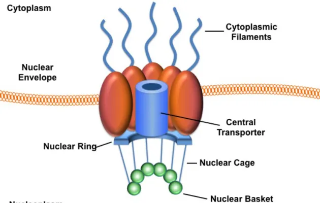
Macromolecular transport between the nucleus and the cytoplasm is routed through NPCs (Figure 1.1). NPCs transport a vast array of molecules across the nuclear envelope, including, proteins, mRNAs, tRNAs, ribosomal subunits, small nuclear ribonucleoprotein (snRNP) complexes, and even DNA in particular instances. The mechanistic complexity of nuclear transport is evidenced by the diverse array of molecules transported by NPCs.
NPCs are large supramolecular structures (~125 MDa in vertebrates) that span the nuclear envelope and thus, connect the cytoplasm to the nucleoplasm (Figure 1.1); (Stoffler et al, 1999). Vertebrate cells have, on average, 2000 NPCs per cell that can individually conduct 1000 translocations per second. The NPC is a cylindrical structure embedded in the lipid bilayer and consists of >30 distinct protein components called nucleoporins (nups). Many of these nups contain FG repeats (phenylalanine-glycine repeats) that are needed to interact with the dual α-helical repeats (HEAT repeats) of transport receptor proteins such as
importin-β (Impβ). The NPC also contains several peripheral structures including, the
nuclear basket and cage, and cytoplasmic filaments. The cytoplasmic ring moiety of the NPC has eight cytoplasmic filaments, while the nuclear ring moiety has eight tenuous filaments that form a distinct nuclear basket. (Figure 1.1).
The central pore of the NPC provides an aqueous channel that smaller molecules (<40 kDa) can passively diffuse through in an energy independent manner. However, the movement of macromolecules (proteins and/or RNAs) is typically energy and signal dependent, and mediated by transport receptors (Mattaj & Englmeier, 1998); (Gorlich & Kutay, 1999).
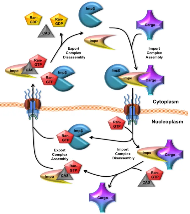
Transport receptors, which are also called karyopherins, recognize specific nuclear localization signals (NLSs) and nuclear export signals (NESs). NLS-containing proteins bind to either the import receptor, Impβ, directly (e.g.
5
NPC, and once in the nucleus, it binds RanGTP, facilitating cargo and/or Impα release. When not bound to Impβ, Impα is thought to fold in on itself through
interactions between its N-terminus and C-terminus, which facilitates cargo release into the nucleus (Fried & Kutay, 2003). Impα is then free to form an export complex with the cellular apoptosis susceptibility protein (CAS) and RanGTP. The Impα/CAS/RanGTP complex and Impβ bound RanGTP are export
competent, and are recycled back to the cytoplasm through the NPC. Once in the cytoplasm, Ran dissociates, allowing Impβ and Impα to participate in additional
Figure 1.2. Classical NLS-dependent import. The import adaptor, Impα, binds to the NLS of a cargo molecule. Subsequently, Impβ binds to Impα to mediated
cargo nuclear import through the NPC. Upon nuclear entry, the import complex is disassembled following RanGTP binding to Impβ, and CAS/RanGTP binding to
7 RanGTP Gradient
RanGTP has an asymmetric distribution across the nuclear envelope, which is essential for the regulation of nucleocytoplasmic transport. RanGTP is enriched in the nucleus, and Ran regulatory proteins maintain the RanGTP energy gradient. Ran’s guanine nucleotide exchange factor (RanGEF) is an exclusively nuclear, chromatin-associated protein that catalyzes the conversion of Ran from its GDP- to GTP-bound form. Ran GTPase activating protein (RanGAP) is exclusively cytoplasmic, and stimulates RanGTP hydrolysis. The strict compartmentalization of RanGEF and RanGAP are essential in maintaining the RanGTP gradient. The NPC itself provides no directional cues for transport. The RanGTP gradient is the sole determinant of the directionality of protein transport, proven by the fact that if you reverse the GTP gradient, protein flow is inverted (Fried & Kutay, 2003).
snRNP Biogenesis
Uridine-rich small nuclear RNAs (U snRNAs) are non-coding RNAs that play many roles in RNA metabolism in the nucleus, most notably in splicing (Mattaj et al, 1993); (Tarn & Steitz, 1997). The Sm-class U snRNAs form the core components of the spliceosome. Two distinct classes of spliceosomes exist: the ‘major’ spliceosome, responsible for >99% of intron splicing in the human genome, and the ‘minor’ spliceosome, which removes the remaining <1% of introns (Levine & Durbin, 2001). The major spliceosome is comprised of the major-class snRNAs U1, U2, U4 and U6, and the minor spliceosome is made up of the minor-class snRNAs U11, U12, U4atac and U6atac. The spliceosomal snRNA, U5, is a component of both spliceosomes (Patel & Steitz, 2003). The major-class snRNAs are ~100 fold more abundant than the minor-class snRNAs, consistent with a greater requirement for the major spliceosome (Zieve & Sauterer, 1990).
9
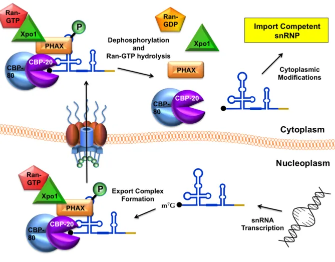
snRNA and Xpo1/RanGTP, and transits through the NPC where it is released into the cytoplasm upon phosphorylation (Figure 1.3); (Ohno et al, 2000).
Figure 1.3. Vertebrate snRNA export pathway. U snRNAs are transcribed and m7G capped in the nucleoplasm. The 5′ cap is bound by cap binding proteins
Once in the cytoplasm, the survival motor neuron (SMN) complex mediates the assembly of snRNPs by loading seven Sm proteins, SmB/B’, SmD1, SmD2, SmD3, SmE, SmF and SmG, onto a conserved motif of the pre-snRNA called the ‘Sm-site (Meister et al, 2002); (Pellizzoni et al, 2002b); (Yong et al, 2004); (Golembe et al, 2005); (Paushkin et al, 2002). This reaction requires energy in the form of adenosine triphosphate (ATP). Although the assembly of the Sm core onto the snRNA can occur spontaneously and non-specifically in vitro, the SMN complex provides specificity and improves the kinetics of this reaction (Pellizzoni et al, 2002b).
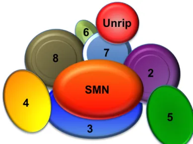
Figure 1.4. Vertebrate SMN complex. The numbered ovals in the complex represent Gemins2-8. Gemins2, 3, 7 and 8 make direct contacts with SMN. Unrip is only present in the cytoplasmic SMN complex. Based on Praveen, 2012.
11
Gemin8 and Unrip (Baccon et al, 2002); (Carissimi et al, 2006a); (Charroux et al, 1999); (Charroux et al, 2000); (Grimmler et al, 2005); (Gubitz et al, 2002); (Pellizzoni et al, 2002a). Unrip is a cytoplasmic specific member (Figure 1.4). The SMN complex serves as a scaffold upon which Sm proteins and snRNA are assembled, and this ensures that the Sm proteins only assemble specifically onto snRNAs. The role SMN plays in snRNP assembly is crucial, because without Sm core assembly, snRNPs are incapable of import, and thus cannot participate in active splicing within the nucleus. Importantly, all of the proteins of the SMN complex are required for snRNP assembly.
The Sm proteins, SmB, SmD1, and SmD3, contain RG rich C-terminal domains. These RG repeats are hypermethylated by the protein arginine methyltransferase 5 (PRMT5) complex, consisting of PRMT5, pICln and WD45 (Mep50); (Brahms et al, 2000); (Brahms et al, 2001); (Friesen et al, 2001); (Meister et al, 2001); (Friesen et al, 2002). These methylation marks enhance the binding of the Sm proteins to SMN, but are not necessary for the snRNP assembly process (Gonsalvez et al, 2008).
hypermethylation (Narayanan et al, 2002). The TMG cap is bound by the import receptor, Snurportin1 (SPN); (Palacios et al, 1997); (Mattaj & De Robertis, 1985); (Hamm et al, 1990); (Fischer et al, 1993). Subsequently, Impβ binds SPN and
imports the partially assembled pre-snRNP, along with the SMN complex, into the nucleus (Figure 1.5); (Palacios et al, 1997); (Huber et al, 1998).
13
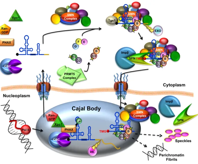
Figure 1.5. Vertebrate snRNP biogenesis overview. RNA polymerase II transcribes the snRNA gene which then undergoes 3’ end cleavage by the integrator complex (Int.) and is bound by the cap binding complex (CBC) and PHAX en route to the Cajal body (CB). Xpo1/RanGTP is recruited to export the pre-snRNA through the NPC. Once in the cytoplasm, the export complex is disassembled, and the SMN complex facilitates assembly of the seven membered ring of Sm proteins onto the pre-snRNA. A subset of Sm proteins are symmetrically dimethylated by the PRMT5 complex. The 3’ end of the snRNA is trimmed by an exonulcease (EXO), and the 7-methyl guanosine (m7G) cap is hyper methylated to a trimethylguanosine (m3G;TMG) cap by trimethylguanosine synthase (Tgs1). The TMG cap of the snRNA is bound by the import adaptor, Snurportin (SPN), and import receptor Importin-β (Impβ). Subsequently, the
Identification of Snurportin
NLS-dependent transport is the most well characterized nuclear import mechanism, but U snRNPs do not appear to have a classical NLS. In contrast to classical NLS-mediated import, U snRNP import does not require Impα. In vitro snRNP import assays led to the conclusion that Impβ was necessary for snRNP import (Palacios et al, 1997). These studies revealed that Impβ alone was unable to support U snRNP import, which suggested that Impβ does not directly recognize U snRNPs. This finding indicated that there was an unidentified import adaptor that played a vital role in cap-dependent snRNP import.
The identification of this cap-dependent snRNP import adaptor, Snurportin (SPN), was an essential element to our understanding of snRNP nuclear import. Prior to SPN identification, we knew that the 5’ TMG caps of U1 and U2 snRNAs were required for snRNP nuclear transport in Xenopus oocytes, suggesting that a cytoplasmic transport factor that bound to TMG caps was yet to be identified. Using this logic, Huber et al. (1998) incubated HeLa cell cytoplasmic lysate with a chemically synthesized, radiolabeled TMG cap oligo and then UV-crosslinked bound proteins. SDS-PAGE analysis revealed a 45 kDa protein band, which was later purified by size exclusion chromatography, followed by affinity chromatography using TMG cap oligo. This suspected snRNP import adaptor was then microsequenced. Several peptide sequences were identified which corresponded with expressed sequence tags (ESTs) of SPN (Huber et al, 1998).
15
This finding supported the conclusion that SPN was a snRNP specific import adaptor, as it specifically bound to the TMG cap. Furthermore, snRNPs lacking the 5’ TMG cap did not interfere with SPN binding to TMG cap oligo alone, and the addition of SPN significantly accelerated U1 import in both Xenopus laevis oocytes and permeabilized HeLa cells. Additionally, they showed that SPN contained an N-terminal Impβ binding domain (IBB), which was needed for SPN interaction with Impβ (Huber et al, 1998).
SPN-mediated snRNP Import
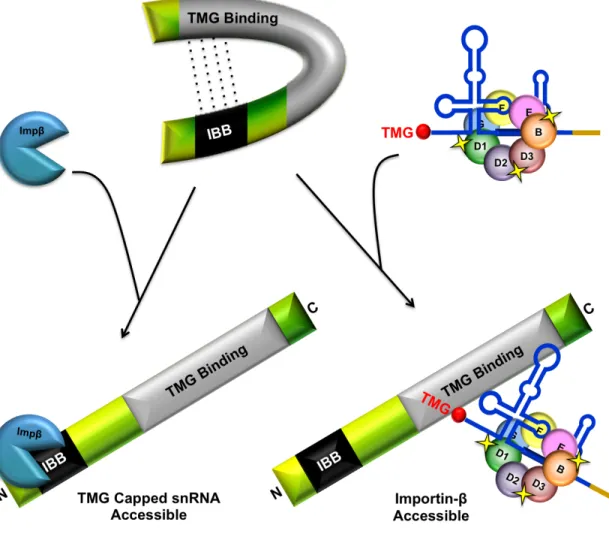
An import complex containing SPN, snRNP cargo, and Impβ facilitates snRNP import (Huber et al, 2002). To form this pre-import complex, SPN must not only bind to the TMG cap of the snRNP, but also simultaneously bind to Impβ. SPN has three functional domains, consisting of an ill-defined Xpo1 binding region, a centrally located TMG cap-binding domain, and an N-terminal IBB motif (Figure 1.6).
These functional domains were better defined by Ospina et al. (2005a). Mutational analysis of SPN revealed specific residues within both the IBB and TMG binding domains that are required for SPN function (Ospina et al, 2005a). Mutation of a single arginine residue within the IBB domain (R27) of SPN disrupted its interaction with Impβ, but preserved its ability to bind to Xpo1 or TMG caps. Interestingly, this Impβ binding point mutant is unable to support snRNP import, but is able to shuttle between the nucleus and the cytoplasm (the reasons for which are discussed under the “snRNP import pathways” section). This study also discovered conserved tryptophan residues outside of the IBB that are required for TMG binding, and that SPN was capable of nuclear import without being bound to snRNP cargo (Ospina et al, 2005a).
17
The precise order of the snRNP import complex formation has yet to be elucidated, but the Sm core is thought to be needed for proper TMG cap formation (Mattaj & Englmeier, 1998); (Luhrmann et al., 1990). We also know that TMG capping must precede SPN binding since SPN specifically recognizes the TMG cap (Figure 1.7). After SPN binding, it is unclear whether additional steps are needed before Impβ binding and cargo import. Furthermore, the SMN complex plays a role in import after Sm core assembly (discussed later), but its possible that it plays additional roles in the snRNP biogenesis pathway that are not fully understood.
snRNP Import Pathways
19
SMN or a component of the SMN complex is thought to be the import adaptor for the Sm core NLS (Narayanan et al, 2002). It has been shown in HeLa cells that SMN is present in an import-competent snRNP complex with Impβ in vivo. Additionally, GST-tagged SMN can interact directly with His-tagged Impβ (Narayanan et al, 2002). These data are consistent with the hypothesis that SMN is the Sm core NLS import adaptor. Moreover, in the absence of import competent SPN, the SMN complex and Impβ can rescue snRNP import in a nuclear transport assay. However, SMN alone with Impβ is not sufficient for TMG cap-independent snRNP import (Ospina et al, 2005a). This observation suggests that other members of the SMN complex are necessary for the nuclear transport of snRNPs using the Sm core NLS import pathway.
When Xenopus egg extract is supplied to somatic cells, U1 and U2 import becomes TMG cap dependent, suggesting that soluble cytosolic factors mediate the TMG cap dependence of U1 and U2 import (Marshallsay and Luhrmann, 1994). The interaction of the U2 snRNP with this cytosolic factor is saturable with TMG cap analogs in oocytes, but not HeLa cells (Fischer et al, 1994). Impβ
depletion from Xenopus egg extract can also significantly inhibit snRNP import, suggesting that either Impβ is required for snRNP import or that the unidentified cytosolic factor is co-depleted with Impβ. Additionally, we know that over expression of an Impβ binding deficient SPN (SPNΔIBB) in Xenopus oocytes
21
Figure 1.8. Vertebrate snRNP Import Pathways. Two independent snRNP import mechanisms exist in vertebrates, both of which require Impβ. The predominant import pathway is mediated by Snurportin (SPN) and requires the TMG cap of the snRNA. This is the only known snRNP import mechanism to exist in the germline, but an additional pathway has been shown to function in somatic cells. In somatic cells, Impβ can import uncapped snRNPs in the absence of SPN through its interaction with the SMN complex.
TMG cap independent import is thought to be due to the direct interaction of SMN with Impβ in the cytoplasm, thus serving as an Sm core NLS receptor,
required for SPN-independent import (Narayanan et al, 2002). Recombinant SPN and Impβ are necessary and sufficient for U1 snRNP import in permeabilized
HeLa cells, and this import is Ran independent for U1 and U5 (Huber et al, 2002). Irrespective of the TMG cap or SPN binding, nuclear import is mediated via Impβ in the vertebrate system, and this observation fails to explain why we
see cell specific differences in snRNP import requirements.
The difference in U1 and U2 versus U4 and U5 TMG cap dependence is not likely to be due to Sm core NLS differences because they compete for the same transport receptor, Impβ. It is possible that the structure and size of the
snRNA accounts for the differences in TMG cap dependence since U1 and U2 are longer than U4 or U5, although assembled U5 snRNP is much larger (Fischer et al, 1994). Understanding how these two pathways function in an in vivo model system will help elucidate the significance of the need for two independent snRNP import pathways.
snRNP translocation through the NPC
23
Neither the translocation of snRNP cargo through the NPC nor its release from the nuclear basket is dependent on Ran. The snRNP cargo must be released into the nucleoplasm where they can undergo further maturation. Disassembly of the snRNP import complex is not fully understood, and the factors involved in this disassembly have yet to be identified. It is not known whether Ran is a requirement; however, we do know that the affinity of Impβ for SPN is reduced upon binding of RanGTP (Paraskeva et al, 1999). RanGTP binding may promote complex disassembly directly or indirectly by destabilizing the import complex.
Import Complex Disassembly
Mitrousis et al. (2008) also found that the Nup153 homology region of SPN was needed for Xpo1 binding. Previous studies suggest that Xpo1 may play a role in complex disassembly and that the binding of SPN to Xpo1 and snRNP cargo may be mutually exclusive (Paraskeva et al, 1999); (Ospina et al, 2005a). The 2.9 Å crystal structure of SPN bound to Xpo1 revealed that SPN binds to Xpo1 in a bipartite manner through both an amino-terminal leucine-rich nuclear export signal (LR-NES) and a nucleotide-binding domain (Dong et al, 2009b). Like the bipartite IBB of SPN, the bipartite Xpo1 binding region increases the affinity of Xpo1 for SPN. This multipartite nature, combining energetically weak and strong epitopes, is also found in nuclear localizations signals. This principal is thought to broaden substrate specificity by amplifying signal diversity and allows for rapid evolution in nuclear trafficking in both directions (Dong et al, 2009a). It is likely that in addition to RanGTP hydrolysis, Xpo1 plays a role in complex disassembly, but additional unknown factors could initiate complex disassembly as well. Whether import complex disassembly is needed for further snRNP modifications is undetermined.
SPN Recycling
25
variety of proteins containing NESs (e.g. PHAX bound pre-snRNAs, Impβ, etc.). Xpo1 binds directly to the leucine-rich NES motif (not clearly defined in SPN), where it can then transport its cargo through the NPC into the cytoplasm. SPN does not contain a consensus NES, and the region mediating the interaction between SPN and Xpo1 has been difficult to determine, even with extensive SPN mutational analysis (Ospina et al, 2005a).
Mutational analysis by Ospina et al. (2005a) uncovered a potential auto-inhibitory interaction within SPN. The N- and C-terminal domains of SPN were found to interact, which suggests that SPN may have an auto-regulatory function similar to that of Impα. It is possible that the binding of Impβ or TMG capped
snRNAs would increase accessibility to snRNP cargo or Impβ, respectively. This
increased binding capacity would facilitate snRNP biogenesis under conditions
when snRNP demand is high (e.g. increased transcription). Alternatively, when
demand for snRNPs is low, this intramolecular interaction could sequester SPN
molecules via steric hindrance. Additionally, it would prevent SPN from aberrantly
binding newly imported snRNPs, and enable the regulation of snRNP biogenesis
through the modulation of import complex formation (Figure 1.9); (Ospina et al,
Figure 1.9. Model of Snurportin auto-regulatory snRNP import function. The N- and C-terminus of Snurportin has been shown to interact. This interaction could sequester SPN ability to bind to snRNPs or Impβ, effectively disrupting snRNP biogenesis. Upon increased demand, the binding of Impβ or snRNP cargo might increase access to snRNP cargo or Impβ, respectively. Adapted from Ospina et al., 2005a.
27
domain of SPN wraps around the nucleotide binding C-terminus (Dong et al, 2009a). This is consistent with the idea that intramolecular interactions within the C- and N-terminus of SPN while bound to Xpo1 inhibit its ability to aberrantly bind TMG caps in the nucleoplasm.
In addition to its role in import complex disassembly, RanGTP also plays an essential role in the export and recycling of SPN. Binding of RanGTP to Xpo1 dramatically increases its affinity for SPN, which facilitates the formation of an export competent complex (Paraskeva et al, 1999). The directionality of transport is dependent on the RanGTP gradient, and export complex formation is likely to only occur in the nucleoplasm where the majority of Ran is in the GTP-bound form.
RanGTP binding increases the affinity of Xpo1 for NESs like the one found in SPN. The 2.9 Å crystal structure of SPN-bound to Xpo1 also revealed that little conformational change is needed for SPN-bound Xpo1 to bind to RanGTP, which explains the high affinity of Xpo1 for the GTPase (Kd~15 nM); (Dong et al, 2009a); (Paraskeva et al, 1999); (Petosa et al, 2004). Xpo1 substrate affinity was further elucidated by Fox et al. (2011). Mutations within the C-terminal helix of Xpo1 did not result in large scale changes in Xpo1 conformation, suggesting that local electrostatic interactions are mediating the NES affinity of Xpo1. In the absence of RanGTP, the Xpo1 NES binding site is in a close conformation, facilitating the release of cargo into the cytoplasm (Fox et al, 2011).
rounds of snRNP import. Disassembly of export complexes is typically catalyzed by RanGAP, which hydrolyzes GTP. GTP hydrolysis decreases the substrate affinity of Xpo1, and low-affinity substrates are released into the cytoplasm. Even after Ran dissociation, high-affinity substrates, like SPN, remain stably associated with Xpo1 (Dong et al, 2009a); (Paraskeva et al, 1999); (Engelsma et al, 2004). Other factors in the cytoplasm are thought to be needed for complete disassembly to occur.
The residues in SPN required for TMG cap binding are also needed for Xpo1 binding (Huber et al, 1998); (Paraskeva et al, 1999). Therefore, Xpo1 and TMG cap binding are mutually exclusive. Additionally, binding of Impβ also decreases the affinity of Xpo1 for SPN; so the combination of TMG capped snRNPs and Impβ effectively dissociate the export complex once it is in the cytoplasm (Dong et al, 2009a).
Nuclear snRNP Modifications and Cajal Bodies
29
be modified throughout the nucleoplasm (Zhao et al, 2002); (Deryusheva & Gall, 2009); (Liu et al, 2009). Even so, CBs can be used as markers of ongoing snRNP biogenesis.
Cajal bodies are a non-membrane bound nuclear suborganelle, and historically, they are identified by the presence of coilin (Andrade et al, 1991). In addition to coilin, SMN has been shown to localize to CBs. SMN also interacts with coilin genetically and physically, but the importance of this interaction has yet to be determined (Tucker et al, 2001); (Hebert et al, 2001). The accumulation of SMN and other snRNP markers, such as SPN, in CBs has been shown to be dependent upon post-translational dimethylation of specific arginine residues within coilin (Hebert et al, 2001); (Boisvert et al, 2002).
Much effort has been focused on understanding the significance of nuclear bodies, yet we still do not know the exact function CBs perform in the cell. However, we do know that CBs contain high concentrations of factors involved in pre-mRNA splicing, ribosome biogenesis, and telomere maintenance (Matera, 1999). Although CBs are not essential structures (Liu et al, 2009), it is likely that they facilitate the pre-assembly of factors that carry out these cell essential functions.
31 Research Objectives
One of the many distinguishing characteristics of eukaryotes is their ability to remove intervening sequences, introns, from pre-messenger RNA (pre-mRNA). This process is known as splicing and it is critical for proper gene function and protein diversity. In eukaryotic cells, the majority of splicing is carried out by the spliceosome.
The spliceosome is a large dynamic complex consisting of five small nuclear (sn) RNAs (U1, U2, U4, U5, and U6) and numerous protein components. snRNAs must be assembled with other proteins to form small nuclear ribonucleoproteins (snRNPs) before assembly into the spliceosome. snRNPs are essential for spliceosome function, so it is important that we understand the complexities of snRNP biogenesis.
The proper targeting of snRNPs to the nucleoplasm is central to snRNP biogenesis. snRNP processing is biphasic, where snRNPs are assembled in the cytoplasm and then must be imported into the nucleus for their assembly into the spliceosome. The transport of large macromolecular molecules such as RNPs is a highly regulated process mediated by import factors. snRNPs contain two independent import signals. One import signal consists of a 5′ RNA cap structure
and is bound by the transport adaptor, snurportin1 (SPN). The Sm core serves as
an alternate import signal and requires the SMN complex.
dependent upon the cytosolic factors present in a particular cell type, and various U snRNPs have different import requirements. Further studies will be needed to fully understand the nature of the Sm core and TMG cap import mechanisms.
All studies done thus far on snRNP import have been conducted in vitro. To fully understand the cell type specific differences we observe, an in vivo model of snRNP import is needed. My work is aimed at developing an in vivo Drosophila snRNP import model so that it may help us to understand the fundamental differences and requirements in these two pathways.
As a first step toward developing an in vivo model system of snRNP nuclear import, I identified and characterized Drosophila Snurportin (dSNUP). Like its human counterpart, dSNUP binds to snRNAs and to dSmB and dSMN in an RNA dependent manner, and localizes to CBs. Surprisingly, dSNUP lacks an IBB and did not bind to Impβ (known as Ketel in flies). Furthermore, Impβ/Ketel
CHAPTER II
Identification and characterization of Drosophila Snurportin reveals a role for the import receptor Moleskin/Importin-7 in snRNP biogenesis1
Overview
Nuclear import is an essential step in small nuclear ribonucleoprotein (snRNP) biogenesis. Snurportin1 (SPN1), the import adaptor, binds to trimethylguanosine (TMG) caps on spliceosomal small nuclear RNAs (snRNAs). Previous studies indicate that vertebrate snRNP import requires importin-β, the
transport receptor that binds directly to SPN1. We have identified CG42303/snup as the Drosophila orthologue of human snurportin1 (SNUPN). Interestingly, the importin-β binding (IBB) domain of SPN1, which is essential for TMG
cap-mediated snRNP import in humans, is not well conserved in flies. Consistent with its lack of an IBB domain, we find that Drosophila SNUP (dSNUP) does not interact with Ketel/importin-β. Fruit fly snRNPs also fail to bind Ketel, however,
the importin-7 orthologue, Moleskin (Msk), physically associates with both dSNUP and spliceosomal snRNPs and localizes to nuclear Cajal bodies.
1 Natalizio AH and Matera AG (2013) Identification and characterization of Drosophila Snurportin
Strikingly, we find that msk null mutants are depleted of the snRNP assembly factor, survival motor neuron (SMN) and the Cajal body marker, coilin is disrupted. Consistent with a loss of snRNP import function, long-lived msk larvae show an accumulation of TMG cap signal in the cytoplasm. These data demonstrate that Ketel/importin-β does not play a significant role in Drosophila
snRNP import and implicate a crucial function for Msk in snRNP biogenesis.
INTRODUCTION
Biogenesis of uridine-rich small nuclear ribonucleoproteins (U snRNPs) is biphasic, taking place in two distinct cellular subcompartments (reviewed in Matera et al., 2007). Small nuclear RNAs (snRNAs) of the Sm-class are transcribed by a specialized form of RNA polymerase II (Hernandez and Weiner, 1986) and then exported to the cytoplasm for assembly into pre-snRNPs by the export adaptor, PHAX (Ohno et al., 2000). Once in the cytoplasm, the survival motor neuron (SMN) complex mediates the assembly of the Sm core RNP by loading seven Sm proteins onto the snRNA (Meister et al., 2001; Pellizzoni et al., 2002).
35
al., 1997). SPN1 contains two coplanar β-sheets linked by two crossing β-strands (Strasser et al., 2005) that selectively bind the TMG cap. Once in the nucleus, snRNPs undergo additional maturation steps within the nucleoplasm and/or in Cajal bodies (Jady et al., 2003). RNP import is a crucial step in the biogenesis of snRNPs, as these factors cannot participate in active splicing without proper import into the nucleus.
U snRNPs do not contain a classical nuclear localization signal (NLS). Instead, U snRNP import depends on two non-canonical signals: the TMG cap and the Sm core (Fischer et al., 1993; Marshallsay and Luhrmann, 1994). SPN1 is the import adaptor for the TMG cap pathway (Huber et al., 1998), whereas the SMN complex (or some component thereof) is thought to function as the import adaptor for the Sm core pathway (Narayanan et al., 2004). Thus, bipartite import signals are thought to ensure that only functional RNPs are imported into the nucleus.
U snRNP import is complicated by the fact that individual U snRNPs have distinct import requirements. Although the TMG cap is required for U1 and U2 snRNP import in frog oocytes, it is neither required in somatic cells nor for U4 and U5 snRNPs in oocytes (Fischer et al., 1991; 1993; Wersig et al., 1992). The observed TMG cap dependence of snRNP import is cell-type specific rather than species specific (Fischer et al., 1994). In digitonin-permeabilized human cells, recombinant SPN and Impβ are necessary and sufficient for U1 snRNP import
(Huber et al., 2002). Moreover, a SPN mutant that is incapable of binding to Impβ
al., 2005). These observations show that the two import pathways are redundant in vitro, but they fail to elucidate the need for two independent snRNP import pathways. An in vivo model system of snRNP import is therefore needed to fully dissect the complex nature of this pathway.
As a first step toward developing an in vivo model system of snRNP nuclear import, we identified and characterized Drosophila Snurportin (dSNUP). We found that, like its human counterpart, dSNUP binds to snRNAs and to dSmB and dSMN in an RNA dependent manner. Surprisingly, dSNUP lacks a discernable IBB and fails to bind to Impβ in vivo and in vitro. Furthermore, fruit fly
Impβ does not interact with snRNAs. We also identified Moleskin (Msk), the
Drosophila orthologue of the vertebrate transport factor importin-7 (Imp7), as the putative snRNP import receptor. Msk localizes to Cajal bodies and physically interacts snRNPs. Additionally, we discovered that Msk null mutant larvae display a significant accumulation of TMG capped RNAs in the cytoplasm of larval Malpighian tubules and reduced levels of snRNP biogenesis markers coilin and dSMN. These results demonstrate a novel and conserved interaction between Snurportin and Msk/Imp7. Implications for studies of vertebrate nuclear import are discussed.
RESULTS
Identification and Characterization of Drosophila Snurportin
37
FlyBase gene model predicts the existence of a dicistronic transcript with two non-overlapping open reading frames (ORFs) present within CG42303. RT-PCR and 5'-RACE data from the modEncode project (Sue Celniker Lab; http://www.modencode.org/celniker/) support the existence of two transcription start sites (Figure 2.1A), one for each ORF.
Figure 2.1. CG42303 is the Drosophila Snurportin orthologue. (A) Cartoon of bicistronic transcripts predicted from dSNUP/DNTTIP1 locus. Translated regions are shown in black and untranslated regions in gray. Black bars indicate regions targeted by dsRNAs or dSPN antibodies. (B) Alignment of N-termini of SPN orthologues. The IBB of hSPN is defined as amino acid residues 26–65, based on similarity with the IBB of importin-α (Huber et al., 1998). Homo sapiens, Xenopus laevis, Caenorhabditis elegans, and Drosophila melanogaster SPN proteins are aligned, with identities in dark gray and similarities in light gray. Asterisk indicates human residue R27, mutation of which abolishes importin-β
binding (Ospina et al., 2005).
(TdIF1 or DNTTIP1), which binds and negatively regulates the activity of terminal deoxynucleotidyltransferase (Kubota et al., 2007). This same genetic architecture exists in all other sequenced Drosophilid genomes, but is not conserved in Anopheles gambiae, Apis mellifera, or Caenorhabditis elegans, as SPN1 and DNTTIP1 homologs are unlinked in these organisms. These data suggest that the two genes have become linked sometime after the Drosophila radiation from other Diptera such as Anopheles or Apis.
Alignment of CG42303 with human SPN1 reveals extensive similarity throughout the length of the two sequences, especially within the TMG cap-binding domain (Huber et al., 1998; Strasser et al., 2005; Ospina et al., 2005, and data not shown). Perhaps the most striking feature is that critical residues known to interact with Impβ in the N-terminal region of human SPN1 are missing
39
Previous gene models had suggested different architectural scenarios for CG42303 and CG42304. One model posited the existence of two completely separate transcripts (CG1247, CG1248), whereas the other (CG32297) suggested there is a single mRNA that generates a fused ORF encoding a predicted protein of ~100 kDa. To examine the specificity of our antibody and to test the various gene models, we designed double stranded RNAs targeted against putative exons 1 and 3 of CG42303 (Figure 2.1A). RNA interference (RNAi) analysis in Drosophila Schneider 2 (S2) cells shows that the 42 kDa band is specifically depleted by dsRNAs targeting either exon 1 or exon 3 (Figure 2.2A). This result supports the prediction that the CG42303 transcript contains a relatively long 3' flanking region, and shows that the 90 kDa band on the western is not a fused SPN1/DNTTIP1-like chimeric protein. We conclude that the exonic organization in the CG42303 gene model is correct. The mRNA encoding the downstream CG42304 protein product is thus likely to originate from an alternative transcription start site (Figure 2.1A).
lysate abolishes these protein interactions, demonstrating that they are RNA-dependent (Figure 2.2E). These results provide strong evidence that CG42303 is the Drosophila orthologue of human SPN1. To avoid confusion with the
41
abbreviations for the Spinophilin gene (Spn) and the spindle gene family (spn-A, spn-B, etc.) in Drosophila, we decided to designate the CG42303 gene as Snurportin (Snup).
43
Drosophila snRNP import is importin-β independent
Studies in vertebrates show that SPN1 interacts with Impβ, and that this
interaction is mediated via the IBB domain (Huber et al., 1998; Huber et al., 2002; Bhardwaj and Cingolani, 2010). The bipartite IBB of SPN1 is contained within residues 1-65 (Mitrousis et al., 2008), and crystal structures reveal that residues 1-16 also contain a nuclear export signal (NES) recognized by the export receptor, Xpo1/Crm1 (Monecke et al., 2009; Dong et al., 2009b). Sequence analysis indicates that dSNUP lacks important residues in the IBB (Figure 2.1B; Huber et al., 2002; Ospina et al., 2005; Mitrousis et al., 2008) suggesting that it might not bind to Impβ. Specifically, a highly conserved
arginine residue, mutation of which disrupts the interaction of SPN1 with Impβ
Figure 2.4.Ketel/Impβ does not interact with Drosophila snRNPs. (A)
Anti-dSNUP Guinea pig IP Western Blot. Anti-dSNUP guinea pig antibody does not co-immunoprecipitate Ketel from cytoplasmic S2 cell lysate. (B) Flag conjugated beads IP-Western Blot. Transfected flag tagged proteins -hSPN and –hIBB-dSNUP, but not -–hIBB-dSNUP, co-immunoprecipitate GFP-Ketel in S2 cell cytoplasmic lysate. The amounts of flag-tagged proteins immunoprecipitated are shown with anti-flag (lower panel). (C) GFP IP-Northern Blot. Transfected GFP-dSNUP co-immunoprecipitates snRNAs U2, U1, and U4, but GFP-Ketel does not from S2 cell cytoplasmic lysate. The amounts of GFP-tagged proteins immunoprecipitated are shown with anti-GFP (lower panel).
45
and U4 snRNAs (Figure 2.4C), GFP-Ketel failed to do so. Thus, neither the RNA nor the protein components of snRNPs interact with Ketel in Drosophila. Taken together with the fact that we were unable to detect Ketel in a complex with dSNUP, these experiments strongly support the interpretation that Ketel does not serve as the snRNP import receptor in Drosophila cells.
Moleskin/Importin-7 interacts with snRNPs and Snurportin
The failure of Ketel/Impβ to associate with either dSNUP or with snRNAs suggests the involvement of another import factor. Because splicing is a cell essential function, we reasoned that potential snRNP import receptors must not only be ubiquitously expressed but also should be able to function independent of Impβ. Interestingly, Paraskeva et al. (1999) originally showed that epitope-tagged human SPN1 co-purifies with three major proteins: Impβ, the export
receptor CRM1, and the transport factor Imp7. The authors went on to show that CRM1 functions as the cytoplasmic recycling factor for SPN1 once it deposits its cargo in the nucleus (Paraskeva et al., 1999). However, the interaction between SPN1 and Imp7 was thought to be indirect due to the fact that Imp7 (formerly RanBP7) was shown to heterodimerize with Impβ (Gorlich et al., 1997). However,
Imp7 also binds directly to the nuclear pore complex (Gorlich et al., 1997) and can transport cargoes independently (Jakel and Gorlich, 1998), thus satisfying an important criterion noted above.
the alleles that were discovered in this suppression screen (msk2, msk4, msk5)
are late embryonic or larval lethal. It is interesting to note that although msk and ketel are both essential genes, there are cell types in which Ketel expression is very low (Flybase; Lippai et al., 2000). On the other hand, Msk is ubiquitously expressed (FlyBase), satisfying the other aforementioned criterion. Thus it is possible that Msk/Imp7 plays a more direct role in snRNP import than previously imagined.
47
Moleskin/Importin-7 localizes to snRNP-rich structures in the nucleus
As a nucleocytoplasmic transport factor, Msk shuttles from the cytoplasm to the nucleus. As such, previous investigations had shown that the subcellular localization of Msk (a.k.a. DIM-7) is dynamic; in certain cells the protein was primarily found in the cytoplasm whereas in others it was predominantly nuclear (Lorenzen et al., 2001; James et al., 2007). Given that Msk forms complexes with snRNP components and biogenesis factors, we carried out immunofluorescence analyses in Drosophila larval and adult tissues. Msk is primarily cytoplasmic within the egg chambers of the ovary (Figure 2.6A), but both nurse and follicle cell nuclei remain largely unstained. However, Msk also shows prominent localization to the nurse cell nuclear periphery and to bright foci within the oocyte germinal vesicle (Figure 2.6A).
49
In Malpighian tubules (Figure 2.6C), we found that Msk is primarily nucleoplasmic and accumulates in bright nuclear foci. The bright Msk foci colocalize with the snRNP core component, dSmB (Figure 2.6C) or coilin (not shown). In S2 cell cultures, only a fraction of the cells display Cajal bodies. However, whenever we observed the bright nuclear foci that stained with anti-Msk, they invariably also stained positive for coilin, the Cajal body marker protein (Figure 2.6D). These results provide strong support for the notion that Msk is involved in import of Sm-class snRNPs.
Moleskin depletion disrupts snRNP biogenesis and import
51
instar. The extended survival of msk mutants suggests that, like Ketel protein (Villanyi et al., 2008), Msk protein also has a long half-life.
Figure 2.7. Moleskin mutant characterization. (A-C) Western Blot of 2nd instar larvae. (A) msk–/– larvae have significantly reduced Msk protein levels. (B) Ketelnull/– larvae have WT levels of Msk. UAS-msk driven by armadillo-Gal4 in the msk–/– background show recovery of Msk protein. (C) msk–/– larvae have significantly reduced levels of dSMN, which can be recovered by UAS-msk driven by armadillo-Gal4. (D) Western Blot of 1st instar larvae. msk–/– larvae have significantly reduced levels of dSMN and coilin by day 1 post egg laying. (E) Western Blot of 2nd instar larvae. UAS-flag-dSMN driven by armadillo-Gal4 in the msk–/–background show flag-dSMN expression. Long and short exposures (top two panels) of the anti-dSMN blot are shown. As described previously (Shpargel et al., 2009), we note that ectopic expression of epitope-tagged dSMN results in stabilization of endogenous dSMN, due to its preferential incorporation into the SMN complex.
Moleskin mutants displayed a slight, but reproducible, cytoplasmic TMG accumulation in the Malpighian tubules (Figure 2.8), suggesting a disruption in snRNP import and/or biogenesis. This accumulation was not simply due to the developmental arrest, as Ketelnull/– mutants do not display this phenotype, and expression of UAS-msk rescues it (Figure 2.8).
53
In an effort to bypass the Msk dependence of this observed snRNP import defect, we generated transgenic flies expressing VFP-dSNUP or VFP-dSNUP from UAS promoters. Because we had previously shown that hIBB-dSNUP forms a complex with Ketel (Figure 2.4B), we hypothesized that its expression might rescue snRNP import in Malpighian tubules. We therefore expressed these transgenes in both wild-type and msk–/– backgrounds. Using
either a ubiquitous tubulin-Gal4 driver or a gut-specific Malpighian tubule driver, we found that expression of VFP-hIBB-dSNUP was dominantly lethal in both backgrounds. It is unlikely that the dominant negative phenotype of the hIBB-dSNUP construct is due to tagging because expression of the control VFP-dSNUP construct had no such dominant effects and was able to rescue VFP-dSNUP RNAi (not shown). The dominant lethality of the hIBB-dSNUP fusion precluded us from testing whether targeting dSNUP to an alternative nuclear import receptor pathway (in this case Ketel) might alleviate the apparent block to snRNP import.
of UAS-msk transgene partially rescues both Msk and dSMN expression (Figure 2.7B and C), as well as development of the organism beyond larval stages (Lorenzen et al., 2001; this work). Additionally coilin and dSMN reduction is detectable by day one post egg laying (Figure 2.7D). Thus, Msk is required for the stability of dSMN and coilin.
55
Figure 2.10. Over expression of flag-dSMN does not rescue Coilin and dSMN reduction in Moleskin mutant Malpighian tubules.
Immunofluorescence in 2nd instar larvae. Long lived msk–/– larvae have reduced dSMN and Cajal bodies (anti-coilin) in Malpighian tubules compared to WT. UAS-msk driven by armadillo-Gal4 in the UAS-msk–/– background show recovery of both coilin and dSMN (UAS-msk; msk–/–). UAS-flag-dSMN driven by armadillo-Gal4 in the msk–/– background fails to rescue coilin or dSMN (UAS-flag-dSMN; msk–/–). Scale bars, 20 µm.
Cajal body disruption or cytoplasmic TMG cap localization (Figure 2.10). Therefore, Msk function in vivo is not limited to the stabilization of dSMN. Taken together with the subcellular localization and biochemical interaction analyses described above, these genetic results provide strong evidence linking Msk to a role in snRNP biogenesis.
DISCUSSION
Vertebrate Imp7 and Impβ form an abundant heterodimeric complex
(Gorlich et al., 1997). Because Impβ is entirely sufficient for snRNP import in vitro
and in Xenopus oocytes (Huber et al., 1998; Palacios et al., 1997), it was assumed that the co-purification of Imp7 with SPN1 in HeLa cells was simply an indirect consequence of its interaction with Impβ (Paraskeva et al., 1999). In this
study, we show that dSNUP is the Drosophila orthologue of human SPN1 and provide convincing evidence that it fails to bind Ketel/Impβ in vitro and in vivo.
Our results strongly favor the interpretation that Drosophila snRNP import utilizes the import receptor Msk/Imp7 in place of Ketel/Impβ. Thus, the physical
interaction between Imp7/Msk and SPN1/dSNUP is conserved in both humans and Drosophila, raising the question of whether Imp7 might play a previously unrecognized role in vertebrate snRNP import.
In mammalian cells, Imp7 has been shown to function as an import receptor for various protein cargoes, independent from its role as an adaptor for Impβ (Jakel et al., 1999; Freedman et al., 2004). Thus, it is possible that Imp7
57
SMN complexes are required for SPN1 independent snRNP import (Narayanan et al., 2004). However, the precise identity of the import adaptor for the Sm-core mediated import pathway is not known. Whether or not the Impβ binding site of
SMN is masked while the protein is in the SMN import complex is also unknown. Several possibilities thus exist in vivo: Impβ may bind directly to SMN, or
indirectly through an unidentified adaptor protein (e.g. Imp7/Msk), or some combination of both scenarios, as they are not mutually exclusive.
Figure 2.11. Models of Imp7’s role in snRNP import. (A) Imp7 and Impβ could
function redundantly as an autonomous snRNP import receptors. (B) Alternatively, Imp7 could function as an Sm core snRNP import adaptor for Impβ.
We envision two models by which Imp7 could function in the nuclear import of snRNPs in vertebrates. In one scenario, Imp7 and Impβ could have
partially redundant functions, wherein they could each independently function as import receptors in single snRNP import events (Figure 2.11A). Alternatively, Imp7 could serve as an import adaptor for Impβ, functioning together in the same
import cycle (Figure 2.11B). Curiously, we find that an unidentified band of the appropriate size co-purifies with the SMN complex in numerous publications (Baccon et al., 2002; Pellizzoni et al., 2002; Yong et al., 2004; Carissimi et al., 2005; 2006a; 2006b). Thus it is possible that Imp7 is the unidentified Sm core import adaptor protein in vertebrates, definitive identification of which remains a subject of considerable interest.
Traditionally, import receptors are thought to be bound immediately by RanGTP in the nucleus; subsequently, the receptors are recycled back into the cytoplasm. However, there is evidence that Imp7 may be a bit different from traditional nuclear import receptors. Unlike Impβ, Imp7 does not require RanGTP
59
Ospina et al., 2005; Shpargel et al., 2005; Liu et al., 2000; Tanackovic et al., 2005).
Supplemental Figure 2.S1. Human Imp7 is enriched in Cajal Bodies of Mammalian Cells. Immunofluorescence in HeLa cells. Imp7 is enriched in a subset of Cajal bodies (marked by arrows) in HeLa cells. Co-stained with anti-SMN.
Navigating the complex nature of snRNP import mechanisms will require precise molecular dissection of the interactions between snRNPs, their transport receptors and their downstream effectors. Our finding that loss of msk function leads (directly or indirectly) to co-depletion of dSMN is particularly significant in this regard. Collectively, our studies provide strong evidence that Ketel/Impβ is not the TMG cap import receptor in Drosophila and that Msk/Imp7 is required for ongoing snRNP biogenesis. Furthermore, we provide important food for thought regarding a potential role for Imp7/Msk in mammalian snRNP import. Imp7/Msk may have different binding capacities than Impβ/Ketel in particular tissues or for