Comparative Study of Cross and Lateral Fixations
on Supracondylar Humerus Fracture among Children
Komang Agung Irianto, Tri Wahyu Martanto, Febrian Brahmana, Laskar PK
Department Orthopedics and Traumatology, Faculty of Medicine, Universitas Airlangga/ Dr. Soetomo Hospital, East Java
ABSTRACT
Background: Management of Gartland type III supracondylar humerus fractures is conducted by open and closed repositioning. An adequate repo-sition and a stable and accurate fixation are des-perately needed to prevent fixation failure, defor-mity, and complication. The study aims to com-pare the clinical and radiological result between crossed and lateral fixation techniques.
Subjects and Method: The study was a retros-pective study toward Gartland type III SCHF children in Dr. Soetomo Hospital, Surabaya, Indonesia from 2013–2016. The dependent vari-able is Supracondylar humerus fracture. Inde-pendent variables is type of fixation option, clini-cal functional test, degrees of satisfactory, and radiology evaluation. The radiology parameter used was Skaggs criteria. An observation was conducted for the occurrence of complication in the form of infection and peripheral nerves injury. All data were analyzed using Kolmogorov Smirnov and Fischer exact test.
Results: The study discovered 28 patients consisted of 20 males and 8 females with age range from 3 – 13 years old with average age in crossed fixation group was 7.6 years and in
lateral fixation was 4.7 years. The injury sides were 46.4% right elbow and 53.5 % left elbow. Among the crossed fixation group there were 54.5 % left elbow and 45.5 % right elbow. Among lateral fixation group there were 50% left side and 50% right side. There was no significant different on clinical functions, radiology as well as complication in the form of infection and peripheral nerves injury.
Conclusion:There is no difference of functional clinical, radiology result as well as post-surgery complication in the form of infection and peri-pheral nerves injury between crossed fixation technique and lateral fixation technique.
Keywords: Supracondylar Humerus Fracture, K-wire fixation, K-wire, Lateral Approach, Com-plication
Correspondence:
Komang Agung Irianto. Department of Orthope-dics and Traumatology, Faculty of Medicine, Uni-versitas Airlangga/ Dr. Soetomo Hospital, Sura-baya. Email: komang168@yahoo.com. Mobile: +62811336080.
Cite this as:
Irianto KA, Martanto TW, Brahmana F, Laskar PK (2020). Comparative Study of Cross and Lateral Fixations 0n Supracondylar Humerus Fracture among Children. Indones J Med. 05(01): 31-37.
https://doi.org/10.26911/theijmed.2020.05.01.05
Indonesian Journal of Medicine is licensed under a Creative Commons Attribution-Non Commercial-Share Alike 4.0 International License.
BACKGROUND
Supracondylar humerus fracture often
hap-pens to children, it is the 2nd most common
fracture after antebrachii fracture. The worl-dwide incidences are around 308/100,000 per year. It is increasing along with the age, and hits the highest point at the age of 5 – 8
years old, afterward it is decreasing after the age of 8 – 15 years old.
which are difficult to be reduced and risk of reduction failure therefore the management approach is relatively challenging for ortho-pedists (Fransworth et al., 1998; Lins et al., 1999; Gartland 1959; Lee, 2000).
Fixation with K-Wire following supra-condylar fracture reduction toward children was firstly introduced by Casino (1960) it is an effective therapy in maintaining reduct-ion. Since then the fixation with K-Wire post supracondylar fracture reduction has become standard therapy for supracondylar humerus fracture among children. However research by Otsuka and Kasser (1997) finding there are complication related to K-Wire fixation including iatrogenic injury of ulnar nerve and reduction failure, that lead to cubitus varus deformity. Research by Gordon et al (2001) and Kalenderer et al. (2008) finding there are an optimal K-Wire configuration gives adequate fracture stability following the re-duction and minimizes the risk of rere-duction failure.
There are several techniques of K- wire fixation configuration for supracondylar hu-merus fracture, among others are crossed and lateral techniques. Crossed technique is biomechanically more stable however, the focal point is directed to the risk of iatrogenic injury of ulnar nerve during K-Wire medial insertion (Rasool, 1998; Wang et al., 2012). The lateral technique may avoid the risk of iatrogenic injury of ulnar nerve, however the configuration may be less stable (Kocher et al., 2007). The study aims to evaluate the effectiveness of fixation of both crossed fixation and lateral fixation techniques from the clinical and radiology results point of view as well as the occurred complication.
SUBJECTS AND METHOD 1. Study Design
This was a retrospection study that aimed to compare clinical functional and radiological
fixation techniques on supracondylar hume-rus fractures among children in Dr. Soetomo Hospital, Surabaya, Indonesia within 3 years, started from 2013 – 2016.
2. Population and Sample
The study sample was all Gartland type III SCHF that undergone closed reposition pro-cedure and K-Wire fixation with C-Arm gui-dance. All procedures were performed by on duty orthopedic surgeons in emergency unit at the period of time.
3. Study Variables
The dependent variable is Supracondylar hu-merus fracture. Independent variables is type of fixation option, clinical functional test, degrees of satisfactory, and radiology evalua-tion.
4. Operational Definition of Variables The option of fixation types was
determi-ned by on duty seniors at the period of time.
Clinical functional test was referred to the passively shifting of conjoint moving space
and carrying angle.
Degrees of satisfactory was degree shift-ing of conjoint movshift-ing space and carryshift-ing
angle.The data scale is categorical with
ex-cellent= 0-5 degrees, good= 5-10 degrees, fair= 10-15 degrees. Shifting of conjoint mo-ving space and carrying angle than 15 degrees
was categorized as poor (Unsatisfactory).
Radiology evaluation was determined in the form of plain image of Baumann angle according to Skaggs criteria. The shifting of Baumann angle < 6 degrees was categorized as none, 6-12 degrees was categorized as mild and major if it was more than 12 degrees.15 Complications in the form of surgical wound infection and peripheral nerves injury were also evaluated.
5. Data Analysis
ESULTS
Figure 1. Study Protocol
RESULTS
The study discovered 28 patients consisted of 20 males and 8 females with the ages ranged from 3 up to 13 years old. The average age in crossed fixation group was 7.6 years and in lateral fixation group was 4.7 years.
Figure 2. Clinical and Radiology Results of Lateral Fixation of Gartland type III SCHF
(A. Pre-fixation plain image, B. Post-fixation plain image, C. Post-Surgical Clinical Image)
Figure 3. Clinical and Radiology Results of Crossed Fixation of Gartland type III SCHF.
A. Pre-fixation plain image, B. Post-fixation plain image, C. Post-Surgical Clinical Image
The time duration between the incidence of injuries and the surgical procedure spanned from about 5 hours up to 30 hours with the average time was 13.4 hours. It is discovered that the duration between the incidence of injuries and the surgical procedure in crossed
fixation was about 5 hours up to 30 hours with the average time was 11.9 hours and in lateral fixation group was about 7 hours up to 26 hours with the average time was 18.7 hours.
Table 1. Satisfactory Degrees measured by using Flynn criteria
Satisfactory Degrees Fixation Configurations Total Crossed Fixation Lateral Fixation
Satisfactory 21 6 27
Unsatisfactory 1 0 1
Total 22 6 28
Satisfactory degree which was measured ba-sed on Flynn criteria discovered the satisfac-tory result on 27 patients and unsatisfacsatisfac-tory result on 1 patient. In crossed fixation group
it was discovered the satisfactory result on 21 patients and unsatisfactory result on 1 pa-tient, whereas in lateral fixation group it dis-covered satisfactory result on all 6 patients. Table 2. Flynn criteria of each treatment group
Treatment
Groups Poor Fair Flynn Grading Good Excellent Total
Crossed Fixation 1 3 12 6 22
Lateral Fixation 0 1 3 2 6
total 1 4 15 8 28
0.786 hence, it can be concluded that there is no significant difference between the two treatment groups concerning the resulted clinical functions.
For the differences of clinical functional re-sult based on Flynn criteria which are divided
into excellent, good, fair, and poor, an analy-sis was conducted by using Two-Sample Kol-mogorov-Smirnov Test and it discovered the value of p was 1.000 which indicated that there was no significant different between the two treatment groups.
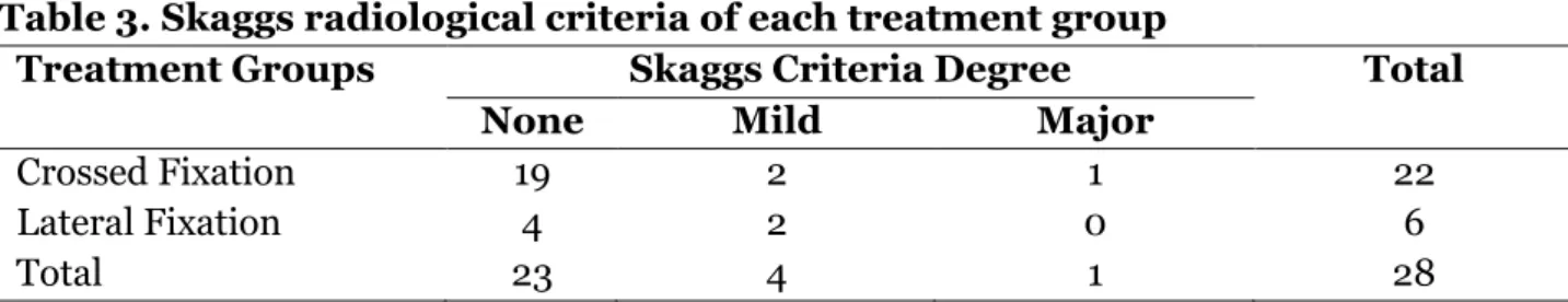
Table 3. Skaggs radiological criteria of each treatment group
Treatment Groups Skaggs Criteria Degree Total
None Mild Major
Crossed Fixation 19 2 1 22
Lateral Fixation 4 2 0 6
Total 23 4 1 28
In radiological evaluation measurement ba-sed on Skaggs criteria which was divided into none, mild, and major, an analysis by using Two-Sample Kolmogorov-Smirnov Test was conducted and obtained the value of p was 0.993. It indicated that there was no signifi-cant different between the two treatment groups. There was no ulnar nerve injury nor post-surgical infections found in all patients both from crossed fixation and lateral fixa-tion groups.
DISCUSSION
The study discovered male patients were 71.4% higher in number than female patients which were only 28.6%. The ratio between male vs. female was 2.5:1. The condition is in line with the previous epidemiology study that there were more male patients of supra-condylar humerus fractures than female pati-ents with the ratio 2:1.16. It was probably be-cause male had a lot more activities and often conducted activities outside compared with female.
The injuries occurred more on left elbows, whereas all patients in the study were right handed, the injury sides were domina-ted by non-dominant side (Kasser, 1992). Meanwhile Balakumar and Madhuri (2012) reported 1.1% ulnar nerve iatrogenic injuries, 2.2% median nerve injuries and 1.1% radial
nerve injuries on both crossed and lateral fixation techniques however, we did not dis-cover peripheral nerve injuries in this study. Awareness and surgeons’ adequate skills factors in the surgical process are the crucial factors in avoiding ulnar nerve iatrogenic injury.
There was no significant difference of patients’ clinical function result which was measured by using Flynn criteria between crossed and lateral fixation technique groups. Configurations, fixation insertion techniques, the size of K-Wire for fixation, post-surgical rehabilitation, patients’ compliance were fac-tors that supported the accomplishment of therapy. Patients’ compliance means all pa-tients visits orthopedic polyclinic after the surgery, perform programs from the poly-clinics, and obtain adequate rehabilitation therapy.
discover between 87.5% up to 100% of satis-factory result (Davis et al., 2000). This study almost obtained data which are approaching the previous studies.
Based on the radiological evaluation measurement by using Skagg criteria toward crossed fixation and lateral fixation it disco-vered that there was no significant difference between the two groups (p=0.993). All pa-tients underwent surgery by using the assis-tance of fluoroscopy (C-Arm) during the sur-gery procedure, hence it obtained optimal re-sult of the surgery in term of radiology.
The weakness of the study was insuffi-cient and imbalanced sample between the two therapy groups. It probably would be better if there are more number of sample and the quantity is comparable between the two groups. There is no difference of radiolo-gy and clinical functions between the crossed and the lateral fixation reduction technique. The study does not discover the surgical risk in the form of ulnar nerve iatrogenic injury and post-surgical infection.
AUTHOR CONTRIBUTION Komang Agung Irianto, Tri Wahyu Martanto, Febrian Brahmana, Laskar PK contributed to the design and implementation of the study, analysis of the results, and writing of the
manuscript.
CONFLICT OF INTEREST There is no conflict of interest in this study.
FUNDING AND SPONSORSHIP This study is self-funded.
ACKNOWLEDGEMENT
The author would like to say thank you to Dr. Soetomo Hospital, Surabaya, Central Java, Indonesia for allowing this study to be carri-ed out and to all study subjects who were wil-ling and cooperative to be part of this study.
REFERENCE
Balakumar B, Madhuri V (2012). A retros-pective analysis of loss of reduction in operated supracondylar humerus frac-tures. Indian J Orthop. 46: 6907. Davis RT, Gorczyca JT, Pugh K (2000).
Su-pracondylar humerus fractures in chil-dren. Comparison of operative treat-ment methods. Clin Orthop Relat Res: 376: 49-55.
Gartland JJ (1959). Management of supra-condylar fractures of the humerus in children. Surg Gynecol Obstet: 145– 154.
Gordon JE, Patton CM, Luhmann SJ, Bassett GS, Schoenecker PL (2001). Fracture stability after pinning of displaced su-pracondylar distal humerus fractures in children. J Pediatr Orthop, 21(3): 313– 318
Kalenderer O, Reisoglu A, Surer L, Agus H (2008). How should one treat iatroge-nic ulnar injury after closed reduction and per-cutaneous pinning of pediatric supracondylar humeral fractures? Inju-ry, 39(4): 463–466. https://doi.org. 10.1016/j.injury.2007.07.016
Kasser JR (1992). Percutaneous pinning of supracondylar fractures of the hume-rus. Instr course lect: 41: 385-390. Kocher MS, Kasser JR, Waters PM, Bae D,
Snyder BD, Hresko MT (2007). Lateral entry compared with medial and lateral entry pin fixation for completely displa-ced supracondylar humeral fractures in children: A randomized clinical trial. J Bone Jt Surg Am, 89(4): 706–712. htt-ps://doi.org.10.2106/JBJS.F.00379 Lee EH (2000). Supracondylar Fractures of
the humerus in Children Back to basics. Singapore Med J; 9: 423-424.
https://doi.org.10.-Otsuka NY, Kasser JR (1997). Supracondylar fractures of the humerus in children. J Am Acad Orthop Surg, 5: 19–26. https://doi.org.10.5435/00124635-19-9701000-00003
Rasool MN (1998). Ulnar nerve injury after K-wire fixation of supracondylar hume-rus fractures in children. J Pediatr Or-thop, 18: 686–690.
Skaggs DL, Cluck MW, Mostofi A (2004). La-teralentry pin fixation in management of supracondylar fractures in children.
J Bone Joint Surg Am, 86(4): 702-707. https:// doi.org. 10.2106/00004623-2-00404000-00006