Tirziu E. et. al./Scientific Papers: Animal Science and Biotechnologies, 2011, 44 (2)
183
Anatomopathological Changes Induced by Mycotoxins
Emil Tirziu, Ileana Nichita, Ciceronis Cumpanasoiu, Daniela Mot, Radu Valentin Gros,
Monica Seres, Magdalena Simona Muntean
Faculty of Veterinary Medicine, 300645, Timisoara, Calea Aradului, 119, Romania
Abstract
Fungi or mycetes represents the biggest group of microorganisms from the entire biologic system (nearly 250.000 species), very widespread in nature. They are inferior vegetal organisms, heterotrophic, lacking chlorophyll or other trophic pigments, which grow up on live organic substrates or on decaying substrates. The intensive researches from the last two decades had proved that only 30 – 40% from the total number of fungi species is capable to synthesize some toxic metabolites, and, among this species, only 60 species had proved to be dangerous for human or animals. Researches about mycotoxins action upon factors that interfere with the natural or acquired immunity are relatively recent and most of them refer to aflatoxins. The immune-suppression phenomena rely on morphological and histological modifications of lymphoid organs, changes of blood parameters, changes of functional capacity of humoral and some cellular factors. The presence of mycotoxins in feed causes major economic losses, either by their direct action (defined by disease state) or indirectly, by affecting the specific and nonspecific resistance of the organism. In the present study we studied the effect of aflatoxins upon the main organs involved in immune response, pathological changes induced by mycotoxins. To determine the influence of mycotoxins on food conversion, weighings were made at the beginning and the end of the experimental period.
Keywords: acquired immunity, animals, histological modifications, mycotoxins.
1. Introduction
Intensive animal husbandry can be adversely affected by feed mycotoxins. Mycotoxins acts upon the animal body in multiple pathways that are not neglected, the most important being nephrotoxic, hepatotoxic, teratogenic, carcinogenic, immuno-suppressive, and/or immunotoxic pathways [1, 2, 3, 4]. These issues are particularly important as it turned out that mycotoxins contained by the animal organisms can reach the human consumer in which can trigger the aforementioned phenomena.
Fungi or mycetes represents the biggest group of microorganisms from the entire biologic system, very widespread in nature. 1They are inferior vegetal organisms, heterotrophic, lacking chlorophyll or other trophic pigments, which grow up on live organic substrates or on decaying
*Corresponding author: Emil Tirziu, tel. 0256277113, fax. 0256277118, [email protected]
substrates [5, 6, 7, 8]. The intensive researches from the last two decades had proved that only 30-40% from the total number of fungi species is capable to synthesize some toxic metabolites, and, among this, only 60 species had proved to be dangerous for human or animals [9, 10].
The researches about mycotoxins action upon factors involved in natural and acquired immunity are relatively new, and most refer to aflatoxins [11]. The immunosuppression phenomena are based on morphological and histological changes in lymphoid organs, blood changes, and changes in functional capacity of humoral and cellular factors [12, 13, 14].
Tirziu E. et. al./Scientific Papers: Animal Science and Biotechnologies, 2011, 44 (2)
184 2. Materials and methods
The investigations were performed on ten 12-week-old piglets divided into two experimental groups:
- Group M (control) consisting of five animals fed with good quality feed and two times subcutaneous vaccinated (14 days apart), with 1 ml vaccine against sheep contagious agalactia; - Group E (experimental) consisting of five animals fed 15 days with feed supplemented with mycotoxins at a dose of 2.5 mg / kg feed.
Mycotoxins used were obtained after cultivation of one Aspergillus flavus strain on a medium containing 5% rice flour and 4% corn flour. Seeded medium was incubated ten days at room temperature, interval considered necessary to produce mycotoxins.
To determine the type of mycotoxin Soxhlet extractor was used. The extract obtained after ethyl ether removal by evaporation on a water bath at 40°C was taken with a solution composed of chloroform and ethanol (9:1).
Purification (for performing chromatography) was carried by filtration through a silica gel column. The extract obtained was evaporated to dryness, after which it was resumed in 0.5 ml washing solution and applied to silica gel plates, previously activated at 105°C.
Reading samples was done after developing and drying the plates at a UV lamp (254 nm). Measurements have shown that 50 ml Aspergillus
flavus culture contain 904 mg of mycotoxin
(ochratoxin and traces of aflatoxin B1). By
homogenization of 275 ml culture in 1 kg feed we get the amount of 2.5 mg ochratoxin/kg feed, which was introduced in the daily ration of the experimental group animals.
3. Results and discussion
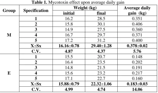
Analyzing the results obtained in experiments about the influence of mycotoxin administered to piglets upon the feed conversion expressed by average daily gain (Table 1), we obtained a 50% reduction in its average.
Average daily gain recorded throughout the experimental period in animals of experimental group was about 183g/day, compared to a gain of 387g/day achieved in the control group animals. Given that the feed with mycotoxin was administered ad libitum, there were large differences in the experimental group (CV=19.06%). Also, a number of authors indicate that the presence of mycotoxins in feed significantly reduces appetite, reducing also the average daily feed consumption (Figure 1).
At the end of the experiment, animals were slaughtered aiming to identify damages caused by the presence of mycotoxin in feed; the major organs involved in mycotoxins metabolism (liver, spleen, kidney) were harvested.
At necropsy of control group animals – vaccinated – we have not found characteristic lesions for functional disorders of major organs and systems; two animals lungs showed small foci of bronchopneumonia.
Table 1. Mycotoxin effect upon average daily gain
Group Specification Weight (kg) Average daily
gain (kg) initial final
M
1 16.2 28.5 0.351
2 15.8 30.1 0.406
3 14.9 27.5 0.360
4 16.7 29.7 0.371
5 17.2 31.2 0.400
X±Sx 16.16±0.78 29.40±1.28 0.378±0.02
C.V. 4.87 4.37 5.76
E
1 15.5 20.7 0.148
2 16.4 23.5 0.202
3 14.8 21.5 0.191
4 15.6 23.2 0.217
5 17.1 22.7 0.160
X±Sx 15.88±0.79 22.32±1.06 0.183±0.03
C.V. 4.99 4.74 14.06
Tirziu E. et. al./Scientific Papers: Animal Science and Biotechnologies, 2011, 44 (2)
185
1 2
3 4
5
E M 0
0,05 0,1 0,15 0,2 0,25 0,3 0,35 0,4 0,45
E M
Figure 1. Average daily gain – comparative evolution in the two groups
Macroscopic lesions found at necropsy of animals from experimental group (mycotoxin/vaccine) confirmed the hepatotoxic and nephrotoxic action of mycotoxins. The necropsy showed pathological changes characteristic for mycotoxins poisoning.
The gastrointestinal tract – on the intestinal
mucosa were found extensive bleeding, erosions and ulcers due to its direct contact with mycotoxins.
On the endocardium were found large bleeding both on surface and in depth.
The liver presented morphological lesions of
extralobular steatosis. The phenomenon has been generalized to four of the piglets in the experimental group and the fifth case was limited to left lateral lobe. On the steatosis background was found the presence of small bleeding foci accompanied by sub-icteric injuries. In one case we observed fibroadesive perihepatitis. The changes suggest that the liver is the target organ for mycotoxins that interferes with the protein, fat and carbohydrates metabolism, having as consequence a reduced ability to detoxify the body.
The kidneys of all piglets in the experimental
group were increased in volume, yellowish red in one case and generalized yellow in other cases. On section, in four cases, lesions of general steatosis were observed, and in one case, early steatosis limited to the cortical area. In most cases bladder edema was observed, probably due to the elimination of toxin and its metabolites in urine.
Spleen – in three cases was increased in volume,
with thin and transparent capsule, and mosaic appearance of splenic parenchyma. On the section
surface, follicular pattern was obviously associated with small foci of necrosis, suggesting hypertrophy, respectively hyperplasia of lymphoid follicles due to cytotoxic action of mycotoxins. In one animal no changes were observed in the spleen, and in another, we noted only the presence of a "pseudo pocket" on the posterior of the organ upper third.
Mesenteric lymph nodes – in three animals from
the experimental group were reduced in volume compared with controls, while two cases had apparently unchanged appearance. The section surface was glossy and wet due a small amount of yellow-citrine liquid.
Thymus, in all five cases, showed no significant
macroscopic changes.
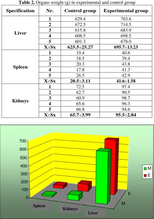
Pathological changes induced by the presence of mycotoxin in the diet influenced also the weight of main organs involved in its metabolism (Table 2, Figure 2).
Spleen shows higher average weight in the
experimental animals than in individual from the control group (up to 50%), which indicates mycotoxin-induced cell changes as a result of cytotoxic effect.
The kidneys of the experimental group individuals
were also increased in volume and weight.
Tirziu E. et. al./Scientific Papers: Animal Science and Biotechnologies, 2011, 44 (2)
186
Table 2. Organs weight (g) in experimental and control group
Specification Nr. Control group Experimental group
Liver
1 629.4 703.6
2 672.5 714.5
3 615.8 683.9
4 608.5 698.5
5 601.3 678.0
X±Sx 625.5±25.27 695.7±13.23
Spleen
1 19.4 40.6
2 18.5 39.4
3 20.3 43.8
4 17.8 41.3
5 26.5 42.9
X±Sx 20.5±3.11 41.6±1.58
Kidneys
1 72.5 97.4
2 62.7 90.5
3 60.9 98.7
4 65.6 96.3
5 66.8 94.6
X±Sx 65.7±3.99 95.5±2.84
Splină
Rinichi
Ficat
M E 0
100 200 300 400 500 600 700
M E
Figure 2. Organs weight (g) in experimental and control group
4. Conclusions
Mycotoxins given in feed to piglets for 15 days at a dose of 2.5 mg/kg feed induce an immune deficiency condition, due their immunosuppressive effect on the specific and non-specific immune effectors.
Anatomopathological examination shows lesions characteristic for chronic intoxication with localization in the organ involved in mycotoxin metabolism and elimination: generalized
hepatosis, gastrointestinal bleeding and ulcers, nephrosis and edema of bladder wall.
Metabolic disorders caused by mycotoxins are also reflected in the degree of food conversion. Average daily gain of animals achieved by the experimental group animals was 50% lower than values recorded in the control group.
Spleen
Kidneys
Tirziu E. et. al./Scientific Papers: Animal Science and Biotechnologies, 2011, 44 (2)
187 References
1. Göbel, Roswitha, Lusky, K., Simultaneous determination of aflatoxins, ochratoxin A, and zearalenone in Grains by new immunoaffinity column/liquid chromatography, Journal of Aoac International, 2004, 87, 2, 411-416
2. Hideko, T., Nobuaki, S., Osamu, M., Hideo, M., Suppressive effect of deoxynivalenol, a Fusarium
mycotoxin, on bovine and porcine neutrophil chemiluminescence: An in vitro study, J. Vet. Med. Sci., 2005, 67, 5, 531-533
3. Probst, C., Njapau, H., Cotty, P. J., Outbreak of an acute aflatoxicosis in Kenya in 2004: Identification of the Causal Agent, Appl. Environ. Microbiol., 2007, 73, 2762-2764
4. Takayama, H., Shimada, N., Mikami, O., Murata, H., Suppressive effect of deoxynivalenol, a Fusarium
mycotoxin, on bovine and porcine neutrophil chemiluminescence: an in vitro study, Journal of veterinary medical science, 2005, 67, 5, 531-533 5. Araguhs, C., Gonzalez-Penas, E., Lopez De Cerain, A., Study on ochratoxin A in cereal-derived products from Spain, Food chemistry, 2005, 92, 3, 459-464. 6. Bennett, J.W., Klich, M., Mycotoxins, Clinic. Microb. Rev., 2003, 16, 3, 497-516
7. Caldas, E.D., Silva, S.C., Oliveira, J.N., Aflatoxinas e ocratoxina A em alimentos e riscos para a saúde humana, Rev. Saúde Pública, 2007, 36(3), 319-323 8. Hedayati, M.T., Pasqualotto, A.C., Warn, P.A., Bowyer, P., Denning, D.W., Aspergillus flavus: human pathogen, allergen and mycotoxin producer, Microbiology, 2007, 153, 1677-1692
9. Döll, S., Dänicke, S., Valenta, H., Flachowsky, G.,
In vitro studies on the evaluation of mycotoxin detoxifying agents for their efficacy on deoxynivalenol and zearalenone, Arch. Anim. Nutr., 2004, 58, 311-324.
10. Janyce, A., Sugui, J.P., Yun, C., Chang, K.A., Zarember, G.N., Galvez,E.M., Müllbacher,A., Gallin, J.I., Simon, M.M., Kwon-Chung, J., Gliotoxin is a virulence factor of Aspergillus fumigatus: gliP deletion attenuates virulence in mice immunosuppressed with hydrocortisone, Eukaryotic cell, 2007, 6, 9, 1562-1569. 11. Obrian, G.R., Wilkinson, J.R., Yu, J., Abbas, H.K., Bhatnagar, D., Cleveland, T.E., Nierman, W., Payne, G.A., The effect of elevated temperature on gene transcription and aflatoxin biosynthesis, Mycologia, 2007, 99, 232-239
12. Kremer, A., Westrich, L., Li, S.M., A 7- dimethylallyltryptophan synthase from Aspergillus fumigatus: overproduction, purification and biochemical characterization, Microbiology, 2007, 153, 3409-3416
13. Smith, C. A., Woloshuk, C. P., Robertson, D., Payne, G. A., Silencing of the aflatoxin gene cluster in a diploid strain of Aspergillus flavus is suppressed by ectopic aflR expression, Genetics, 2007, 176, 2077-2086