http://www.ijorl.com
Case Report
Squamous cell carcinoma of tympano-mastoid region:
a series of six cases
Surender Kumar*,
Uma Garg, Naveen Sharma, Neha Salaria,
Deepak Verma
INTRODUCTION
The malignancies of tympanomastoid region are very rare entity. Squamous cell carcinoma being the most common malignancy in head and neck region, is also most common variety found in temporal bone tumours.1 Other varieties are adenocarcinoma, adenocystic carcinoma, mucoepidermoid carcinoma, ceruminous carcinoma and rhabdomyosarcoma. The incidence of primary temporal bone tumours ranges between 0.8-1.0 per 1,00,000 per year and approximately 60-80-% are squamous cell carcinoma.2 Overall squamous cell carcinoma accounts approximately 1.5% of tympanomastoid tumours.3,4 Metastatic lesions in tympanomastoid region are very rare and are usually from breast, lung and kidney tumours.5 The tumours of tympano-mastoid region are more common in elderly persons in 6th and 7th decade. Males are more commonly affected than females. Various predisposing factors have been proposed like chronic
suppurative otitis media, prior exposure to radiations in nasopharyngeal, intracranial and head and neck cancers and ultraviolet radiations. The presentation of these tumors are usually like chronic suppurative otitis media (CSOM) which makes the diagnosis very difficult and even after diagnosis, treatment is very challenging and unrewarding with a very high recurrence and mortality rate. So here, we present six cases of tympanomastoid malignancies to discuss the presentation, diagnostic difficulties and modalities of management of these types of patients.
METHODS
A retrospective study was conducted among patient with tympanomastoid malignancy presented from 2011 to 2017. Total six patients whose details are described in Table 1 were included.
ABSTRACT
The malignancies of tympanomastoid region are very rare entity. These tumours are more common in elderly persons in 6th and 7th decade. Males are more commonly affected than females. Chronically discharging ears are considered as risk factor which may be due to metaplasia in the middle ear mucosa following prolonged chronic infection. The most common symptoms are long standing blood tinged ear discharge, severe nocturnal pain, rapidly growing polypoidal or granulomatous mass in EAC or middle ear, peripheral facial palsy and painless ulceration over pinna or EAC. CT scan for bony erosion and MRI for soft tissue involvement and neural invasion are investigations of choice. Tissue biopsy is must for histopathological examination and confirmation of diagnosis. Surgery and chemo-radiotherapy are the mainstay of treatment. Surgery includes wide local excision, lateral temporal bone resection, subtotal temporal bone resection and total temporal bone resection.
Keywords: Squamous cell carcinoma, Tympanomastoid malignancy, Temporal bone resection, Chemo-radiotherapy
Department of ENT & HNS, Bhagat Phool Singh Government Medical College, Khanpur Kalan, Sonepat, Haryana, India
Received: 14 March 2018
Revised: 09 April 2018
Accepted: 11 April 2018
*Correspondence:
Dr. Surender Kumar,
E-mail: dr.morodia@gmail.com
Copyright: © the author(s), publisher and licensee Medip Academy. This is an open-access article distributed under
the terms of the Creative Commons Attribution Non-Commercial License, which permits unrestricted non-commercial use, distribution, and reproduction in any medium, provided the original work is properly cited.
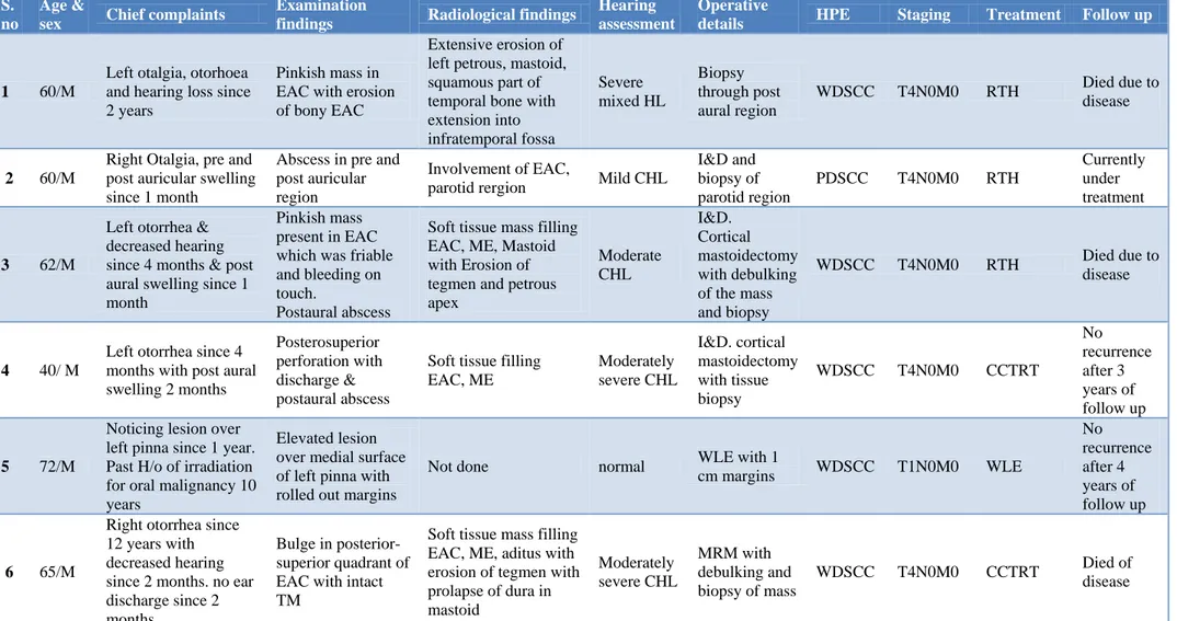
Table 1: Showing details of the all the patients.
S. no
Age &
sex Chief complaints
Examination
findings Radiological findings
Hearing assessment
Operative
details HPE Staging Treatment Follow up
1 60/M
Left otalgia, otorhoea and hearing loss since 2 years
Pinkish mass in EAC with erosion of bony EAC
Extensive erosion of left petrous, mastoid, squamous part of temporal bone with extension into infratemporal fossa Severe mixed HL Biopsy through post aural region
WDSCC T4N0M0 RTH Died due to
disease
2 60/M
Right Otalgia, pre and post auricular swelling since 1 month
Abscess in pre and post auricular region
Involvement of EAC,
parotid rergion Mild CHL
I&D and biopsy of parotid region
PDSCC T4N0M0 RTH
Currently under treatment
3 62/M
Left otorrhea & decreased hearing since 4 months & post aural swelling since 1 month
Pinkish mass present in EAC which was friable and bleeding on touch.
Postaural abscess
Soft tissue mass filling EAC, ME, Mastoid with Erosion of tegmen and petrous apex Moderate CHL I&D. Cortical mastoidectomy with debulking of the mass and biopsy
WDSCC T4N0M0 RTH Died due to
disease
4 40/ M
Left otorrhea since 4 months with post aural swelling 2 months
Posterosuperior perforation with discharge & postaural abscess
Soft tissue filling EAC, ME Moderately severe CHL I&D. cortical mastoidectomy with tissue biopsy
WDSCC T4N0M0 CCTRT
No recurrence after 3 years of follow up
5 72/M
Noticing lesion over left pinna since 1 year. Past H/o of irradiation for oral malignancy 10 years
Elevated lesion over medial surface of left pinna with rolled out margins
Not done normal WLE with 1
cm margins WDSCC T1N0M0 WLE
No recurrence after 4 years of follow up
6 65/M
Right otorrhea since 12 years with decreased hearing since 2 months. no ear discharge since 2 months
Bulge in posterior-superior quadrant of EAC with intact TM
Soft tissue mass filling EAC, ME, aditus with erosion of tegmen with prolapse of dura in mastoid
Moderately severe CHL
MRM with debulking and biopsy of mass
WDSCC T4N0M0 CCTRT Died of
disease
Case no: 1
A 60 years old male patient, labourer by occupation presented to ENT OPD with history of (h/o) left ear discharge and hearing loss since 2 years. The discharge was scanty, blood tinged and foul smelling. There was h/o pain in left ear which was more during night and was not relieved with use of medications. There was no h/o facial weakness, giddiness, tinnitus. Patient was being treated for chronic suppurative otitis media for long time but he didn’t get any improvement. On examination, there was a pinkish mass noted in external auditory canal (EAC) with extensive erosion of bony EAC (Figure 1 A). There was no associated lymphadenopathy. The patient was admitted and investigated. On contrast enhanced CT (CECT) scan, there was extensive erosion /lysis of left side petrous, mastoid and squamous part of temporal bone and sphenoid wing posteriorly with soft tissue component. The soft tissue was extending into infratemporal fossa and posterior fossa. There was subtle erosion of basal turn of cochlea, semicircular canals, carotid canal, condyle of mandible and zygomatic arch (Figure 2). Patients was planned for biopsy under local anaesthesia through post aural region. After elevating the tissue postauraly, there was big defect noted in the mastoid cortex (Figure 1, B and C). Middle ear and mastoid cavity was filled of pinkish mass which was very friable and bled on touch. Multiple biopsies taken (Figure 1 D) and sent for histopathological examination (HPE) which came out to be well differentiated squamous cell carcinoma. The diagnosis was made as squamous cell carcinoma of left temporal region T4N0M0 Stage IV. Patient was planned for concurrent chemoradiotherapy which he could not complete and expired within 2 months of treatment.
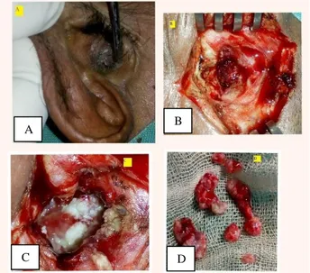
Figure 1: (A) Pinkish mass noted in EAC; (B and C) defect present in mastoid cortex and cavity filled with
pinkish mass with overlying whitish discharge; (D)tissue for biopsy.
Figure 2 (A-D): CECT (axial, coronal and virtual 3D reconstruction images) suggestive of extensive erosion of petrous, squamous and mastoid part of temporal
bone with cochlea, semicicular canals, ossicles and carotid with lytic destruction of mandibular condyle
and zygomatic arch.
Case no: 2
A 60 years old male patient farmer by occupation presented to ENT OPD with h/o of noticing swelling in right pre-auricular and infra-auricular region since 15 days. Patient also complained of pain in right ear which was dull aching and persistent through out the day. There was no history of ear discharge, decreased hearing, giddiness or facial weakness in past. On examination, there was swelling measuring 4 3 cm in right pre-auricular region which was firm in consistency, tender with minimal fluctuation. There was local rise of temperature and blanching present. There was another swelling present in right infra-auricular region which was measuring 1 1 cm soft in consistency and fluctuant. On examination of ear, there was a smooth bulge present on anterior wall of right external auditory canal which was tender. Rest of the ear examination was normal.
Figure 3: Showing incision and drainage site of right preauricular region.
Patient was admitted and empirically started on intravenous antibiotics. Ultrasound scan (USG) and high resolution CT scan (HRCT) temporal bone was done. USG suggestive of abscess in right pre-auricular and infra-auricular region. However HRCT temporal bone
A
D C
B
A B
revealed no abnormality in middle ear and inner ear. Rest of the findings were suggestive of abscess only. Patient was taken for incision and drainage under local anaesthesia and pus was drained out. With regular dressing and intravenous antibiotics, patient’s condition improved. Patient was discharged on oral antibiotics with an advice of regular dressings. However patient reported back with continuous discharge from incision and drainage site (Figure 3). Patient was readmitted and started with antibiotics. CECT right parotid region was done which showed irregular minimally enhancing soft tissue thickening with superficial ulceration involving the scalp in right temporal region with infiltration of adjacent part of temporalis and masseter muscles and inferiorly showing ill-defined fat planes with upper border of superficial lobe of parotid gland. Subtle erosion of base of zygomatic process with extension into bony EAC. Keeping the possibility of malignancy, biopsy was taken
from EAC and parotid region and sent for
histopathological examination. HPE report suggestive of poorly differentiated squamous cell carcinoma. Patient was diagnosed as SCC temporal T4N0M0 Stage IV and sent for concurrent chemo-radiotherapy for further management.
Case no: 3
A 62 years old male presented to ENT OPD with a complaint of left ear discharge for past 3-4 months. The discharge was scanty, foul smelling and serosanguinous. This was also associated with decreased hearing. There was no history of tinnitus, vertigo, facial weakness, headache or earache. Patient had also noted post-aural swelling since one month. On examination, there was pinkish mass present filling the external auditory canal which was friable and bled on touch. The post aural swelling was approximately 1 1 cm, firm with some fluctuant areas, tender, fixed to the underlying structures and warm on palpation. There was no associated cervical
lymphadenopathy. Pure tone audiometry showed
moderate conductive hearing loss in left ear with normal hearing in right ear. The patient was diagnosed as a case of complicated chronic suppurative otitis media with post aural abscess. Patient was planned for incision and drainage, as indicated. The pus was sent for microbiological culture and sensitivity which didn’t show any growth. High resolution computed tomography scan of temporal bone showed a poorly enhancing soft tissue mass involving external auditory canal, middle ear and mastoid. There was erosion of tegmen tympani and significant destruction of petrous temporal bone. The patient was planned for mastoid exploration under general anaesthesia. Intraoperatively, there was pinkish growth noted filling the EAC, middle ear and eroding the mastoid cortex. The mass was debulked keeping the possibility of malignancy and sent for histopathological examination. The histopathological examination showed the mass to be well differentiated squamous cell carcinoma with nests of squamous cell showing dysplasia, along with keratin pearls and individual cell keratinisation. On the basis of clinical, radiological and histopathological findings, patient was diagnosed as case
of squamous cell carcinoma of left temporal bone stage IV (T4N0M0).
Case no: 4
A 40 years old male presented to ENT OPD with a complain of left ear discharge since 4 months and swelling in left post aural region since 2 months. The discharge was scanty, foul smelling, yellowish. It was also associated with decrease hearing. The post aural abscess was drained under local anaesthesia. There was no cervical lymph node. HRCT temporal bone was done which suggested moderately enhancing mass filling the middle ear and mastoid. Patient was planned for mastoid exploration under general anaesthesia. Intra op, pinkish mass noted eroding the mastoid cortex, posterior canal wall and filling the external auditory canal. No ossicles were found. The mass was debulked and sent for histopathological examination. Biopsy report came as well differentiated squamous cell carcinoma grade 1. Patient was staged as T4N0M0 Stage IV and sent for post op radiotherapy.
Case no: 5
A 72 years old male presented to ENT OPD with h/o noticing lesion in left pinna since 5-6 years. Patient had received radiotherapy for oral malignancy around 10 years ago. He noticed this lesion after 3 years of post-radiotherapy. Biopsy was already taken in private hospital which was suggestive of squamous cell carcinoma. Patient was planned for excision under local anaesthesia. Specimen sent for histopathological examination which came positive for squamous cell carcinoma. Patient was observed for 5 years and no recurrence was noted.
Case no: 6
A 65 years male patient presented to ENT OPD with right ear discharge since 12 years which was foul smelling, scanty, purulent associated with decreased hearing from right ear since 2 months and occasional tinnitus. There was no ear discharge since 2 months. On examination there was bulge EAC in posterior-superior quadrant while TM was intact. Patients was also known case of bronchial asthma. Patient was planned for mastoid exploration under general anaesthesia. Intraoperatively, there was pinkish mass present in middle ear which bled on touch, eroding posterior wall of EAC, aditus, extending into antrum. The dural plate found dehiscent and duramater exposed and bulging inferiorly. The mass debulked and sent for histopathological examination. The report suggestive of well differentiated squamous cell carcinoma. Patient was planned for post-operative radiotherapy.
DISCUSSION
mastoid, petrous bone and metastatic lesions are very rare entity. The incidence has been reported very low by many studies which ranges between 0.03-1.0 per 100000 per year and it is more common in males in age group of 6th and 7th decade. In present study all the six patients were male and 5 out of 6 patients were of more than 60 years of age. Only one person was 40 years old. Many etiological factors have been reported like h/o previous radiation exposure, ultraviolet radiation and chronic otitis media.6 Chronically discharging ears are considered as risk factor in many studies which may be due to metaplasia in the middle ear mucosa due to chronic infection. In our study out of 6 patients, only one patient (case no 6) had long standing ear discharge for more than 12 years and his ear was dry for 2 months at the time of presentation and ear drum was also intact on examination. Rest of the three patients had very short history of otorrhea of less than 6 months. Previous h/o of radiation exposure is a well-known risk factor. In this study, one patient (case no 5) had treatment for oral malignancy 10 years back and he developed lesion over pinna after 3 years of his radiotherapy. Some studies have also considered agents such as chlorinated disinfectants or human papillomavirus in cases of carcinomas associated with inverted papillomas as possible carcinogens.7-9
The clinical features are very similar to chronic otitis media rendering diagnosis very difficult. The most common symptoms are long standing blood tinged ear discharge, severe nocturnal pain, rapidly growing polypoidal or granulomatous mass in EAC or Middle Ear, peripheral facial palsy and painless ulceration over pinna or EAC. A high index of suspicion in such cases should render a clinician for thorough clinical examination and investigation to rule out malignancy. Radiological investigations play a very important role in diagnosis in suspicious cases. CT scan for bony erosion and MRI for soft tissue involvement and neural invasion are investigations of choice. Tissue biopsy is must for histopathological examination and confirmation of diagnosis.
Table 2: Showing T staging in modified Pittsburgh staging system.
T1 Tumour limited to EAC without bony erosion or evidence of soft tissue involvement
T2
Tumour with limited EAC bone erosion (not full thickness) with limited (0.5cm) soft tissue involvement.
T3
Tumour eroding the osseous EAC (full thickness) with limited (0.5cm) soft tissue involvement or tumour involving the middle ear, mastoid or both.
T4
Tumour eroding the cochlea, petrous apex, medial wall of the middle ear, carotid canal, jugular foramen, duramater or with extensive soft tissue involvement (>0.5cm), such as
involvement of temporomandibular joint or stylomastoid foramen or with evidence of facial paresis.
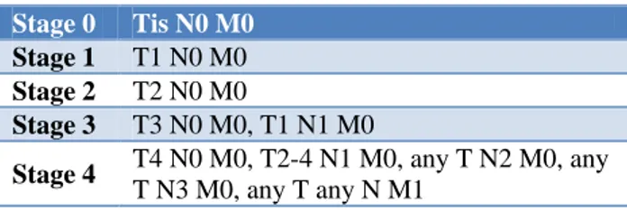
Table 3: Showing modified Pittsburgh staging system.
Stage 0 Tis N0 M0
Stage 1 T1 N0 M0
Stage 2 T2 N0 M0
Stage 3 T3 N0 M0, T1 N1 M0
Stage 4 T4 N0 M0, T2-4 N1 M0, any T N2 M0, any
T N3 M0, any T any N M1
The staging of these tumours are very difficult as there is no universally acceptable system. The most commonly used staging system is modified Pittsburgh staging system. (Table 2 and 3).
Tumours of the pinna and the EAC are considered to be more aggressive and have a higher risk of recurrence and lymph node metastasis, most probably due to the presence of the fusion of multiple embryonic planes in this region, which may facilitate tumour dissemination.
10-12
Surgery and chemo-radiotherapy are the mainstay of treatment. Surgery includes wide local excision (WLE), lateral temporal bone resection (LTBR), subtotal temporal bone resection (STBR), total temporal bone resection (TTBR). En block resection of the tumour along with total parotidectomy should be considered as first line management. Surgical resection is contraindicated in cases of intracranial extension, cavernous sinus thrombosis, unresectable cervical nodal disease, poor general condition of the patient and distant metastasis. Primary radiotherapy is reserved for locally advanced tumour (T3, T4), close or positive margins, perineural invasion and cervical node metastasis. Chemotherapy can also be considered in cases with post-surgical residual disease or recurrence, distant metastasis and conditions where surgery and radiotherapy are contraindicated.
Regarding clinically negative neck nodes, treatment is still controversial. Some authors recommend ipsilateral selective neck dissection in cases of N0 due to higher chances of micrometastasis in locally advanced tumours.
With all treatment options available, prognosis is very poor and mortality rate is very high.
CONCLUSION
Temporal bone tumours are rare but very aggressive and difficult to treat. Diagnosis becomes very difficult as the clinical features are most of the time similar to chronic suppurative otitis media. Strong suspicion is required for timely diagnosis and treatment to improve survival.
REFERENCES
1. National Cancer Institute. “Cancer facts. Head and neck cancer: questions and answers.” Available at: http://www.cancer.gov/cancertopics/factsheet/Sites-types/head-and-neck/. Accessed on 3 February 2018.
2. Lionello M, Stritoni P, Facciolo MC, Staffieri A, Martini A,Mazzoni A, et al. Temporal bone carcinoma. Current diag-nostic, therapeutic, and prognostic concepts. J Surg Oncol. 2014;110:383-92.
3. Gidley PW, Roberts DB, Sturgis EM. Squamous cell carcinoma of the temporal bone. Lary-ngoscope 2010;120:1144-51.
4. Shu MT, Lee JC, Yang CC, Wu KC. Squamous cell
carcinoma of the middle ear. Ear Nose Throat J. 2012;91:14.
5. Cureoglu S, Tulunay O, Ferlito A, Schachern PA, Paparella MM, Rinaldo A. Otologic manifestations of metastatic tumors to thetemporal bone. Acta Otolaryngol. 2004;124:1117-23.
6. Elsürer C, Senkal HA, Zayyan E, Yilmaz T, Kaya S. Bilateral external auditory canal squamous cell carcinoma: a case report. Eur Arch Otor-hinolaryngol. 2007;264:941-5.
7. Monem SA, Moffat DA, Frampton MC. Carcinoma
of the ear: a case report of a possible association with chlorinated disinfec-tants. J Laryngol Otol. 1999;113:1004-7.
8. Gaio E, Marioni G, Blandamura S, Staffieri A. Inverted papiloma involving the temporal bone and its association with squamous cell carcinoma: critical analysis of the literature. Expert Rev Anticancer Ther. 2005;5:391-7.
9. Marioni G, Altavilla G, Busatto G, Blandamura S, De Filippis C, Staffieri A. Detection of human papillomavirus in temporal bone inverted papilloma by polymerase chain reaction. Acta Otolaryngol. 2003;123:367-71.
10. Niparko JK, Swanson NA, Baker SR, Telian SA, Sullivan MJ, Kemink JL. Local control of auricular, periauricular, and external canal cutaneous malignancies with Mohs surgery. Laryngoscope. 1990;100:1047-51.
11. Gal TJ, Futran ND, Bartels LJ, Klotch DW. Auricular carcinoma with temporal bone invasion: outcome analysis. Otolaryngol Head Neck Surg. 1999;121:62-5.
12. Gaudet JE, Walvekar RR, Arriaga MA, Dileo MD, Nuss DW, Pou AM, et al. Applicability of the Pittsburgh staging system for advanced cutaneous malignancy of the temporal bone. Skull Base. 2010;20:409-14.
Cite this article as: Kumar S, Garg U, Sharma N,