Original Research Article
Cytopathological study of salivary gland lesions by fine needle
aspiration cytology
Priyanka Desai, Bhavna Gamit, Neha S. Shahu*, Bhavika Dholiya
INTRODUCTION
Fine needle aspiration cytology is an inexpensive, minimally invasive, outpatient, routinely used diagnostic procedure. The foundation of this current procedure was established by “Karolonska Group” between 1974-1976.1
As compared to biopsy methods, FNA is a very smooth, cheaper, outpatient procedure helping clinician to save his time for earlier diagnosis and treatment. Significant complication is rare.2,3 FNA of salivary gland is very
easier to perform as the site is very superficial. Though
anatomical structure of the gland is very simple, it is subjected to a diverse and heterogenous range of tumors.4,5
Among all the salivary gland tumors, the pleomorphic adenoma is the most common tumor type which accounts for 50% of all tumors. Warthin’s tumor is the second most common among benign tumors whereas mucoepidermoid carcinoma is the most common malignant tumor. Most canalicular adenomas and polymorphous low-grade adenocarcinoma arise from Department of Pathology, Government Medical College, Surat, Gujarat, India
Received: 15 October 2019
Revised: 08 November 2019
Accepted: 15 November 2019
*Correspondence:
Dr. Neha S. Shahu,
E-mail: shahu581993@gmail.com
Copyright: © the author(s), publisher and licensee Medip Academy. This is an open-access article distributed under the terms of the Creative Commons Attribution Non-Commercial License, which permits unrestricted non-commercial use, distribution, and reproduction in any medium, provided the original work is properly cited.
ABSTRACT
Background: Fine needle aspiration cytology is an inexpensive, minimally invasive, outpatient diagnostic procedure. FNA of salivary gland is easier to perform as the site is superficial and repeat FNAC can be perform. As compared to biopsy methods, FNA is a very smooth, cheaper, outpatient procedure helping clinician to save his time for earlier diagnosis and treatment. Though anatomical structure of the gland is very simple, it is subjected to a diverse and heterogenous range of tumors.
Methods: The present study on “Cytomorphological features of salivary gland lesion by FNAC” was carried out on department of pathology from June 2015 to June 2017. 65 patients with salivary gland lesions who were sent to pathology department for FNAC were aspirated and correlated histopathologically.
Results: Benign salivary gland lesions contribute to majority of cases (54%). Pleomorphic Adenoma was the most common benign salivary gland lesions and Mucoepidermoid Carcinoma was most common malignant lesion. Parotid gland was the most commonly involved in benign tumors and submandibular gland was commonly involved by malignant tumors. Commonly affected age group by benign salivary gland lesion was 31-40 years and those with malignant salivary gland lesion was 41-50 years.
Conclusions: FNA cytology provides useful information for the management of salivary gland lesions and prevents unnecessary surgery in cases of nonneoplastic lesions and identification of malignancy helps the surgeon in deciding type and extent of surgery.
Keywords:Cytohistological correlation, Fine needle aspiration cytology,Salivary gland neoplasms, Salivary gland lesions
minor salivary glands whereas nearly all warthin’s tumor
occurs in parotid gland or peri parotid lymphnodes.6
METHODS
The present study on “Cytomorphological study of salivary gland lesions by FNAC” was carried out in department of pathology from June 2015 to June 2017. 65 patients with salivary gland lesions who were sent to pathology department for FNAC were aspirated and subsequently examined and some of them were correlated histopathologically.
Inclusion criteria
• All neoplastic and non- neoplastic lesion of salivary gland referred by clinician for FNAC from June 2015 to June 2017 were included in the study.
Exclusion criteria
• Swelling with scant or inadequate aspirate on smear • Swelling diagnosed to be of any other origin than
salivary gland
The clinical data pertaining to patient’s age, sex and anatomical site were recorded. The patient’s consent was taken before the procedure. Aspirations were performed using a 22 gauze needles and smears are prepared on clean glass slides. The air dried and ethanol fixed smears were stained with May Grunwald’s Giemsa, papanicolaou and hematoxylin and eosin stain respectively.7
The histopathological findings of respective cases wherever available are noted. Thus, the cytological and histological diagnosis was arrived independently at different times and correlated to obtain the accuracy of FNAC in salivary gland lesions. To measure the performance of FNAC author took into account true positive, true negative, false positive and false negative cases and calculated various parameters as follows:
Sensitivity = (True positive)/ (True positive + False negative).
Specificity = (True negative)/ (True negative + False positive).
RESULTS
The objective of this study was to study cytomorphological features of salivary gland lesions on FNAC and to evaluate the accuracy of FNAC by correlating cytology findings with histopathology whenever possible.
Benign salivary gland lesions contribute to majority of cases accounting 54%, malignant lesions contribute to 25% and non- neoplastic lesions contribute to 21% cases (Table 1).
Table 1:Distribution of salivary gland lesion.
Lesion No. of cases Percentage
Non neoplastic 14 21.8%
Benign 35 53.8%
Malignant 16 24.6%
Total 65 100
Of non-neoplastic lesions, sialadenitis contributes to majority of cases accounting for around 50% of cases (Table 2).
Table 2: Distribution of various non-neoplastic lesions.
Lesion No. of cases Percentage
Sialadenitis 7 50%
Mucocele 2 14.2%
Sialadenosis 2 14.2%
Other non-neoplastic
salivary gland lesion 3 21.4%
Total 14 100%
Figure 1: Sialadenitis Fragments of duct epithelium with small round nuclei and purulent
background (MGG, 10X).
Fine needle aspiration cytology smears show many clusters of ductal cells, scanty acinar cells and inflammatory cells in a background of mucinous material. The sparse number of acinar cells was usually due to chronic destruction of such cells (Figure 1).
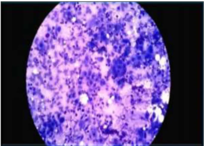
Benign salivary gland lesions were most common as compared to malignant one. Among benign lesions, Pleomorphic Adenoma was the most common contributing lesion (Table 3).
individual cells were round to oval with moderate to
scanty cytoplasm (Figure 2).
Table 3: Distribution of neoplastic salivary gland lesions.
Lesions No. of
cases %
Benign
Pleomorphic adenoma 30 58.8%
Warthin’s tumor 01 2%
Oncocytoma 01 2%
Benign salivary gland
neoplasm 03 5.9%
Malignant
Mucoepidermoid
carcinoma 10 19.6%
Acinic cell carcinoma 03 5.9% Adenoid cystic
carcinoma 01 2%
Carcinoma ex
pleomorphic adenoma 01 2% Adenocarcinoma NOS 01 2%
Total 51 100%
Figure 2: Pleomorphic adenoma poorly cohesive epithelial cells with fibrillar chondro-myxoid stroma
staining brightly magenta (MGG, 10x).
Among malignant lesions, mucoepidermoid carcinoma was most common which accounted for 19.6% of all neoplastic cases (Table 3).
Fine needle aspiration cytology smears show predominantly three types of cells: (1) squamoid cells, (2) intermediate cells and (3) mucus secreting cells.
The background of the tumor was dirty with mucus and debris. The squamoid or epidermoid cells were polygonal in shape with orangeophilic cytoplasm in papanicolaou’s stain. The intermediate cells were smaller in size, columnar to polygonal and had enlarged hyperchromatic nuclei with high nucleo cytoplasmic ratio. The mucus secreting cells were round with abundant vacuolated cytoplasm. The nuclei were eccentrically placed and indented and often give signet ring
like appearance. The nuclei of mucus secreting cells of MEC show low nucleus-to-cytoplasm (N/C) ratio and almost no nuclear enlargement (Figure 3,4).
Figure 3: Mucoepidermoid carcinoma cell clusters probably of intermediate cells in dirty appearing
mucus, debris, inflammatory cells and macrophages (MGG,10X).
Figure 4: Mucoepidermoid carcinoma pleomorphic, clearly malignant cells, some cells showing squamous
differentiation (MGG, 40X).
One case of Warthin tumor was diagnosed showing oncocytic cells arranged in monolayered sheets along with lymphoid population and background of proteinaceous and cell debris (Figure 5).
Three cases of Acinic cell carcinoma had been reported. The smears showing cells arranged in three-dimensional clusters and sheet. The individual cells are polygonal with abundant finely vacuolated cytoplasm. Nuclei are rounded, monomorphic, centrally placed with a small distinct nucleolus (Figure 6).
basophilic granular cytoplasm and uniform round central
nuclei with finely granular chromatin (Figure 7).
Parotid gland was the most commonly involved gland in benign tumors, the most common of which being pleomorphic adenoma.
Figure 5: Warthin’s tumor monolayered sheets of uniform oncocytic epithelial cells with small bland nuclei and lymphocytes in background (PAP 10X).
Figure 6: Acinic cell carcinoma abundant cell material in clusters and forming micro acinar
groupings (MGG,10X).
Figure 7: Oncocytic adenoma: multi-layered aggregates of cohesive oxyphil cells with abundant
eosinophilic granular cytoplasm (MGG,10X).
Submandibular gland was most commonly involved gland by malignant tumor, the most common of which being MEC.
Most common age group affected in non-neoplastic lesion was 31-40 years of life. The most commonly affected age group by pleomorphic adenoma was 31-40 years of life and overall most commonly affected age group by benign salivary gland lesion was 31-40 years of life.
Most commonly affected age group by MEC was 41-50 years of life and overall most commonly affected age group by malignant salivary gland lesion was 41-50 years of life.
Overall most commonly affected age group in salivary gland lesions was 31-40 years of age. In this study for non-neoplastic lesion male preponderance is seen with male: female ratio of 1.6:1. For neoplastic lesion, female preponderance is seen with male: female ratio of 0.9:1 (Table 4).
Table 4: Age wise distribution of the non-neoplastic and neoplastic salivary gland lesion (age in years).
Age groups ( in years)
Lesions 0-10 11-20 21-30 31-40 41-50 51-60 61-70 >70
Non-neoplastic 2 0 1 6 3 0 1 1
Pleomorphic adenoma 0 1 8 9 4 5 2 1
Warthin’s tumor 0 0 0 0 0 0 0 1
Oncocytoma 0 0 0 0 0 1 0 0
Benign salivary gland neoplasm 0 0 0 1 1 0 1 0
MEC 0 0 1 2 4 1 1 1
Acinic cell carcinoma 1 0 1 0 0 1 0 0
Adenoid cystic carcinoma 0 0 1 0 0 0 0 0
Ca ex PA 0 0 0 0 0 0 1 0
Adenocarcinoma NOS 0 0 0 0 0 0 0 1
Total 65 cases were examined cytologically out of which
14 cases was non neoplastic, among which sialadenitis were most common and treated conservatively and cured. 30 cases of pleomorphic adenoma were diagnosed cytologically out of which 6 cases were available for histopathological examination and all of them were confirmed histopathologically. One case of oncocytic adenoma was diagnosed cytologically which was available for histopathological examination and
confirmed histopathologically. Out of 16 malignant cases, 10 cases of Mucoepidermoid carcinoma were diagnosed on FNAC, 2 cases were examined histopathologically out of which one is confirmed histologically. And one case was diagnosed as Warthin’s tumor histopathologically, 3 cases were diagnosed as Acinic Cell Carcinoma by FNAC from which 2 cases were available for histopathological examination and both of them confirmed histopathologically (Table 5).
Table 5: Histopathological correlation of salivary gland lesions diagnosed by FNAC.
Lesions No. of cases Histopathology available Confirmed Not confirmed
Pleomorphic adenoma 30 06 06 00
Warthin’s tumor 01 - - -
Oncocytoma 01 01 01 00
Benign salivary gland neoplasm 03 - - -
Mucoepidermoid Carcinoma 10 02 01 01
Ca Ex PA 01 00 - -
Acinic cell carcinoma 03 02 02 00
Adenoid Cystic Carcinoma 01 00 - -
Adenocarcinoma NOS 01 00 - -
Out of 7 benign lesions which were available for histopathological examination, all of them were confirmed histopathologically. So diagnostic accuracy for benign lesions is 100%. Out of 4 malignant lesions which were available for histopathological examination, 3 of
them were confirmed histopathologically. So, diagnostic accuracy for malignant lesion is 75%. Thus, out of total 11 cases available for histopathological examination, 10 were confirmed histopathologically. So ovaerall diagnostic accuracy of FNAC for neoplastic salivary gland lesion is 90.9% (Table 6).
Table 6: Accuracy of FNAC of various salivary neoplasm.
Lesion No. of cases diagnosed on FNAC which were
received in histopathology HPE diagnosis confirmed Diagnostic accuracy
Benign 07 07 100%
Malignant 04 03 75%
Total 11 10 90.9%
Table 7: Sensitivity and specificity of FNAC.
Malignant neoplasm on HPE
Benign neoplasm on
HPE Total
Malignant neoplasm on FNAC 03(TP) 01(FP) 04
Benign neoplasm on FNAC 00(FN) 07(TN) 07
Total 03 08 11
Sensitivity of FNAC test in diagnosis of neoplasm TP/TP+FN*100 (03/03+0) x100 100% Specificity of FNAC test in diagnosis of neoplasm TN/TN+FP*100 (07/07+01) x100 87.5%
Thus, in this study, true positive cases were 3, true negative cases were 7 and false positive case was 1. No
DISCUSSION
The fine needle aspiration cytology is an easy, cost effective and highly reliable technique for pre-operative diagnosis of salivary gland tumors in hands of experienced cytopathologists.8 Although the salivary
gland tumors account for less than 3% of all head and neck tumors, its superficial location, easy accessibility and high diagnostic accuracy make FNA a popular method for evaluating salivary gland tumors.9 Surgeon
can plan the treatment according to preoperative diagnosis and avoid unnecessary surgeries in cases where it is not required.10 The usefulness of FNAC as primary
diagnostic procedure was studied here into patient with different types of salivary gland lesions over a time period from June 2015 to June 2017.
In this study ratio of benign and malignant tumors is 2.3:1 which is correlated with the study by Roma et al, (3.8:1), Ganguly et al, (4.1:1), Junnudevi et al, (2.1:1), Suhela et al, (2.5:1) and Konnamalla et al, (2.7:1) (Table 8).
Table 8: Comparison of benign: malignant tumor ratio with other studies.
Study B:M ratio
Roma et al11 3.8:1
Ganguly et al8 4.1:1
Jannudevi et al12 2.1:1
Suhela et al13 2.5:1
Koonamalla et al14 2.7:1
This study 2.3:1
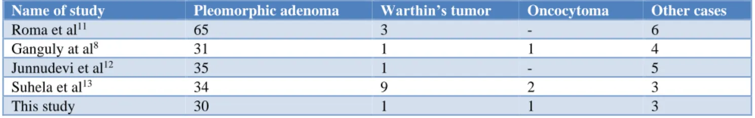
Table 9: Comparison of number of cases of various benign tumors with other studies.
Name of study Pleomorphic adenoma Warthin’s tumor Oncocytoma Other cases
Roma et al11 65 3 - 6
Ganguly at al8 31 1 1 4
Junnudevi et al12 35 1 - 5
Suhela et al13 34 9 2 3
This study 30 1 1 3
Most common benign neoplasm found was pleomorphic adenoma which was consistent with the above studies (Table 9). Pleomorphic adenoma is the most common tumor of the salivary gland and constitutes about 60% of all tumors of the salivary gland.
About 80% cases of PA develop in parotid gland, 10% in submandibular gland and the rest 10% cases in the paranasal sinuses, oral cavity, upper respiratory tract and skin.
It commonly presents as a solitary well-defined, slow growing and painless swelling. Mean age of the patient is around 46 years and females are commonly affected than male. Pleomorphic adenoma is also known as mixed tumor as it contains both epithelial and also mesenchymal or stromal material. The tumor possibly develops from the pluripotent reserve cells of the intercalated duct.
Table 10: Comparison of frequency of various malignant lesions with other studies.
Number of cases
Study MEC ACC AdCC Ca Ex PA Adenocarcinoma NOS Other cases
Roma et al11 8 1 4 1 1 4
Ganguly at al8 6 1 - 1 - 1
Junnudevi et al12 19 3 4 1 - -
Suhela et al13 9 3 5 1 - -
This study 10 3 1 1 1 -
Pleomorphic adenoma may show mild atypical cells in one-fifth of the cases. In absence of necrosis and significant changes in nuclear size, shape and chromatin pattern, these atypical cells should be ignored. However, the presence of necrosis should raise the possibility of carcinoma in PA.3 Most common malignant neoplasm of
salivary gland was Mucoepidermoid carcinoma which was correlated with the other studies (Table 10).
children to old, and mean age of the patient is 45 years.
There is mild female predilection of this tumor. About 50% of MEC arises from the major salivary gland and rest of the tumor arises from buccal mucosa and palate.
The patients usually present with painless, firm, fixed, mass. Mucoepidermoid carcinoma is divided on histology as low, intermediate and grade tumor. The high-grade MEC is aggressive compared to low-high-grade MEC.
Because of the presence of heterogenous population of cells, there is high risk of misdiagnosis. Inflammatory cells and macrophages, if numerous, may mask neoplastic epithelial cells, resulting in an erroneous diagnosis of an inflammatory lesion.
Aspirates from high-grade tumors, recognised as a carcinoma, may be difficult to differentiate from other high-grade carcinomas such as salivary duct carcinoma and adenocarcinoma, not otherwise specified.
Low grade MEC is the common source of under diagnosis. It may often be mistaken as Warthin tumor as seen in one case. Bland nuclear morphology, presence of oncocytes and reactive lymphoid cells favors a diagnosis of Warthin’s tumor.
Extensively keratinised malignant squamous cells when present, the possibility of squamous cell carcinoma, especially secondary involvement from an upper aerodigestive tract or lung tumor should be considered.
Rarely, aspirates from chronic sialadenitis may mimic a mucoepidermoid carcinoma. In sialadenitis, the ductal cell groups show straight edges with branching and contain scant cytoplasm with uniform nuclei. Squamous metaplasia may occur, but epithelial heterogeneity is not seen in sialadenitis.3
A single case of Warthin’s tumor reported in 80 years old man in FNAC. Cytological smear of it was showing oncocytic cells arranged in monolayered sheets, confused with oncocytoma, but presence of lymphoid population and background of protenaceous and cell debris helped in correct diagnosis.
Three cases of Acinic cell carcinoma had been reported. It is the malignant neoplasm of the epithelial cell with serous acinar cell differentiation and constitutes only 1-3% of the malignant salivary gland tumors. Females are slightly more affected. The peak incidence of the tumor is in third decades of life. About 80-90% of tumor arises in the parotid gland and a small group of tumor develops from the minor salivary glands. The patient classically presents as a single, mobile and painless mass.
Fine needle aspiration cytology smear is usually rich in cells. The cells are arranged in three-dimensional clusters and sheets in a clean background. Occasionally, follicular
or micropapillary structures may be noted. The cells may also be arranged around thin capillaries.
The individual cells are polygonal with abundant finely vacuolated cytoplasm. Nuclei are rounded, monomorphic, centrally placed with a small distinct nucleolus. The cytoplasmic vacuole is subtle and better seen in MGG stain. The vacuoles contain PAS positive diastase-resistant zymogen granules. The cytoplasmic vacuole is one of the important finding and should not be overlooked.3
A single case of oncocytoma was diagnosed in 50-year-old female on FNAC. FNAC smears of this showed presence of oncocytic epithelial cells which were arranged in acini, at places in papillary fragments and singly. Oncocytic cells were having abundant deeply basophilic granular cytoplasm and uniform round central nuclei with finely granular chromatin. The absence of lymphocytes and cellular debris helped in differentiating it from warthin’s tumor. Bland nuclear morphology, presence of oncocytes and reactive lymphoid cells favor a diagnosis of Warthin’s tumor.
Parotid gland was the most commonly involved gland followed by submandibular gland and minor salivary glands which was consistent with the other studies as shown above (Table 11).
Male: female ratio in all salivary gland neoplasm was 0.91:1 suggesting slightly female preponderance with the study of Roma et al,While in studies of Ganguly et al, Junnudevi et al, Suhel et al, male preponderance was seen.8,11-13
Table 11: Site wise distribution of salivary gland tumor.
Study name Parotid (%) Submandibular (%) Minor salivary gland (%)
Roma et al11 70 24 5
Junnudevi at al8 61.9 32.15 4.78
Suhela et al12 89.5 10.50 -
Vaishali et al13 80 16.67 3.33
This study 52.9 41.17 5.8
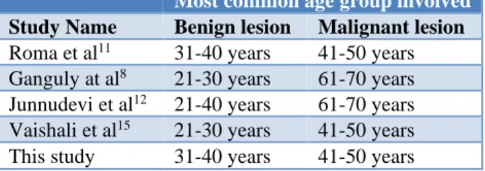
Common age group affected in benign lesion was 31-40 years followed by 21-30 years which is correlated with the study of Roma et al, and junnudevi et al.11,12 In the
study of Ganguly et al, and Vaishali et al, the most common age group affected by benign lesions was 21-30 years followed by 31-40 years (Table 12).8,15
The most common age group affected by malignant lesion was 41-50 years in this study which is correlated with the study of Roma et al, and Vaishali et al.11,15 In the
health awareness in this study group or may be due to
different sample size, different geographical factors.
Table 12: Comparison of commonest age group involved with other study.
Most common age group involved
Study Name Benign lesion Malignant lesion
Roma et al11 31-40 years 41-50 years
Ganguly at al8 21-30 years 61-70 years
Junnudevi et al12 21-40 years 61-70 years
Vaishali et al15 21-30 years 41-50 years
This study 31-40 years 41-50 years
Diagnostic accuracy for various neoplastic lesions by FNAC was 90.9% which is close to the diagnostic accuracy seen in above mentioned studies (Table 13).
Table 13: Comparison of diagnostic accuracy for neoplastic lesions of salivary gland by FNAC in
various studies.
Study Diagnostic accuracy
Roma et al11 96%
Junnudevi at al12 90.9%
Koonamalla et al14 93.8%
Vaishali et al15 96.66%
Sukesh et al16 89.47%
This study 90.9%
Table 14: Comparison with other studies for sensitivity, specificity and diagnostic accuracy of
salivary gland tumors by FNAC.
Study Diagnostic
accuracy Sensitivity Specificity
Roma et al11 96% 96.87% 100%
Junnudevi
at al12 90.9% 100% 87.5%
Koonamalla et
al14 93.8% 96.9% 96.8%
Vaishali et al15 96.66% 80.00% 100%
This study 90.9% 100% 87.5%
Diagnostic accuracy ranges from 90% to 97% in different studies carried out in country. Diagnostic accuracy was 90.9% which was correlated with the above studies.
The sensitivity ranges from 80 to 100% in different studies. In present study, sensitivity was 100% which was correlated quite well with the above studies.
The specificity ranges 87 to 100 % in different studies. In this study, specificity was 87.5% which is correlated with study of Junnudevi et al, and slightly lower than the specificities in other studies discussed above (Table 14).12
CONCLUSION
FNAC results had given us overall accuracy rate among neoplasm of 90.9%, sensitivity of 100%, and specificity of 87.5%. Taking into account the heterogeneity of salivary gland masses giving rise to varied and overlapping cytomorphological features and inevitable sampling errors, the diagnostic accuracy, sensitivity and specificity was appreciable. No major complications had been encountered in the present series.
FNAC of salivary gland tumors is advantageous for both clinicians and patients because of its immediate results, economy and accuracy. Inflammatory lesions like sialadenitis and lymphoepithelial cysts could be diagnosed with fair ease on FNAC and thus avoiding the psychological impact of malignancy on patient’s mind; thus, saving time and effort.
FNAC has got significant diagnostic value in differentiating benign from malignant lesions providing us valuable information for planning of subsequent therapeutic management and avoiding unwanted surgeries.
The FNAC procedure is safe, non-hazardous, minimally invasive, rapid, simple, repeatable, fairly reliable, outpatient-based procedure, carried out without advance preparation or anaesthesia and provide diagnosis within hours.
Funding: No funding sources Conflict of interest: None declared Ethical approval: Not required
REFERENCES
1. Welton TS. Biographical brevities: Stenson’s duct. Am J Surg. 1931;14:501.
2. Sidawy MK, Ali SZ. Fine Needle Aspiration Cytology. Foundations in Diagnostic Pathology. 1st
ed. Churchill Livingstone; 2007:1-34.
3. Dey P. Fine Needle Aspiration Cytology: Diagnostic cytology. 1st Ed. Jaypee
Brothers;2014.298-319.
4. Speight PM, Barrett AW. Salivary gland tumours. Oral Dis. 2002 Sep;8(5):229-40.
5. Mendenhall WM, Riggs CE Jr, Cassisi NJ: Treatment of head and neck cancers. In: DeVita Jr, Hellman S, Rosenberg SA, eds.: cancer: Principles and Practice of Oncology. 7th Ed. Philadelphia, Pa:
Lippincott Williams and Wilkins;2005:662-732. 6. Barnes L, Eveson JW, Reichart P, Sidransky D.
World Health Organization Classification of tumors: Pathology and genetics of head and neck tumours. 3rd Ed, Volume 9. IARC Press: Lyon 2005.213.
Oral Maxillofacial Pathol: JOMFP.
2017;21(2):203-10.
8. Ganguly S, Gupta R, Bagde R, Bhardwaj AK, Tiwari AK. Salivary gland lesions: A retrospective study of 100 cases of salivary swelling presenting for FNAC at cytopathology department, CIMS, Bilaspur. J Evidence Based Med Healthcare. 2015;2(44):7896-909.
9. Dalve KT, Swami SY, Rutuja LU, Narhire VV, Bakshi AP. Study of FNAC of salivary gland lesions in a tertiary care hospital. J Diagnostic Pathol Oncol. 2016;1(2):24-8.
10. Kambale T, Iqbal B, Patil A, Kumar H. Diagnostic role of FNAC in Salivary gland lesions and its histopathological correlation. Ind J Pathol Oncol. 2016;3(3):372-5.
11. Rajdeo RN, Shrivastava AC, Bajaj J, Shrikhande AV, Rajdeo RN. Clinicopathological study of salivary gland tumors: An observation in tertiary hospital of central India. Inter J Rese Med Sci. 2015;3(7):1691-6.
12. Devi J, Taludkar KL. Salivary gland neoplasms: A clinicopathological study of 84 cases. IAIM. 2015;2(4):70-7.
13. Rachakonda S, Gattu V. Study of morphological subtypes of major salivary gland tumours. PIMR.2017;5(3):24-8.
14. Devi KR, Toopalli K, Shravan KO. Cytohistological Study of Salivary Gland Lesions. Scholar J Appl Med Sci. 2016;4(7):2338-42.
15. Anand VH, Prajapati D, Dave KK. FNAC and histopathology of salivary gland tumours. SEAJCRR.2014;3(1):609-18.
16. Bhagavath P. Role of FNAC in diagnosing salivary gland lesions. Inter J AJ Institute Med Sci. 2012;1(2):118-24.