Jyothi V S et al JMSCR Volume 06 Issue 09 September 2018 Page 881
Systemic Co Morbidities, Visual Defect and Severity of Diabetic
Retinopathy in Diabetics at Presentation
Authors
Jyothi V S
1, Padmasree K M
2, Ranjini Kotancheri
31
Senior Resident, Dept of Ophthalmology, Government Medical College, Kottayam, Kerala, India 2
Assistant Professor, Dept of Ophthalmology, Government Medical College, Kottayam, Kerala, India 3
Associate Professor, Dept of Ophthalmology, Government Medical College, Calicut, Kerala, India. Corresponding Author
Padmasree K M
Assistant Professor, Dept of Ophthalmology, Government Medical College, Kottayam, Kerala, India- 686008
Phone No.:- 9495346541, Email: drpadmasree@gmail.com
Abstract
Aim: To assess systemic co morbidities, visual defect and severity of DR at presentation in diabetics in a
tertiary care center.
Methods: A cross sectional study conducted for a period of 18 months in 130 newly detected Diabetic Retinopathy cases (DR) in a tertiary care center. They were evaluated with a proforma for risk factors and staging of retinopathy was done with International Clinical Diabetic Retinopathy Disease Severity Scale.
Results and Discussion: The duration of diabetes had significant association with stage of DR at presentation (p=0.048). Co morbidities like hypertension, dyslipidemia, CVA, CAD had no significant association with the sage of DR (P=0.001). Previous cataract surgery had no association with the stage of DR at presentation. Patients with co-existing diabetic nephropathy had more prevalence of DR and they had more severe presentation at the beginning (p=0.004).
Conclusion: The study found significant association between stage of diabetic retinopathy and age at diagnosis, mean duration of diabetes, co morbid nephropathy and low BMI in advanced cases. The study opened up a window to intensify the need for early diagnosis and management of DR cases, because it was found that there is progression of stage of DR, and deterioration of visual acuity as the time passed by.
Keywords:diabetic retinopathy, risk factor, clinical profile, visual outcome.
Introduction
Diabetic Retinopathy (DR), a long-term complication of Diabetes Mellitus, is asymptomatic in its most treatable stages when the condition would be neglected due to ignorance or carelessness. Vision Threatening Diabetic Retinopathy (VTDR) is a major cause of blindness
among the working age group1,2,3,4. The estimated high prevalence of diabetes by 2030 in India is a matter of concern considering the potential of vision loss associated with increase in incidence of diabetic retinopathy. However, progression of diabetic retinopathy can be delayed by early detection of diabetes, proper screening to detect
www.jmscr.igmpublication.org Impact Factor (SJIF): 6.379
Jyothi V S et al JMSCR Volume 06 Issue 09 September 2018 Page 882 retinopathy in early stages and timely treatment of
the same, along with control of other contributing factors. Thus, timely detection and treatment can significantly reduce visual impairment, and thus improve quality of life. So, there is a need to assess the stage of retinopathy at diagnosis and the different factors contributing to the development of retinopathy. Although many epidemiologic studies conducted worldwide have clearly outlined the clinical-epidemiological profile of DR at presentation this study will give regional data which would be helpful to reassess the current status, identify any changing trends, create awareness among the study population and would serve as a baseline data for future research.
Materials and Methods
The present study was conducted with an objective of (1) To assess visual defect, systemic comorbidities and severity of DR at the time of presentation (2) To describe the clinical profile of DR at the time of diagnosis. Sample size was calculated from a study conducted by Singh P et al5 assessing prevalence of DR in diabetic patients of Vindhya region; 69.44% of DR cases were in pre-proliferative stage. A cross sectional study of 127 newly diagnosed diabetic retinopathy cases who attended OPD formed the sample. The study was conducted for a period of 18 months at a tertiary care center. Diabetic retinopathy cases (1) on treatment and follow up, (2) with significant hazy media preventing staging of retinopathy and (3) Diabetics with other retinal disorders like age related macular degeneration, hypertensive
retinopathy, venous occlusions, primary open angle glaucoma, optic atrophy etc. were excluded from the study.
Procedure
After getting Institutional review board clearance, those who gave informed consent were evaluated by the principal investigator with the help of a structured proforma for socio demographic and disease related variables. Height and weight of the subjects were measured. Blood pressure recorded in the right arm in the sitting posture after taking rest for a period of fiveminutes. Comprehensive ocular examination was done. Fundus evaluation with indirect ophthalmoscopy with 20D lens and slit lamp biomicroscope using 90D lens was done. ICDRS classification were used to classify Diabetic retinopathy and Diabetic Macular Edema.
Fundus Fluorescein Angiography and Optical Coherence Tomography were used in cases with macular edema, suspicious macular ischemia, doubtful proliferative retinopathy and also to rule out other diagnosis.
Data was analyzed with IBM SPSS version 16 software. The study variables were assessed using chi-square test (for qualitative variables) and t test/ANOVA (for quantitative/continuous variables). The level of statistical significance was taken as p value less than 0.05.
Results
Results are presented in Table 1.
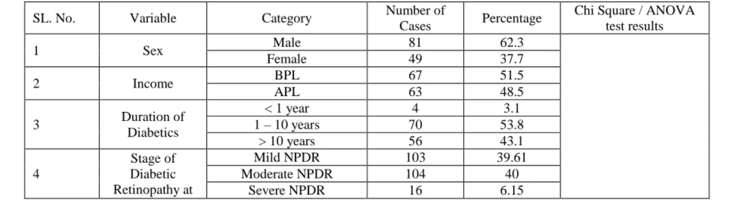
Table 1 Incidence of Variables, Categories, Number of cases, Percentage, Chi square and ANOVA Test Results
SL. No. Variable Category Number of
Cases Percentage
Chi Square / ANOVA test results
1 Sex Male 81 62.3
Female 49 37.7
2 Income BPL 67 51.5
APL 63 48.5
3 Duration of
Diabetics
< 1 year 4 3.1
1 – 10 years 70 53.8
> 10 years 56 43.1
4
Stage of Diabetic Retinopathy at
Mild NPDR 103 39.61
Moderate NPDR 104 40
Jyothi V S et al JMSCR Volume 06 Issue 09 September 2018 Page 883
presentation PDR 37 14.23
5 Stage of DME
at presentation
No 137 58
Mild 58 22.3
Moderate 60 23.07
Severe 5 1.92
6 Status of Iris
(NVI)
Present 12 4.6
Absent 248 95.4
7 Status of Lens
Normal 166 63.84
Cataract 74 28.46
IOL 20 7.69
Chi Square Test Results
8
Nephropathy
Mild NPDR 1 9.1
X2= 13.25 P=0.004
P<0.05
Moderate NPDR 2 18.2
Severe NPDR 4 36.4
PDR 4 36.4
No Nephropathy
Mild NPDR 48 40.3
Moderate NPDR 46 38.7
Severe NPDR 8 6.7
PDR 17 14.3
9
History of Cataract Surgery
Mild NPDR 25
X2= 12.02 P=0.061
p>0.05
Moderate NPDR 62.5
Severe NPDR 12.5
PDR 0
No History of Cataract Surgery
Mild NPDR 43.4
Moderate NPDR 40.7
Severe NPDR 1.8
PDR 14.2
10
Hypertension
Mild NPDR 32.7
X2=3.553 P=0.314 P<0.05
Moderate NPDR 48.2
Severe NPDR 3.44
PDR 15.5
No Hypertension
Mild NPDR 48.61
Moderate NPDR 38.88
Severe NPDR 2.77
PDR 9.72
11
Dyslipidemia
Mild NPDR 33.33
X2=5.781 P=0.123 P<0.05
Moderate NPDR 41.02
Severe NPDR 2.56
PDR 23.07
No Dyslipidemia
Mild NPDR 45.05
Moderate NPDR 43.95
Severe NPDR 3.29
PDR 7.69
12 Activity
Active 6.2 X2= 13.25
P=0.004 p>0.05
Moderate 84.6
Sedentary 9.2
ANOVA Test results
Mean ANOVA
13 Visual Acquity
Mild NPDR 0.0611
P= 0.00 P<0.5
Moderate NPDR 0.075
Severe NPDR 0.3143
PDR 0.7875
14 Mean BMI
Mild NPDR 24.55
P=0.001 P<0.5
Moderate NPDR 25.36
Severe NPDR 26.95
PDR 23.2
15 Mean Hb
Mild NPDR 13.43
P=0.00 P<0.5
Moderate NPDR 14.04
Severe NPDR 13.73
PDR 12.32
16 Mean HbA1C
Mild NPDR 6.708
P=0.00 P<0.5
Moderate NPDR 7.283
Severe NPDR 7.3
Jyothi V S et al JMSCR Volume 06 Issue 09 September 2018 Page 884
Discussion
The global increase of diabetes has a significant impact on the prevalence of diabetic complications among which diabetic retinopathy takes an important place. DR is a leading cause of acquired blindness in working age adults and has been estimated to represent 12% of blindness in developed countries. In the study the stage of diabetic retinopathy at which patients present to us for the first time was found out and the associated risk factors of DR were also assessed. The present study indicates that factors like age, duration of DM, nephropathy, and BMI are influencing DR. Male gender was observed to be associated with the presence of DR, but not its severity. In this study out of 130 patients 81 (62.3%) were males and 49 (37.7 %) were females. Similar observations were made by Pradeepa et. al.6 in an urban Indian population. In a study conducted by Rani P K et al7 in Southern districts of Tamil Nadu male gender was considered as a risk factor associated with development of any DR. The age group of present study ranged from 25 to 72 years with the maximum patients (37.7%) coming under the range of 60 to 70 years. Results indicates that increasing age was significantly associated with increasing risk of diabetic retinopathy(p<.05). Of the 130 patients 4 (3.1%) had duration of DM less than 1 year, 70 (53.8%) had duration of DM between 1 to 10 years and 56(43.1%) had duration more than 10 years. The relation between duration of DM and development of DR was statistically significant both in the 1 to 10 years group (p=.04) and more than 10 years group (p=.03).It shows that duration of diabetes is an important risk factor for development of diabetic retinopathy. Similar findings were seen in the studies conducted in Andhra Pradesh by Sannapaneni Krishnaiah et al8 and also by Rani P K et al7.
According to the income status patients were divided into two categories APL and BPL; APL were48.5% and BPL were 51.5%. The present study indicates that socio economic strata is not influencing DR occurrence. The study sample included 6.2 % of physically active subjects, 84.6
% of moderately active subjects, and 9.2 % with sedentary lifestyle. DR was found to be highest in moderately active group irrespective of its stages. Present study further substantiates the fact that DR is a complication found in active working population. This finding is contradicting with that of Anna Praidou et al9 which indicates that lack of physical activity is a risk factor in the development of DM as well as DR.
At presentation out of 260 eyes of 130 cases 39.61% (103 eyes) showed mild NPDR changes, 40% (104 eyes) showed moderate NPDR changes, 6.15% (16 eyes) showed severe NPDR changes and 14.23% (37 eyes) showed PDR changes. Sannapaneni Krishnaiah et al. ‘s8 population-based study in the state of Andhra Pradesh in India found out that the diabetic retinopathy is more in mild NPDR (51.3%),then moderate NPDR (35.9%), andproliferative retinopathy (2.6%) stages. Narendran et al’s10 population-based prevalence survey of DR in Palakkad district of Kerala also showed similar finding. NPDR (94.1%) was the most common form of retinopathy seen. So, the stage of DR at presentation is comparable to the findings obtained in other similar studies.
The relation between comorbidities like hypertension, dyslipidemia, neuropathy, nephropathy, CAD, CVA etc and stage of DR was assessed. Only nephropathy showed a statistically significant relation with diabetic retinopathy (p=.004).72.8% of patients with nephropathy had severe stages of retinopathy at presentation. Hypertension was considered as established risk factor for the progression of DR according to a study by Krishnaiah8 in the state of Andhra Pradesh. But in the current study no significant association could be found between hypertension and DR (p>.05). Narendran et al in Palakkad district of Kerala also presented a similar finding (p=.658).
Jyothi V S et al JMSCR Volume 06 Issue 09 September 2018 Page 885 stage of DR showed a statistically significant
relation (p=.001). The PDR stage showed a mean BMI of 23.2. The present study is more in accordance with Rani PK7 which projects Lean BMI as a significant risk factor associated with development of retinopathy. This inverse relation between BMI and DR could be due to the catabolic effect of lack of insulin over a long duration of hyperglycemia, resulting in lean individuals. There are evidences that South Asians have abdominal obesity despite normal BMIs, the so called “Asian Indian phenotype”; which may explain the paradox11. Further, it is interesting to note that in Caucasians, high BMI is observed in DM subjects, but in Asian population subjects with DM are lean12.This may be linked to the fact that Asians with type 2 DM show lesser insulin secretion, but greater insulin sensitivity as compared to Caucasian diabetic patients.13,14. But in another study by Narendran11 in Southern India, BMI was found to be not associated with DR (p=0.317)
The status of iris and lens at presentation were assessed in the study and 4.6% of eyes had neovascularization of iris and 95.4% had no neovascularization of eyes at the time of presentation. The status of lens was assessed and 166 eyes (63.84%) were normal, 74 eyes (28.46%) had cataract and 20 eyes (7.69%) had intraocular lens implanted. The association between cataract surgery and stage of retinopathy was assessed and found to be not significant in the study. However, cataract surgery was found to have a significant correlation with progression of diabetic retinopathy in a study conducted by Chew EY, et al15 in the National Institute of Health, Bethesda. The proportion of DME cases at presentation was found out from different stages of DR and it was found to be no DME in 52.69%, mild DME in 22.3 %, moderate DME in 23.07 % and severe DME in 1.92%. Similar finding of diabetic maculopathy was observed in a study carried out in Department of Ophthalmology of Douala General Hospital, Cameroon by Ahmadou M Jingi et al.16. The mean visual acuity at presentation was
assessed for different stages of retinopathy and was found to be .0611 for mild, .0750 for moderate, .3143 for severe NPDR and .7875 for PDR cases which showed a statistically significant decrease in mean visual acuity as DR progressed with a p value of p=.000.Similar risk association of DR with moderate visual impairment was found in accordance with studies conducted by Rani PK and Raman R7.
Mean hemoglobin and HbA1C levels in the blood was calculated at the time of presentation and its association with stage of DR was found out. Mean Hb at presentation was 13.43mg/dl for mild, 14.04mg/dl for moderate, 13.73mg/dl for severe NPDR and 12.32mg/dl for PDR cases which had a significant p value (p= .00) with mean hemoglobin least for severe cases. Such a significant association was not found in reviewed studies. Nephropathy may be the contributory factor for this association . Mean HbA1C levels also showed a significant relation with the stage of DR with p value <.05. The mean values of HbA1C were 6.708 for mild NPDR, 7.283 for moderate NPDR, 7.3 for severe NPDR and 8.457 for PDR cases. The mean HbA1C were lesser for mild cases and it increased as the stage of DR worsened. A significant association was found between HbA1C levels and DR in a study conducted by Axer- Siegel and Herscovici in Israel17.
Conclusion
Jyothi V S et al JMSCR Volume 06 Issue 09 September 2018 Page 886 retinopathy. Other factors like dyslipidemia,
hypertension, CAD, CVA was found to have no significant association. In our study it was found that a lean BMI has significant association with advanced stages of DR, which was shown in few studies conducted in India and is a significantly different finding from western countries were the patient is more obese. This interesting disparity was found to be due to relative insulin deficiency in Asian population compared to those in the west which leads to an increased catabolism and low BMI. Only few had neovascularization of iris (4.6%) at presentation, which is an indication of less severity at the beginning. It was found that there is no significant association between cataract surgery and DR. It was found that visual acuity is worsening with progression of retinopathy. So, it is possible to halt the progress of retinopathy and resulting visual impairment with early identification and treatment. Almost 50% had DME at presentation. These patients can be properly assessed and treated resulting in better prognosis. The study opens our view towards the need for regular screening, early detection and management of retinopathy for preventing blindness.
Source of support: Nil
References
1. Wild S, Roglic G, Green A, et al. Global prevalence of diabetes: estimates for the year 2000 and projections for 2030. Diabetes Care 2004;27:1047–53.
2. Eye Diseases Prevalence Research Group. Causes and prevalence of visual impairment among adults in the United States. Arch Ophthalmol 2004; 122:477– 85.
3. Eye Diseases Prevalence Research Group. The prevalence of diabetic retinopathy among adults in the United States. Arch Ophthalmol 2004;122:552– 63.
4. Ramachandran A, Jali MV, Mohan V, et al. High prevalence of diabetes in an urban
population in South India. BMJ 1988; 297:587–90.
5. Singh P., Wadhwani, E., Gupta R., et al. Prevalence of Diabetic Retinopathy in Diabetic Patients of Vindhya Region. International Journal of Scientific and Research Publications 2012;2 (4) 1-3 6. Pradeepa R Anitha B, et al.Risk factors for
diabetic retinopathy in a South Indian diabetic population –the Chennai Urban Rural Epidemiological Study (CURES) Eye Study4. Diabetes Med 2008;25:536-42.
7. A Rani P K Raman R, Chandrakantan A et al. Risk factors for diabetic retinopathy in self reported rural population with diabetes. J Postgrad Med 2009;55:92-96. 8. Sannapaneni Krishnaiah, Taraprasad Das,
Praveen K Nirmalan et al. Risk factors for diabetic retinopathy: Findings from The Andhra Pradesh Eye Disease Study. Clinical Ophthalmology 2007;1(4):475-482.
9. Praidou, Anna & Harris, Martin &Niakas, Dimitris &Labiris, Gregorios.(2016) Physical activity and its correlation to diabetic retinopathy. Journal of Diabetes and its Complications. 31.10.1016 / j.jdiacomp.2016.06.027.
10.V Narendran,R K John,A Raghuram et al. Diabetic retinopathy among self reported diabetics in Southern India:a population based assessment. British Journal of Ophthalmology2002;86:1014-1018.
11.Raji A,Seely EW et al.Body fat distribution and insulin resistance in healthy Asian Indians and Caucasians. J Clin Endocrinol Metab 2001;86:5366-71 12.Deurenberg P, Yap M et al.Body mass
index and percent body fat :A meta-analysis among different ethnic groups .Int J Obes Relat Metab Disord 1998;22:1164. 13.Yoshiike N, Matsmura Y et al. Descriptive
Jyothi V S et al JMSCR Volume 06 Issue 09 September 2018 Page 887 from the National Nutrition Survey
1990-1994.Int J Obes Relat Metab Disord 1998; 22:684-7.
14.Sone H, Ito H et al. Japan Diabetes Complication Study Group: Obesity and type 2 diabetes in Japanese patients. Lancet 2003;361:85
15. Chew EY, Benson WE et al. Results after lens extraction in patients with diabetic retinopathy :Early Treatment Diabetic Retinopathy study report number2.Arch Ophthalmol 1999;117(12)1600-6
16.Ahmadou M Jingi, Jean Jacques N Noubiap et al. Epidemiology and treatment outcomes of diabetic retinopathy in a diabetic population from Cameroon. BMC Ophthalmology 2014,14:19.