Original Research Article
Dissection of posterior triangle and lower deep jugular lymph node is
mandatory in therapeutic neck dissection as a part of treatment for
squamous cell carcinoma of oral cavity with clinically N
1neck
Shilpa R.
1*, Azeem Moyihuddin
2INTRODUCTION
Oral cancer is the sixth most common cancer in the world and is largely preventable. 1 It accounts for approximately 4% of all cancers and 2% of all cancer deaths world-wide. 2 Besides paan chewing, the effects of tobacco use and alcohol drinking are clear risk factors for oral cancer in India and elsewhere.3-5 Among Indian men, the attributable oral cancer risk due to smoking, alcohol and paan chewing is over 80% and among women in India,
paan chewing alone explains almost all (over 90%) the oral cancer risk.6-8
Malignancy within oral cavity is potentially devastating due to the associated morbidity. Therefore early detection and appropriate treatment of cancers remains the most effective weapon against cancers of the oral cavity. A critical prognostic factor in head and neck cancer is spread of disease to regional lymph node.
It was Crile in 1906, who described a systematic operative procedure for removal of cervical lymphatics
ABSTRACT
Background: In India oral cancer is the commonest malignant neoplasm, accounting for 20-30% of all cancers. Southern India presents the highest oral cancer incidence rates among women worldwide.
Methods: This study was conducted in R. L. Jalappa Hospital and Research Centre and SDU Medical College Kolar, Karnataka. Thirty patients having oral squamous cell carcinoma with clinically N1 neck undergoing modified radical
neck dissection between December 2010 and June 2012 were enrolled in the study. The objective of study was to determine whether dissection of posterior triangle and lower deep jugular lymph node is mandatory in therapeutic neck dissection as a part of treatment for squamous cell carcinoma of oral cavity with clinically N1 neck.
Results: Out of 24 patients, 16 patients underwent wide excision with hemimandibulectomy. In these 16 cases, 2 patients had reconstruction with double flap while rest 14 cases with island pectoralis major myocutaneous flap. Out of remaining 8 patients, 2 patients underwent marginal mandibulectomy. In all these 8 patients, reconstruction was done using nasolabial flap in 1 patient, buccal pad of fat in 2 patients, masseter flap in 1 patient and forehead flap in 4 patients. In carcinoma anterior 2/3rd tongue, all 6 patients underwent hemiglossectomy with simultaneous modified radical neck dissection.
Conclusions: It was concluded that during neck dissection, it may be oncologically safe to avoid level IV and level V clearance in buccal mucosa squamous cell carcinoma with N1 neck.
Keywords: Modified radical neck dissection, Functional neck dissection, Squamous cell carcinoma, Posterior triangle lymph nodes, Lower deep jugular
1Department of ENT, Koppal Institute of Medical Sciences, Koppal, Karnataka, India 2
Department of ENT, Sri Devaraj Urs Academy of Higher Education and Research, Kolar, Karnataka, India
Received: 18 August 2019
Revised: 01 October 2019
Accepted: 03 October 2019
*Correspondence:
Dr. Shilpa R.,
E-mail: itsshilpahere@gmail.com
Copyright: © the author(s), publisher and licensee Medip Academy. This is an open-access article distributed under the terms of the Creative Commons Attribution Non-Commercial License, which permits unrestricted non-commercial use, distribution, and reproduction in any medium, provided the original work is properly cited.
termed „Radical neck dissection‟ which was standardised by Martin (Father of modern head and neck surgery) and his associates.9,10
As described by Crile in 1906, radical neck dissection (RND) required complete removal of lymph nodes from level I to V along with sternocleidomastoid (SCM) muscle, internal jugular vein (IJV) and spinal accessory nerve (SAN). By sacrificing the SAN, patients suffered from the “Eleventh nerve syndrome” or “Shoulder syndrome” characterized by shoulder droop, winged scapula, weak abduction, inability to shrug and dull ache with pain localized to shoulder.18-22 Earlier efforts to treat this problem involved rehabilitation of the functionally impaired extremity, the results of which were not very encouraging. This led to modifications to the classic RND.11
Pioneers like Suarez from Argentina, Ballantyne from North America and Bocca from Italy, began to explore surgical alternatives that would be oncologically sound but preserve important functional and anatomical structures in the neck. These variations in the surgical procedures were categorised as modified radical neck dissection (MRND) or functional neck dissection (FND).
12
The basis of modified radical neck dissection was that the whole lymphatic system of the neck lies within fascial compartments which can be removed without sacrificing the non-lymphatic structures. By 1980‟s, several concepts played an important role in the emergence of selective neck dissection which preserves the SAN, SCM and IJV and resects only those nodal levels most likely to be involved with tumour, based on location of the primary tumour as lymph node metastasis in neck has a predictable pattern. However selective neck dissection (SND) is used only in N0 neck and its use in N1 neck is
still under evaluation.
In our country, most of the patients who undergo MRND or SND are manual labourers. Hence the integrity of the spinal accessory nerve is all the more important as the shoulder dysfunction caused by the damage to the nerve will directly affect the day to day earnings of the patient.
Hence this cross-sectional study was undertaken to determine whether dissection of posterior triangle and lower deep jugular lymph node is mandatory in therapeutic neck dissection as a part of treatment for squamous cell carcinoma of oral cavity with clinically N1
neck.
METHODS
This prospective study was conducted in R. L. Jalappa Hospital and Research Centre and SDU Medical College Kolar, Karnataka. Thirty patients having oral squamous cell carcinoma with clinically N1 neck (single ipsilateral
lymph node less than 3cms in diameter) undergoing
modified radical neck dissection in R. L. Jalappa Hospital and Research Centre between December 2010 and June 2012 were enrolled in the study. Permission for the study was obtained from the college authorities prior to commencement. Written informed consent taken for inclusion in the study, surgical excision of primary tumour, modified radical neck dissection and histopathological examination.
Inclusion criteria
Patients with histologically proven oral squamous cell carcinoma with clinically single ipsilateral lymph node less than 3 cms in greatest diameter (N1) were included in
the study.
Exclusion criteria
Patients with no palpable lymph nodes (N0 neck) with
oral squamous cell cancers, oral squamous cell cancer patients treated by other methods such as radiotherapy or chemotherapy, patients with oral cancer with advanced cervical lymph node metastasis (N2, N3), patients with
non-squamous malignancies of oral cavity, patients unfit for surgery (neck dissection) and patients not willing for surgical treatment
Following surgical excision of the primary lesion along with simultaneous neck dissection (modified radical), contents of posterior triangle and lower deep jugular lymph nodes along with other dissected lymph nodes was sent for histopathology after marking the various lymph node levels
Pathological assessment of metastatic nodes: Lymph nodes were identified by visual inspection and palpation and were dissected out from the fixed gross specimen in each of the five anatomic levels. All nodes were measured and processed routinely. Histological assessment was made on a single hilar section with examination of step serial sections in selected nodes.
Metastasis to posterior triangle and lower deep jugular lymph nodes and their size were documented. In addition documentation of other criteria in the primary tumor which affect lymph node metastasis like T-stage, histological grade and presence of other positive lymph nodes was done.
Data analysis
The data was analysed using Microsoft Excel and presented in numbers and percentage.
RESULTS
In our study, the majority of primary tumours were buccal mucosa tumours (24). We had 6 anterior 2/3rd tongue tumours. The primary tumour staging included 17 T2 lesions (57%) (Table 2).
Table 1: Demographic data of patients.
Demographic No. of patients %
Sex
Male 4
Female 26 86.66
Age (in years)
40-50 14 46.66
51-60 12 40
61-70 04 13.33
Table 2: Distribution of primary tumour.
Site of primary Buccal mucosa Tongue
N (%) N (%)
No. of patients (n=30) 24 (80) 6 (20)
3 T3 lesions (10%), 10 T4 lesions (33%). All patients in
our study selected were having N1 neck. In our study, in
patients with buccal mucosa carcinoma, fourteen patients had T2 disease, nine patients had T4 and one patient had
T3 disease. In patients with carcinoma lateral border
tongue, three patients had T2 disease, two patients had T3
and one patient had T4 disease.
Table 3: Surgery done.
Treatment of the primary tumour Number of cases
N (%)
Buccal mucosa carcinoma Wide excision 24 (80)
Tongue carcinoma Hemi glossectomy 6 (20)
Neck dissection MRND (functional neck dissection) 28 (93.33)
MRND (sternomastoid sacrificed) 2 (6.66)
Reconstruction in buccal mucosa carcinoma
Nasolabial flap 1
PMMC+DP 2
Buccal pad of fat 2
Masseter flap 1
Forehead flap 4
Island PMMC 14
Hemimandibulectomy 16
Marginal mandibulectmy 2
In patients with carcinoma lateral border tongue, three patients had T2 disease, two patients had T3 and one
patient had T4 disease.
In buccal mucosa carcinoma, out of 24 patients, 16 patients underwent wide excision with hemimandibulectomy. In these 16 cases, 2 patients had reconstruction with double flap (DP+PMMC) while rest 14 cases with island pectoralis major myocutaneous (PMMC) flap. Out of remaining 8 patients, 2 patients underwent marginal mandibulectomy. In all these 8 patients , reconstruction was done using nasolabial flap in
1 patient, buccal pad of fat in 2 patients, masseter flap in 1 patient and forehead flap in 4 patients. In carcinoma anterior 2/3rd tongue, all 6 patients underwent hemiglossectomy with simultaneous modified radical neck dissection (Table 3).
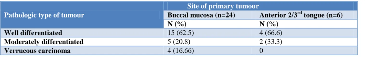
Histopathologic examination of the tumour specimens in our study revealed squamous cell carcinoma in 26 patients and verrucous variant of squamous cell carcinoma in 4 patients. Majority of our patients belonged to well differentiated squamous cell carcinoma (19 out of 30) (Table 4).
Table 4: Pathologic distribution of primary tumour.
Pathologic type of tumour
Site of primary tumour
Buccal mucosa (n=24) Anterior 2/3rd tongue (n=6)
N (%) N (%)
Well differentiated 15 (62.5) 4 (66.6)
Moderately differentiated 5 (20.8) 2 (33.3)
Verrucous carcinoma 4 (16.66) 0
Among 13 pathologically proven metastatic cases, 10 patients with buccal mucosa carcinoma had lymph nodes showing squamous cell deposits at level I and II. None of
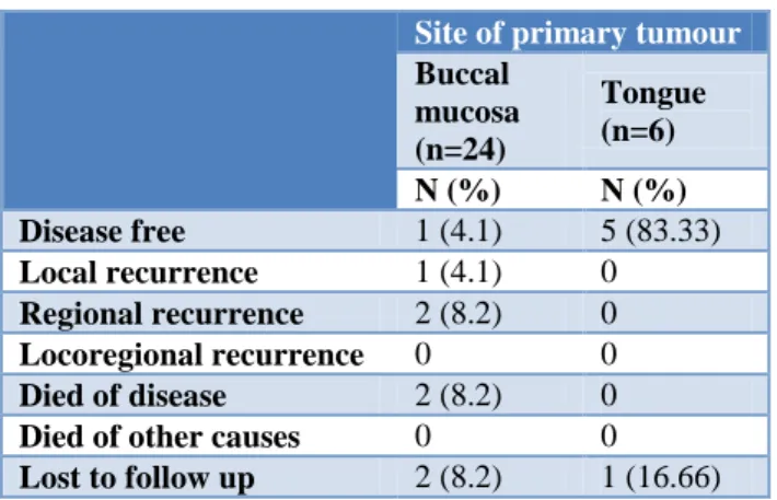
Within 6 months from the date of surgery, 3 patients had recurrence. Out of 3 recurrences, 1 patient had local recurrence while 2 patients (T3 tongue tumour and T4
buccal mucosa cancer) had regional recurrence at nodal level II. 1 patient (regional recurrence in buccal mucosa cancer patient) was salvaged by radical neck dissection out of 3 recurrences while 2 patients died of disease (Table 5).
Table 5: Showing post op follow up of patients.
Site of primary tumour
Buccal mucosa (n=24)
Tongue (n=6)
N (%) N (%)
Disease free 1 (4.1) 5 (83.33)
Local recurrence 1 (4.1) 0
Regional recurrence 2 (8.2) 0
Locoregional recurrence 0 0
Died of disease 2 (8.2) 0
Died of other causes 0 0
Lost to follow up 2 (8.2) 1 (16.66)
DISCUSSION
Out of 30 patients, four were males (13%) and the majority (i.e., twenty six) was females (87%). This shows that oral cancers are more common among females in this region. This can be attributed to the habit of chewing tobacco, beetle nuts and keeping a cud in the mouth. In literature, Southern India presents the highest oral cancer incidence rates among women worldwide and the highest in India overall.1,6-8 These very high incidence rates in Indian population reflect the continued prevalence of pan chewing in India, a habit which is equally common in both genders.2,6-8
Owing to their addiction to chewing beetle nuts and tobacco and keeping a cud, buccal mucosa cancer is by far the most common malignancy in this region.
In buccal mucosa carcinoma, out of 24 patients, 16 patients underwent wide excision with hemimandibulectomy. In these 16 patients, 2 patients had reconstruction with double flap (DP+PMMC) while rest 14 cases with island pectoralis major myocutaneous (PMMC) flap. Out of remaining 8 patients, 2 patients underwent marginal mandibulectomy. In all these 8 patients, reconstruction was done using nasolabial flap in 1 patient, buccal pad of fat in 2 patients, masseter flap in 1 patient and forehead flap in 4 patients. In carcinoma anterior 2/3rd tongue, all 6 patients underwent hemiglossectomy with simultaneous modified radical neck dissection.
Hemimandibulectomy was done whenever tumour was involving posterior most region of buccal mucosa or involving the bone. Marginal mandibulectomy was done
when tumour was reaching lower alveolus without infiltrating bone. Hemiglossectomy was done in 6 patients of tongue cancer as they were well lateralised not extending to midline or base tongue.
Table 6: Literature reports on prevalence of level IV and V lymph node metastasis in N1 neck.
Various studies Buccal mucosa cancer
Level IV (%) Level V (%) Shah et al14 3 0
Woolgar15 0 0
Our study 0 0
Table 7: Various studies for discussion.
Various studies Anterior 2/3rd tongue cancer
Level IV (%) Level V (%)
Shah et al14 15 7
Byer et al16 15.8 0
Nitya et al17 15.2 0
Our study 16.6 0
Prognosis depends on tumour primary site, nodal involvement, depth of tumour, and the status of the surgical margins. Also, the cumulative effects of tobacco, betel nut and alcohol decrease the survival rate. TNM system is a good indicator of tumour prognosis.13 Prognosis is better in early cancers, particularly those that are well-differentiated.
Most of the patients in this study had well differentiated squamous cell carcinoma (60%). This is because majority of the patients had buccal mucosa cancer. 10% showed moderately differentiated squamous cell carcinoma. 13.3% showed verrucous carcinoma. Recurrence was associated with moderately differentiated tumours.
Considering the above observations and results of our study, metastasis to posterior triangle nodes in oral cancer with N1 neck was 0% in buccal mucosa and tongue.
However, metastasis at level IV in tongue cancers with N1 neck is higher (16.6%) in our study. It was not an
isolated skip metastasis and this patient was also found to have metastasis at level II.
CONCLUSION
It was concluded that during neck dissection, it may be oncologically safe to avoid level IV (supraclavicular) and level V (posterior triangle) clearance in buccal mucosa squamous cell carcinoma with N1 neck. However, tongue
dissection (MRND) in carcinoma tongue. In our study, since the sample size is too small, it may be feasible after multi institutional studies with greater number of patients to formulate a definite protocol on posterior triangle and supraclavicular node clearance in oral cancers with clinically N1 neck.
ACKNOWLEDGEMENTS
The authors would like to express their profound gratitude to all the participants.
Funding: No funding sources Conflict of interest: None declared
Ethical approval: The study was approved by the Institutional Ethics Committee
REFERENCES
1. Franceschi S, Bidoli E, Herrero R, Muoz N. Comparison of cancers of the oral cavity and pharynx worldwide: etiological clues. Oral Oncol. 2000;36:106-15.
2. IARC. Monographs on the Evaluation of Carcinogenic Risk of Chemicals to Humans. Tobacco Habits other than Smoking; Betel-quid and Areca-nut Chewing, and Some Related Nitrosamines. Lyon: International Agency for Research on Cancer; 1985: 37.
3. Znaor A, Brennan P, Gajalakshmi V, Mathew A, Shanta V, Varghese C, et al. Independent and combined effects of tobacco smoking, chewing and alcohol drinking on the risk of oral, pharyngeal and esophageal cancers in Indian men. Int J Cancer. 2003;105:681-6.
4. WCRF/AICR (World Cancer Research Fund and American Institute/Cancer Research). Food, Nutrition and the Prevention of Cancer A Global Perspective. Washington, DC: American Institute of Cancer Research; 1997: 96-106.
5. Balaram P, Nandakumar A, Rajkumar T, Vaccarella S, Herrero R, Nandakumar A, et al. Oral cancer in southern India: the influence of smoking, drinking, paan chewing and oral hygiene. Int J Cancer. 2002;98:440-5.
6. Sunny L, Yeole BB, Hakama M, Shiri R, Sastry PS, Mathews S, et al.Oral Cancers in Mumbai, India: a
fifteen years perspective with respect to incidence trend and cumulative risk. Asian Pacific J Cancer. 2004;5:294-300.
7. Thorat RV, Panse NS, Budukh AM, Dinshaw KA, Nene BM, Jayant K.Prevalence of Tobacco Use and Tobacco-dependent Cancers in Males in the Rural Cancer Registry Population at Barshi, India. Asian Pacific J Cancer Prev. 2009;10:1167-70.
8. Kalyani R, Das S, Bindra S, Kumar HML. Cancer profile in Kolar: a ten years study. Indian J Cancer. 2010;47:160-5.
9. Carl ES, Alessandra R, Ferlito A. Crile‟s neck dissection. Laryngoscope. 2007;117:1974 -7. 10. Martin H, Del Valle B, Ehrlich H, Cahan WG. Neck
dissection. Cancer. 1951;4(3):441-99.
11. Harish K. Neck dissections: radical to conservative. J Surg Oncol. 2005; 3:1-13.
12. Bocca E, Pignataro O, Sasaki TC. Functional neck dissection. Arch Otolaryngol. 1980;106:524-7. 13. Head and neck sites. In: Green FL, Page DL,
Fleming ID, eds. The AJCC Cancer Staging Manual. 6th ed. New York: Springer Verlag; 2002: 17-31.
14. Shah JP. Patterns of cervical lymph node metastasis from squamous cell carcinomas of the upper aerodigestive tract. Am J Surg. 1999;160:405-9. 15. Woolgar JA. Histological distribution of cervical
lymph node metastases from intraoral or oropharyngeal squamous cell carcinomas.Br J Oral Maxillofac Surg. 1999;37:175-80.
16. Byers RM, Weber RS, Andrews T, McGill D, Kare R, Wolf P. Frequency and therapeutic implications of “skip metastases” in the neck from squamous carcinoma of the oral tongue. Head Neck. 1997;19(1):14-9.
17. Nitya CS, Pandey M, Naik BR, Ahamed M. Patterns of cervical metastases from carcinoma of the oral tongue. World J Surg Oncol. 2003;1:1-6.
Cite this article as: Shilpa R, Moyihuddin A.Dissection of posterior triangle and lower deep jugular lymph node is mandatory in therapeutic neck dissection as a part of treatment for squamous cell carcinoma of oral cavity with clinically N1 neck. Int J Otorhinolaryngol Head Neck