International Research Journal of Pharmaceutical and Biosciences(IRJPBS) 4 (7) 1
RESEARCH ARTICLE
International Research Journal of Pharmaceutical and Biosciences
Pri
-
ISSN: 2394 - 5826http://www.irjpbs.com e-ISSN: 2394 - 5834
In-vitro Antidiabetic and Anti-Microbial activity of Silver
nanoparticles synthesized using Medicinal plant -
Aegle
marmelos
R.Bhavani, V.Sivakumar and D.Prabakaran
PG and Research Department of Biochemistry, Adhiparasakthi College of Arts and Science, Kalavai – 632 513. Vellore district, Tamil Nadu, India.
Article info Abstract
Article history: Received 03 JUNE 2018 Accepted 10 JULY 2018
*Corresponding author:
Copyright 2018 irjpbs
The advance and very applicable technology is nanotechnology and it was derived from the term of nano it is the billionth of meter or 10-9 m. It
played an important role in many of the recent trends related to human life improvement. There were many fields which interact with the nanotechnology and resulted to the good need for the human beings. In this study, we report the synthesis of silver nanoparticles by using reduction of silver nitrate. The biosynthesized nanoparticles were characterized by using UV - visible spectrophotometer, the surface plasmon resonance observed at 420 nm to 450 nm due to the presence of silver nanoparticles. The spherical shaped and 50–100 nm sized nanoparticles were viewed by Scanning electron microscope, Atomic force microscopic image of silver nanoparticles were analysed and to investigate the in-vitro antidiabetic potentials and anti-microbial activity of the silver nanoparticles synthesized from Aegle marmelos leaf extract. In-vitro antidiabetic activity was analyzed through Alpha-amylase inhibitory test, the silver nanoparticle exhibited 100µg/mL maximum inhibitory effect on the enzyme in a dose-dependent manner. The anti-microbial activity of the silver nanoparticles was investigated the
Klebsiella planticola having a maximum zone of inhibition at 50 μL of
AgNPs solution when compared to another organism. It is one of the simple and ecofriendly method,it has cost effective and there is no side effect.
International Research Journal of Pharmaceutical and Biosciences(IRJPBS) 4 (7) 2 INTRODUCTION
Diabetes remains to be one of the most prevalent chronic disorders in both developing and developed countries [1]. Type 1 diabetes is characterized by impaired insulin secretion of the
pancreatic β cells, whereas type 2 diabetes, which accounts for more than 90% of diabetic
cases, is characterized by insulin resistance and progressive β-cell dysfunction. Although many
hypoglycemic agents have been developed into the market, their various side effects greatly limit their wide application in the clinic [2]. Thus, there is still a need for more effective and
safe oral antidiabetic agents.Botanicals are a valuable source of therapeutics for metabolic
disease including diabetes. Because various kinds of chemical constituents present in botanicals act on a variety of targets by different modes and mechanisms, they can exert distinctively therapeutic effects in diabetes and/or its complications. Due to relatively easy accessibility and availability of dietary botanicals compared to prescription pharmaceuticals, scientific research supporting the efficacy and safety of botanical therapies is of paramount priority [3].
The advance and very applicable technology is nanotechnology and it was derived from the
term of nano, it is the billionth of a meter or 10-9 m. The Nano comes ultimately from the
Greek word for dwarf and is also related to the Spanish word Nino [4]. It played an important role in many of the recent trends related to human life improvement. There is a many of the fields were interact with the nanotechnology and resulted to the good need for the human beings. The medical applications such as treatment and disease diagnosis are coming under the nanomedical technology [5]. The present study, we investigated the antidiabetic
potentials and anti-microbial activity of the silver nanoparticles synthesized from Aegle
marmelos leaf extract.
MATERIALS AND METHODS Chemicals
Nutrient broth, Nutrient agar and silver nitrate were purchased from Hi-Media, Mumbai. Glucose test kits were purchased from Beijing BHKT clinical reagent Co., Ltd Beijing, China). All the experiments were performed by using double distilled water.
Plant material
The leaf of Aegle marmelos was collected in August 2015, Adhiparasakthi college Agricultural
college medicinal garden, kalavai, and identified by professor Jayaraman, Plant Anatomy Research Center, Chennai, Tamilnadu (Voucher specimen no. PARC-2017/2443) has been deposited in the herbarium of the center. The experiment was conducted using the stored plant materials.
Preparation of Aegle marmelos leaf extract
5 g of fresh leaves were surface sterilized using Tween 20 and double distilled water. Then the leaves were cut into fine pieces and dispersed in 100 ml distilled water and boiled for 10 min
International Research Journal of Pharmaceutical and Biosciences(IRJPBS) 4 (7) 3 reduced pressure and finally vacuum dried at temperature 30°C and the pressure 760 torr to 1 bar. The yield of the aqueous extract was 12.5% w/w [6].
Synthesis of Silver nanoparticles
For silver nanoparticles synthesis, 1 mM silver nitrate solution was prepared in 90 ml of
distilled water and the solution was taken in 250 ml Erlenmeyer flask. About 10 ml of plant extract added to silver nitrate solution and kept the flask at room temperature. A control was also maintained without the addition of leaf extract. The colour changes observed visually and the synthesis of silver nanoparticles at different time intervals were monitored by UV-vis spectrophotometer of the solution [7].
Characterization of silver nanoparticles
About 10 ml of leaf extract was added to 90 ml of silver nitrate solution. The colour changes measured by UV –vis spectrophotometer at specific time intervals of 30 min, 1 h, 2 h, 4 h, 6 h, and 12 h.
Characterization studies UV–vis spectrophotometer
The reduction of silver ions was monitored by using double beam UV-vis spectrophotometer (Perkin Elmer, Singapore) of the reaction medium in the wavelength range of 300-700 nm with 1000 mm quartz cell. The resolution of the vis spectrophotometer was 1 nm. The UV-vis spectra of the resulting solution were recorded. The graph of wavelength on X-axis and absorbance on Y-axis was plotted.
Scanning Electron Microscope
The morphology and size of the silver nanoparticles were found by Scanning Electron Microscope (Philip model CM 200). The thin films of the sample were prepared on a carbon coated copper grids by just dropping a very small amount of the sample, the extra solution was removed by using blotting paper and the grids were allowed to dry by putting it under the mercury lamp for 5 min and the images were recorded.
Energy Dispersive X-ray analysis
Elemental analysis of silver was carried out by EDAX (Philips XL-30). The aqueous suspension of silver nanoparticles sample was prepared for the analysis of EDX by the drop-coating method. EDX analysis was carried out in the spot profile mode with a beam diameter of 1 μm at several places on the sample.
Transmission Electron Microscope and SAED Pattern
International Research Journal of Pharmaceutical and Biosciences(IRJPBS) 4 (7) 4 diluted sample was placed onto carbon coated copper grid. The extra solution was removed by using blotting paper. After that, the liquid fraction was allowed to dry at room temperature.
In vitro - Antidiabetic assay (α-Amylase inhibition assay)
Alpha-amylase inhibition was determined by quantifying the amount of maltose liberated during the experiment. Different concentration of nanoparticles (25, 50, 75, 100, 125 µL) were
pre-incubated with 100 µL of α-amylase solution (1 U/mL) at room temperature for 30
minutes. 100µL of starch solution (1%w/v) was further added to it and the mixture was incubated at room temperature for 10 minutes. 100µL of 96 mM (3, 5- dinitrosalicylic acid solution) DNSA reagent was added to it to stop the reaction and the solution was heated in a water bath for 5 minutes. Control was maintained where the equal quantity of enzyme extract was replaced by sodium phosphate buffer maintained at a pH value of 6.9. Reading was measured at 540 nm. The experiment was performed in triplicate. Acarbose was used as a positive control [8].
% inhibition was calculated using the formulae-
% inhibition =𝐶 − 𝑇𝐶 ∗ 100
Where, C= control, T= test sample.
Antibacterial activity of biosynthesized silver nanoparticles
The silver nanoparticles synthesized using Aegle marmelos was tested for antimicrobial
activity by agar well diffusion method against pathogenic bacteria Bacillus subtilis, Klebsiella
planticola, Pseudomonas sp, Streptococcus sp and Staphylococcus aureus. Luria Bertani Agar
medium was used to cultivate bacteria. The fresh overnight culture of each strain was swabbed uniformly onto the individual plates using sterile cotton swabs. 5 wells were made on each Luria Bertani Agar plates. Then the centrifuged silver nanoparticles (10, 20, 30, 40 and 50 µL) were poured into each well on all plates and incubate for 24 hr at 37°C. After incubation, the different levels of zonation formed around the well was measured [9].
RESULTS AND DISCUSSION
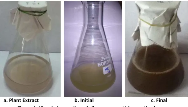
Plant-mediated synthesis of silver nanoparticles - Visual observation
Fig. 1b. shows the appearances of yellowish brown colour suggests the formation of silver
nanoparticles through the leaf extract of A. marmelos. The brown colour formation started in
2 hr and attained dark brown colour after 6 hr of incubation. The appearance of precipitation notices at the reaction vessel after 6 h of incubation indicates that the reaction was
completed. Similarly, the colour change from yellow to brown within 6 hr using the leaf
International Research Journal of Pharmaceutical and Biosciences(IRJPBS) 4 (7) 5
a. Plant Extract b. Initial c. Final Figure 1: Visual observation of silver nanoparticles synthesis process
UV-vis spectroscopic analysis
The synthesis process was started at the 2nd hour and the very good peak was observed at the
6th hour. The good peak was found at 420 nm indicates the synthesis of silver nanoparticles at
4th and 6th hour absorbance. After that, the peak was shifted to 440 nm shows changes in the
nanoparticles reaction mixture. It may be the agglomeration may lead to the big size of nanoparticles [10].
Figure 2: UV-vis spectroscopic analysis of Silver nanoparticles synthesized by A. marmelos
0 0.5 1 1.5 2 2.5
360 410 460 510
A
bs
or
bance
Wavelength (nm)
International Research Journal of Pharmaceutical and Biosciences(IRJPBS) 4 (7) 6 Atomic force microscopy
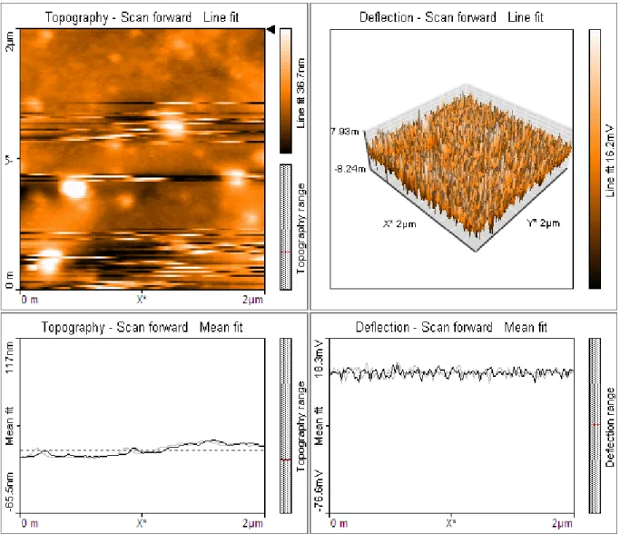
Figure 3: AFM image of Silver nanoparticles synthesized by A. marmelos
Figure 3 shows the atomic force microscopic image of silver nanoparticles synthesized using medicinally important plant A. marmelos. The spherical shaped particles were observed in this image and in the background some light colour background also observed may be the phytochemicals of plant extract.
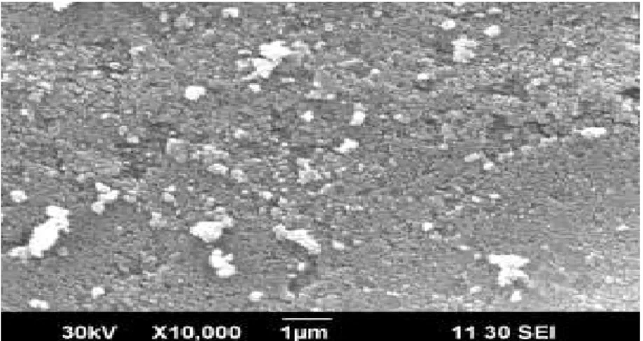
Scanning electron microscope
The SEM image shows the silver nanoparticles synthesized using A. marmelos and the size of
International Research Journal of Pharmaceutical and Biosciences(IRJPBS) 4 (7) 7
Figure 4: SEM image of Silver nanoparticles synthesized by A. marmelos
In vitro antidiabetic activity Alpha-amylase inhibitory assay
Alpha-amylase is the main enzyme involved in the breakdown of polysaccharides and release of sugar in the main blood stream which further leads to increased blood glucose level and ultimately diabetes. The inhibitory effect of this enzyme can prove to have a potential therapeutic effect on diabetes. Figure 5 represents the % inhibition effect of silver nanoparticle on the enzyme. Silver nanoparticle exhibited an inhibitory effect on the enzyme in a dose-dependent manner and when the dose was increased to 100µg/mL, it resembled the % inhibition exhibited by standard Acarbose. It could be interpreted from the results that silver nanoparticle could be used as an alternative to Acarbose because of the potential inhibitory effect that it has on the enzyme.
Figure 5: In vitro antidiabetic activity of silver nanoparticles 0
20 40 60 80 100 120
10 20 40 60 80 100
%
In
h
ib
it
ion
Concentration (
μg/mL)
Alpha Amylase Inhibitory Assay
International Research Journal of Pharmaceutical and Biosciences(IRJPBS) 4 (7) 8 Antibacterial activity of silver nanoparticles
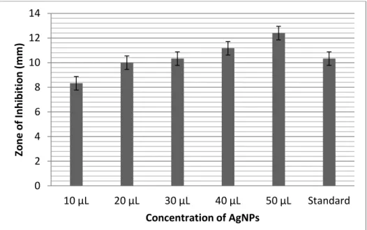
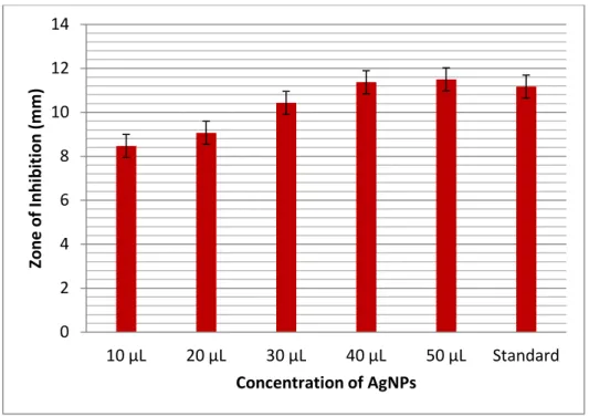
The antibacterial activity of silver nanoparticles against different bacterial pathogens like
Bacillus subtilis, Klebsiella planticola, Pseudomonas sp, Streptococcus sp. and Staphylococcus
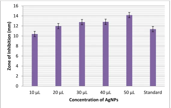
aureus were performed. The zone of inhibition against the pathogens is shown in Table 1 and
figure 6-10. The silver nanoparticles are having a very good zone of inhibition against most of
the pathogens. Klebsiella planticola having a maximum zone of inhibition at 50 μL and
Streptococcus sp has a minimum zone of inhibition in maximum volume of AgNPs solution.
Based on this study we will use the silver nanoparticles much biomedical application related to clinical diseases caused by pathogenic microorganisms.
Figure 6: Antibacterial activity of silver nanoparticles synthesized using A. marmelos against Bacillus subtilis
Table. 1 Antibacterial activity of AgNPs synthesized from A. marmelos leaf extract Conc. of
AgNPs
Bacillus subtilis
Klebsiella planticola
Pseudomonas sp.
Streptococcus sp.
Staphylococcus aureus
10 μL 08.33±0.167 10.40±0.100 10.00±0.000 08.47±0.034 08.50±0.289 20 μL 10.00±0.501 11.97±0.261 10.13±0.467 09.07±0.067 09.17±0.167 30 μL 10.33±0.441 12.77±0.234 10.67±0.334 10.43±0.434 10.67±0.334 40 μL 11.17±0.334 12.83±0.167 12.50±0.289 11.37±0.134 11.83±0.273 50 μL 12.40±0.100 14.17±0.334 13.33±0.167 11.50±0.289 12.07±0.115 Standard 10.33±0.467 11.370.501 12.07±0.167 11.17±0.289 12.77±0.000
0 2 4 6 8 10 12 14
10 μL 20 μL 30 μL 40 μL 50 μL Standard
Zo
ne
o
f I
nhi
bi
tio
n (
m
m
)
International Research Journal of Pharmaceutical and Biosciences(IRJPBS) 4 (7) 9 Figure 7: Antibacterial activity of silver nanoparticles synthesized using A. marmelos against
Klebsiella planticola
Figure 8: Antibacterial activity of silver nanoparticles synthesized using A. marmelos against Pseudomonas sp
0 2 4 6 8 10 12 14 16
10 μL 20 μL 30 μL 40 μL 50 μL Standard
Zo
ne
o
f I
nhi
bi
tio
n (
m
m
)
Concentration of AgNPs
0 2 4 6 8 10 12 14 16
10 μL 20 μL 30 μL 40 μL 50 μL Standard
Zo
ne
o
f I
nhi
bi
tio
n (
m
m
)
International Research Journal of Pharmaceutical and Biosciences(IRJPBS) 4 (7) 10 Figure 9: Antibacterial activity of silver nanoparticles synthesized using A. marmelos against
Streptococcus sp
Figure 10: Antibacterial activity of silver nanoparticles synthesized using A. marmelos against Staphylococcus aureus
Conclusion
In this present investigation, we used the most applicable and ancient traditional and
medicinal plant Aegle marmelos for the green synthesis of silver nanoparticles. The
characterization results are showing the synthesized nanoparticles are very good quality. The 0
2 4 6 8 10 12 14
10 μL 20 μL 30 μL 40 μL 50 μL Standard
Zo
ne
o
f I
nhi
bi
tio
n (
m
m
)
Concentration of AgNPs
0 2 4 6 8 10 12 14 16
10 μL 20 μL 30 μL 40 μL 50 μL Standard
Zo
ne
o
f I
nhi
bi
tio
n (
m
m
)
International Research Journal of Pharmaceutical and Biosciences(IRJPBS) 4 (7) 11 UV-vis spectrophotometer exhibit the peak at 420 nm clearly indicates the silver nanoparticles surface Plasmon resonance. The microscope like atomic force microscope and scanning electron microscope clearly indicate the silver nanoparticles are spherical and many
nanoparticles are get agglomerated in nature due to the Aegle marmelos plant phyto
compounds. Finally, the in-vitro antidiabetic activities of silver nanoparticles are highly important in this study. Based on these results, silver nanoparticles may be used for the anti-diabetic drug in future. The silver nanoparticles are very much used in controlling disease causing and disease spreading microorganisms. The silver nanoparticles synthesized using
Aegle marmelos showing very good growth inhibition of disease-causing pathogens. It is one
of the simple and ecofriendly methods because when compared to the chemical and physical method it has cost effective and there is no side effect.
REFERENCES
1. Baynes, J.W., Role of oxidative stress in development of complications in diabetes, Diabetes, 1991, 40, 405–412.
2. Bonnefont Rousselot, D., J.P. Bastard, M.C. Jaudon, J. Delattre, Consequences of the diabetic status on the oxidant/antioxidant balance, Diabetes Metab., 2000, 26, 163–176.
3. Sivakumar.V and Dhana Rajan. M. S. Hypoglycemic and antioxidant activity of Tinospora
cordifolia in experimental diabetes. IJPSR, 2011; Vol. 2(3): 608-613.
4. B. N. Taylor (ed.), The International Systems of Units (SI), United States Department of Commerce National Institute of Standards and Technology, Washington, DC, NIST Special Publication; (2001) 330.
5. Mohammed AE, Al-Brahim JS. Synthesis, characterization and evaluation of antimicrobial
potency of silver nanoparticles using Ziziphus spina-christi L. leaf extract. J Pure Appl
Microbiol 2014; 8(5): 3903-8.
6. Sivakumar.V and Rajeshkumar. S. (2015). Protective effect of Andrographis paniculata on
hyperglycemic mediated oxidative damage in renal tissues of diabetic rats. The Journal of Phytopharmacology; 4(6): 287-294.
7. Saxena A, Tripathi RM, Singh RP. Biological synthesis of silver nanoparticles by using onion
Allium cepa) extract and their antibacterial activity. Dig J Nanomater Biostruc 2010; 5: 427-
32.
8. Sadowski Z. Biosynthesis and application of silver and gold nanoparticles. In: Perez DP,
editor. Silver nanoparticles. Rijeka: InTech; 2010.
9. Kudle KR, Donda MR, Alwala J, Koyyati R, Nagati V, Merugu R, et al. Biofabrication of silver
nanoparticles using Cuminum cyminum through microwave irradiation. Int J Nanomater
International Research Journal of Pharmaceutical and Biosciences(IRJPBS) 4 (7) 12
10. P Daisy K Saipriya Biochemical analysis of Cassia fistula aqueous extract and
phytochemically synthesized gold nanoparticles as hypoglycemic treatment for diabetes
mellitus, Research J. Pharm. and Tech. 9(8): August. 2016.
11. Sharma VK, Yngard RA, Lin Y. Silver nanoparticles: green synthesis and their antimicrobial