Dr P Sharmas Vali et al JMSCR Volume 06 Issue 10 October 2018 Page 1244
Original Article
A Study of the spectrum of Urinary Tract infections in patients with
Adult Polycystic Kidney Disease – An old problem with a better outcome
Authors
Dr P Sharmas Vali
1, Dr Kiranmai Ismal
2, Dr Manisha Sahay
31
Associate Professor, Dept of Nephrology, Osmania General Hospital, Hyderabad
2,3
Professor, Dept of Nephrology, Osmania General Hospital, Hyderabad, Telangana Corresponding Author
Dr P Sharmas Vali
Associate Professor, Dept of Nephrology, Osmania General Hospital, Hyderabad-35, Telangana, India Email: [email protected], Mob: 9397399953
Abstract
Background: Autosomal Dominant Polycystic kidney disease (ADPKD) is the most common hereditary cause of End stage renal disease (ESRD). Urinary tract Infections (UTI) are common in these patients and include a spectrum of both Lower tract and upper tract UTI.
Methods: This retrospective study which included patients of ADPKD who manifested UTI between June 2012 to May 2017. Clinical, Laboratory, imaging and microbiological data was analyzed. Few patients had more than one episode of UTI and there by the total number of UTI episodes were 52.Out of the 58 episodes of UTI, Lower tract UTI and Upper tract UTIs were 13 and 45 episodes. Among the total 45 episodes of upper UTI, 17 were Acute Pyelonephritis and 28 were Cyst infections. 40 % of UTI episodes necessitated Hospitalization.
Conclusions: Cyst infection is the most common form of UTI followed by acute pyelonephritis and Lower tract UTI. Majority of the UTI episodes were caused by E.coli. Around 40 % of patients with UTI mandated hospitalization but no episode of death attributable to UTI was noted.
Keywords: Autosomal dominant polycystic kidney disease (ADPKD), Urinary Tract infection (UTI), Acute Pyelonephritis, Cyst infection, Extra renal manifestations.
Introduction
Autosomal dominant polycystic kidney disease (ADPKD) is the most common monogenic renal disorder and it is credited as the commonest hereditary disorder causing End Stage Renal Disorder (ESRD)1. It has an incidence of around 1 in 500 to 1in 1,0002. In this condition there would be progressive formation of multiple fluid laden
cysts in the renal parenchyma. ADPKD essentially involves adults and is the consequence of mutations in the PKD1 or PKD2 genes, which encode the proteins polycystin-1 and polycystin-2, respectively. By any account, at least 50 % of individuals with ADPKD progress to ESRD usually by sixth decade of life3.
Patients of ADPKD usually seek medical
www.jmscr.igmpublication.org Impact Factor (SJIF): 6.379
Index Copernicus Value: 79.54 ISSN (e)-2347-176x ISSN (p) 2455-0450
Dr P Sharmas Vali et al JMSCR Volume 06 Issue 10 October 2018 Page 1245 attentionin view of enlarged abdomen,
haematuria, infected renal cysts and pain abdomen. Cysts can occur in multiple Sub diaphragmatic organs with Kidney and Liver being the most common sites of cyst formation. Cysts are linked with various attendant complications Viz bleeding into cyst, cyst infection and obstruction. Obstruction due to cyst enlargement is particularly Concertful in Liver4. Infections are an important cause of morbidity and mortality in ADPKD patients5,6. Urinary tract infections (UTI) and especially Cyst infections constitute important events in the clinical course of ADPKD patients and they can be at times be recurrent. The current study is intended to study the clinical profile, Microbiology and outcome of UTI in patients with ADPKD.
Material and Methods
This is a retrospective study which included ADPKD patients who experienced either one or more episodes of UTI. The records of such patients between June 2012 to May 2017 were analysed. The inclusion criteria included ADPKD patients who experienced one or more episodes of UTI. Patients who were diagnosed to have acquired cystic kidney disease were excluded. A diagnosis of lower tract UTI was empirically made when patient presented with new onset dysuria and with microscopic pyuria. A diagnosis of Acute pyelonephritis was made when a patient presents with dysuria, flank pain, fever and microscopic or gross pyuria. A diagnosis of Cyst infection was made when the combination of fever, new onset tender nodular areas over the abdomen and C-Reactive protein level of more than 5 mg/dl.
Data regarding Clinical profile, laboratory parameters, imaging studies and Microbiological investigations, Antimicrobial regimen and the response to treatment was recorded and analysed. Urine culture and US Scan abdomen was performed in every patient while Blood Culture was performed in select patients in whom there is suspicion of Upper tract infection or if there is
undue delay in response despite administering appropriate antibiotics as per urine microbiology reports. CT scan Abdomen was performed when UTI does not improve at expected pace, to rule out obstruction due to Nephrolithiasis, when there is a suspicion of perinephric abscess or when the patient presents with a combination of UTI and acute abdomen. Further, the basic laboratory investigations (Renal Function tests, Liver Function Tests, Complete blood count) were performed. Detailed analysis of the Microbiological investigations to incorporate the Microbiological spectrum and antibiotic sensitivity pattern was carried out. IBM SPSS version 22 was used for statistical analysis. Descriptive analysis was carried out by the mean and standard deviation for quantitative variables, frequency, and proportion for categorical variables. Data was also represented using appropriate diagrams.
Results
Dr P Sharmas Vali et al JMSCR Volume 06 Issue 10 October 2018 Page 1246 aneurysms as there was negative family of either
Cerebral aneurysms or sudden death in any of the family members of the study subjects.
Nephrolithiasis was noted in 12 patients of those presented with UTI (26.7%) and in 2 patients obstruction was evident due to ureteric calculi, which mandated Lithotripsic destoning.
All patients were started on empirical antibiotic regimen consisting of one or more antibiotics, out of which one antibiotic would be a fluoroquinolone antibiotic with good cyst penetration capability (either Ciprofloxacin or Levofloaxacin). Antibiotic therapy was modulated basing on the culture reports. All episodes of UTI included in the study recovered and no deaths were noted.
Table 1: Clinical Presentations in Patients of ADPKD with UTI
S.No Clinical Symptom Percentage
1. Dysuria 94 %
2. Flank Pain 42 %
3. Haematuria 27%
4. Fever 82 %
5. New onset abdominal
Painful nodule
38 %
6. Vomiting and Nausea 64 %
7. Diarrhoea 44 %
Discussion
UTI which constitute a spectrum with regard to their anatomical location and severity are very common in ADPKD and can present in 30-50 % of ADPKD patients in their life time7.UTI is more common in females akin to UTI in the general population8.UTI in ADPKD can present as both Lower tact UTI and Upper tract UTI. As the name implies Lower tact UTI indicates Cystitis while the two major types of Upper tract UTI are Acute Pyelonephritis and Cyst infection. Majority of the infections are ascending in nature and the major predisposing factor for UTI in ADPKD being structurally abnormal kidneys and Nephrolithiasis. In the current study, 47.9% of the study population had experienced one or more episodes of UTI. Among those patients who had UTI, 64.5% were females, though males have outnumbered females with respect to the total number of ADPKD patients (those with UTI and without UTI) whose files were analysed in the study period. This denotes the clear cut preponderance of UTI in ADPKD woman similar to UTI occurring in general population.
Family History is usually present in majority of the patients though a typical family history might be absent in up to 25 % of patients9. In this study, 93.6% of total ADPKD patients reported positive family History.
Dr P Sharmas Vali et al JMSCR Volume 06 Issue 10 October 2018 Page 1247 other cause of Chronic kidney disease.
Hypertension is noted in around 50-70 % of ADPKD patients and it might manifest even before the onset of decline of renal function10. The average ages of diagnosis of ADPKD is 27 while the age of detecting Hypertension in ADPKD is around 31 years11. In the current study, Hypertension is noted in 61 % of the study subjects, implying that the incidence of Hypertension is uniform across the various geographical regions.
In ADPKD, both lower and Upper tract UTIs are common and contribute to mortality and morbidity5.However, there is paucity of data regarding the distribution of various forms of UTI in ADPKD patients with the exception of Cyst infections. In the current study, Out of the 58 episodes of UTI, 22.4 % (13 episodes) were of lower tract UTI and 77.58 % (45 episodes) were of Upper tract UTI. Among the upper tract UTIs, approximately one third were Acute Pyelonephritis episodes and two thirds were Cyst infections. Pyelonephritis and cyst infections contributed for 29.3 % (17 episodes) and 48.2 %(28 episodes) of total UTI episodes. Out of the total 58 episodes of UTI, in 23 episodes patients necessitated admission in view of severe nature of UTI. This indicates that almost 40 % of episodes of UTI in ADPKD cases required hospitalization. The reasons for hospitalization included persistent high grade fever, inability to maintain optimal hydration, presence of obstruction and the impending multiorgan nature of Sepsis syndrome. This rate of hospitalization is higher than the need for hospitalization in general population with acute pyelonephritis and this would portend the complex nature of UTI in ADPKD patients.
The incidence of Cyst infection in ADPKD is reported as around 0.01 episode per patient with an estimated microbiological evidence in 75 % of cyst infections. Escherichia coli was reported to be responsible for cyst infection in around three fourths of such cyst infections. 10 % of hospitalizations in ADPKD patients are accountable for cyst infection. The diagnosis of
cyst infection constitutes a diagnostic challenge and is based on clinical features such as features of sepsis on the back drop of new onset tender nodules over the abdomen. Definitive diagnosis utilizing cyst aspiration of pus and the microbiological testing of cyst fluid is not always practically feasible. The inability of CT and MR imaging to differentiate between Cyst infection and Cyst haemorrhage adds to the complexity of diagnosis of cyst infection4. Renal cyst infection in ADPKD can be at times be a challenging task as the presentations are protean in nature and can even present like a mass lesion mimicking a malignant lesion12.
Francois et al described the criteria of Cyst infection as the combination of four parameters Viz. fever, new onset tender nodular areas over the abdomen and C-Reactive protein level of more than 5 mg/dl and absence of intra cystic density of more than 25 Hounsfield units on CT imaging of kidneys13.
In the current study, Cyst infections were the most common form of UTI by accounting to 48.2 % of total UTI episodes. Further cyst infections contributed for almost two thirds of upper tract UTIs. Further out of 28 cases of Cyst infections, 18 cases necessitated hospitalization (64.28 % of Cyst infection cases).
Urine culture is usually sterile in Cyst infections as Cysts larger than 2 Cm in size usually gets detached and establish no communication with the kidney tubule14. Therefore either Blood culture or microbiological study of the fluid aspirated from the cyst in question would result in bacteriological yield. It is prudent to note that demonstration of microbiological positivity in cyst aspirate, though considered gold standard is not always practically feasible15.
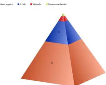
Dr P Sharmas Vali et al JMSCR Volume 06 Issue 10 October 2018 Page 1248 of UTI) and in 42.8 % cases (12 episodes of UTI)
of Cyst infection. In cases where in urine C&S was positive, E.coli was isolated in 73.3% of cases (22 out of 30 total episodes with Urine Culture positivity), while Klebsiella and Enterococcus faecalis were isolated in 16.6 % (5 episodes of Culture positive UTI) and 10 % of cases (3 episodes of culture positive UTI).
Conventionally Nephrolithiasis is described in around 20 % of individuals with ADPKD and majority of the stones were either Calcium oxalate or uric acid. In the current study, Nephrolithiasis was noted in 12 patients of those presented with UTI (26.7%) And in 2 patients obstruction was evident due to ureteric calculi, which mandated Lithotripsic destoning15.
Extra renal manifestations are the rule in ADPKD and the major organs involved being Liver, Heart, Brain. Liver cysts are noted in around 80 % of individuals and liver cysts might appear 10 years after the onset of renal cysts16. Hepatic cysts unlike renal cysts will not impede hepatic functions except when they cause obstruction to the biliary system. 25 % can have cardiac abnormalities and 8 % can have Intra cranial aneurysms17. In the current study, hepatic cysts were noted in all the individuals of ADPKD who presented with UTI. Mitral valve prolapse was noted in 17 % and mild Mitral Regurgitation was noted in 6% of individuals. None had aortic valve lesions. None of the patients had been screened for intra cranial aneurysms as none of them had family history of neither the Sudden death nor the intra cranial aneurysm.
Infection is considered as an important causes of death in ADPKD patients. Death rate due to infection was more than 30 % prior to 1975 but the rate has declined to around 15 % in the first decade of Twenty First Century5,6. But in the current study, though around 40 % of UTI episodes necessitated Hospitalization, mortality due to Sepsis was documented in none. This could be because of the wide spread awareness about the potential seriousness of UTI and infections in ADPKD and due to our institutional policy of
administering intra venous antibiotics empirically without delay at the first suspicion of UTI in ADPKD patients.
Conclusion
ADPKD is a ciliopathy and an important cause of ESRD globally. Even though ESRD is manifested after fifth decade of life, the course of ADPKD patients is punctured with multiple renal and extra renal complications. One such Renal complication is Urinary Tract infection. In the current study Cyst infection is the most common form of UTI followed by acute pyelonephritis and Lower tract UTI. Majority of the UTI episodes were caused by E.coli. Around 40 % of patients with UTI mandated hospitalization but no episode of death attributable to UTI was noted, indicating the importance of initiation of intravenous antibiotic at the earliest suspicion of UTI in ADPKD patients.
References
1. Patel, Vishal; Chowdhury, Renuka; Igarashi P. Advances in the Pathogenesis and Treatment of Polycystic Kidney Disease. Curr Opin Nephrol Hypertens. 2010;18(2):1-12.
doi:10.1097/MNH.0b013e3283262ab0 2. Ong ACM, Devuyst O, Knebelmann B,
Walz G. Autosomal dominant polycystic kidney disease: The changing face of clinical management. Lancet. 2015;385(9981):1993-2002.
doi:10.1016/S0140-6736(15)60907-2 3. Igarashi P, Somlo S. Genetics and
pathogenesis of polycystic kidney disease.
J Am Soc Nephrol. 2002;13(9):2384-2398. doi:10.1097/01.ASN.0000028643.17901.4 2
4. Sallée M, Rafat C, Zahar JR, et al. Cyst infections in patients with autosomal dominant polycystic kidney disease. Clin J Am Soc Nephrol. 2009;4(7):1183-1189. doi:10.2215/CJN.01870309
Dr P Sharmas Vali et al JMSCR Volume 06 Issue 10 October 2018 Page 1249 Gabow PA. Causes of death in autosomal
dominant polycystic kidney disease. J Am
Soc Nephrol. 1995;5(12):2048-2056.
http://cod.
6. Rahman E, Niaz FA, Al-suwaida A, et al. Polycystic Kidney Disease : A Single Center Study. Nephrology. 2009;20(5): 806-810.
7. Hwang JH, Park HC, Jeong JC, et al. Chronic asymptomatic pyuria precedes overt urinary tract infection and deterioration of renal function in autosomal dominant polycystic kidney disease. BMC Nephrol. 2013;14(1):1-9. doi:10.1186/1471-2369-14-1
8. Sklar AH, Caruana RJ, Lammers JE, Strauser GD. Renal infections in autosomal dominant polycystic kidney disease. Am J Kidney Dis. 1987;10(2):81-88.
9. Keenan D, Maxwell AP. Optimising the management of polycystic kidney disease.
Practitioner. 2016;260(1790):2,13-16. 10.SCHRIER TE and RW. Hypertension in
Autosomal-Dominant Polycystic Kidney Disease: Early Occurrence and Unique Aspects. J Am Soc Nephrol. 2001;12(1):194-200.
doi:10.1037/h0046469
11.Tkachenko O, Helal I, Shchekochikhin D, Schrier RW. Renin-angiotensin-aldosterone system in autosomal dominant polycystic kidney disease. Curr Hypertens Rev. 2013;9(1):12-20. doi:10.2174/1573402111309010003 12.Kamboj M, Zeng X, Koratala A. Renal
cyst infection: a diagnostic dilemma. Clin
case reports. 2018;6(4):762-763.
doi:10.1002/ccr3.1441
13.Jouret F, Lhommel R, Devuyst O, et al. Diagnosis of cyst infection in patients with autosomal dominant polycystic kidney disease: Attributes and limitations of the current modalities. Nephrol Dial
Transplant. 2012;27(10):3746-3751.
doi:10.1093/ndt/gfs352
14.Grantham JJ. The etiology, pathogenesis, and treatment of autosomal dominant polycystic kidney disease: Recent advances. Am J Kidney Dis. 1996;28(6): 788-803. doi:10.1016/S0272-6386(96)90378-9
15.Rizk D, Chapman AB. Cystic and inherited kidney diseases. Am J Kidney Dis. 2003;42(6):1305-1317.
16.Irazabal M V., Torres VE. Complications of polycystic kidney disease. Polycystic
Kidney Dis From Bench to Bedside.
2015;51(1):149-163. doi:10.2217/EBO.12.123