THE EFFECT OF A HIGH FAT DIET ON THE BRAIN FOLLOWING HSV-1 INFECTION
by
Simone Nicole Reaves
Honors Thesis Department of Nutrition
University of North Carolina at Chapel Hill 2015
Approved:
Abstract
Children from low socioeconomic status have an increased risk of developing anxiety. Additionally, both obesity and herpes simplex virus 1 (HSV-1) infection have an increased prevalence among children from lower socioeconomic status, and may be environmental factors that contribute to the development of anxiety disorders. This study aimed to determine if a high fat diet following HSV-1 infection increased inflammation in the hippocampus and altered the phenotypes of monocytes in the blood. Female C57BL/6 mice were placed on a 10% LF diet for one week prior to intranasal HSV-1 or mock infection. Mice were randomized to either the 45% HF diet or remain on the LF diet 14 days post infection. Mice were sacrificed at either 4-5 months or 9 months, and hippocampus samples were obtained. RNA extraction, reverse transcription and qPCR were performed to examine expression of IL-6, IL-1β, TNFα, MHCII and CCL2. Flow cytometry was utilized to determine the percentage of CCR2+ and Cx3CR1+ monocytes in the blood. The infected LF and HF groups saw a significant decrease in IL-6 expression with age. Expression of the inflammatory monocyte CCR2+ was highest in the lymphocytes of young, LF uninfected mice, and significantly lower in the older, HF infected group. In conclusion, the level of inflammation seen in these female mice was lower than that seen in the larger study using male mice, suggesting that other factors, such as weight,
Chapter 1: Study Aims and Hypotheses
Children from low socioeconomic backgrounds have a greater risk of developing anxiety and learning problems19, but the biological factors that contribute to this increased risk are not well understood. Early exposure to certain environmental factors that affect the brain may play a role in the development of these mental health disorders. Two examples of these factors are the high rates of herpes simplex virus (HSV)-1 and the prevalence of obesity in children from low socio-economic backgrounds20,21. We have developed a mouse model that mimics this
environment. My project is part of a larger study to test the hypothesis that obesity (induced by a diet high in saturated fatty acids) in mice latently infected with HSV-1 results in
neuroinflammation and increased anxiety in these mice.
The main study focuses on the response in male mice, here we utilize female (C57BL/6) mice; this is unique in that females are more prone to anxiety and also constitute an understudied population (both in rodent and human studies). In order to study the effect of a high fat diet on perpetuating neuroinflammation induced by HSV-1 infection, mice were randomly assigned to HSV-1 infection or mock infection. The mice were allowed to recover from the infection (and the virus became latent). After 14 days, the mice were then randomized to a high-fat or a low-fat diet. After the mice were sacrificed, blood samples and brain tissue samples were analyzed to investigate differences in the immune response among the mice.
Neuroinflammation is associated with increases in anxiety and is a product of cytokines made by microglia in the brain. In addition, recent evidence suggests that inflammatory
Specific Aim 1: To determine if HF diet following HSV-1 infection increases
inflammation in the hippocampus of the brain, an area that is key to the development of anxiety o qRT-PCR: IL-6, IL-1β, TNFα, MHCII and CCL2 (a chemokine that attracts
CCR2+ monocytes)
Specific Aim 2: To determine if HF diet following HSV-1 infection alters the monocyte phenotypes in the blood
1. Genotyping mice: CCR2 and Cx3Cr1 heterozygous
2. Flow cytometry: CCR2 and Cx3Cr1 expression monocytes to quantify “inflammatory” monocytes.
Hypothesis: After HSV-1 infection, mice on high-fat diet will have an increased inflammatory response in the brain and increased CCR2+ monocytes in the blood, which are associated with increased anxiety behaviors.
The techniques used for this study are RNA extraction, reverse transcription and qPCR to examine inflammation. DNA extraction and PCR were utilized to genotype the
Chapter 2: Introduction
With a 12-month prevalence of 18.1%, anxiety and anxiety related disorders are the most common mental health problem in the United States25. The burden and impairment caused by anxiety and anxiety disorders is significant and comparable to other chronic medical illnesses, such as diabetes26. Additionally, anxiety disorders are highly comorbid with other mental
illnesses26. Genetic factors play a role in the development of anxiety disorders, and heritability is estimated to be around 30%26. However, environmental factors also have a significant role in the development of anxiety26.
Previous studies have suggested a correlation between mental disorders and obesity27. Additionally, recent literature has suggested that high fat diets and obesity can contribute to cognitive impairment in both rodents and humans10,12-24. Recent evidence has shown that obesity results in a state of chronic inflammation. Specifically, it has been shown that diet-induced obesity causes a shift in the state of macrophages in adipocytes to a more inflammatory state6. Furthermore, obese individuals have higher circulating levels of inflammatory cytokines such as TNFα, IL-1β and IL-6, which are thought to contribute to this chronic inflammation2,8. A
The relationship between diet and neuroinflammation also warrants more attention. It has been shown that in instances of diet-induced obesity, hypertriglyceridemia contributes to
cognitive impairment10. Additionally, a diet high in saturated fat or hydrogenated fats has a
positive correlation with risk of developing Alzheimer’s disease11.
A better understanding of the implications of diet-induced obesity on neural
inflammation is relevant today as obesity becomes increasingly common in our society.
Furthermore, both obesity and HSV-1 seropositivity are prevalent in low socioeconomic (SES) conditions. A recent study found that obese individuals were significantly more likely to be
HSV-1 positive4. A separate study found a correlation between fat mass and HSV-1 seropositivity in middle aged men5. One study found an association between HSV-1
seropositivity and impaired cognitive function among children ages 6-1629.
This study will examine the relationship between diet-induced obesity, inflammation, and anxiety. Recently, childhood obesity has decreased in children among higher socioeconomic
classes, but increased in children from low socioeconomic status20. Additionally, children ages 6-16 in lower socioeconomic classes have a higher prevalence of HSV-1 infection28. Both obesity
and HSV-1 infection have been implicated in cognitive impairment. Therefore this mouse model mimics the daily environment of children living in poverty in order to analyze the effects of obesity and HSV-1 infection on the development of anxiety behavior. As part of a larger study,
this study focused on examining inflammation in the hippocampus, an area of the brain
important to the development of anxiety. The expression of proinflammatory cytokines 6,
Chapter 3: Methods
Animals
In all of these experiments, weanling female C57Bl/6 CCR2RFP/+/CX3CR1eGFP/+
heterozygous mice were placed on a 10% LF diet and allowed to acclimate for one week prior to
intranasal HSV-1 or mock infection. Fourteen days post infection (p.i.), mice were randomized
to either the 45% HF diet or remain on the LF diet. The mice were sacrificed at either 4-5 months
or 9 months. The mice sacrificed at 4 and 5 months were categorized as “young,” and the mice
sacrificed at 9 months were categorized as the “old” group.
Genotyping
DNA was extracted from tail snips of mice bred to be heterozygous
(CCR2RFP/+Cx3Cr1GFP/+) using the Qiagen DNeasy Quick-StartProtocol. The DNA extraction
was performed according to manufacturer’s instructions for the DNeasy Blood and Tissue Kit,
which included overnight lysing (in a 56°C water bath) of the tail snips in 180 µL Buffer ATL
and 20 µL proteinase K. Next, 200 µL of Buffer AL and 200 µL of 96% ethanol was added. The
600 µL mixture was placed in a 2 mL collection tube, which was centrifuged at 8000 rpm for 1
minute. The flow-through and collection tube were discarded. The spin column was placed into a
new 2 mL collection tube and 500 µL Buffer AW1 was added. The sample was centrifuged for 1
minute at 8000 rpm, and the flow-through and collection tube were discarded. The spin column
was then placed into a new 2 mL collection tube, and 500 µL Buffer AW2 was added. The
samples were centrifuged for 3 minutes at 14000 rpm and the flow-through and collection tubes
were discarded. The spin columns were transferred to new 1.5 mL microcentrifuge tubes, and the
minute at room temperature, and then were centrifuged for 1 minute at 8000 rpm. This last step
(DNA elution) was repeated using 100 µL DEPC H2O to increase DNA yield.
PCR was used to amplify the DNA. The CCR2 primer set (wild-type, mutant, and
common) was from Bioneer, as well as the Cx3Cr1 wild-type primer set (wild-type and common
primers). The Cx3Cr1 mutant primer set (mutant and common primers) was from Invitrogen.
One µL of DNA, 0.2 µL of each primer, 0.2 µL dNTP mix, 1 µL 10x Thermopol Buffer, and 0.5
Taq polymerase was used. The cycler program was set to denature the DNA at 94 °C, allow
annealing at 50 °C, and then elongate the DNA at 72°C. Gel visualization (1% Ethidium
Bromide gel) was performed using 1:8 dilutions of the amplified DNA.
RNA Isolation
Brain tissue samples were homogenized in 1mL of TRIzol, and then incubated for 5
minutes at room temperature. Next, 0.2 mL of chloroform was added and tubes were shaken
vigorously by hand for 15 seconds. Samples were incubated for 2-3 minutes at room
temperature, and then the samples were centrifuged at 4°C for 15 minutes at 12,000 x g. The
aqueous phase was removed from the samples and placed in a new tube. The RNA was then
isolated by adding 0.5mL of 100% isopropanol to the aqueous phase, and then incubating the
samples at room temperature for 10 minutes. The samples were then centrifuged at 12,000 x g
for 10 minutes at 4°C. The supernatant was then removed from the tube, leaving an RNA pellet.
The pellet was washed with 1 mL of 75% ethanol. The samples were briefly vortexed, and then
centrifuged at 7500 x g for 5 minutes at 4°C. The wash was discarded, and the RNA pellet was
allowed to air dry, and then was resuspended in 40 µL H2O.
The previously isolated RNA (1 µg in 8 µL water), 1 µL of 10x Reaction Buffer, and 1 µL Sigma DNase I, Amplification Grade (1 unit/ µL) were added to an RNase-free PCR tube. The solution was mixed gently, and then incubated for 15 minutes at room temperature. Next, 1 µL of Stop Solution was added, and the samples were heated at 70°C for 10 minutes to denature the DNase I and RNA. The solution was chilled on ice, and then 4 µL 5x iScript reaction mix, 1 µL iScript reverse transcriptase, and 4 µL nuclease-free H2O was added to the solution. The
reaction mix was incubated for 5 minutes at 25°C, 30 minutes at 42°C, and then for 5 minutes at 85°C.
qPCR
For the purposes of this thesis, only qRT-PCR data from the hippocampus were analyzed and presented. time qPCR was performed using the synthesized cDNA samples. The Real-Time PCR System used was Bio-Rad® CFX96™ and the thermal cycling protocol consisted of polymerase activation and DNA denaturation at 95°C for 30 seconds, amplification at 95°C for 5 seconds (for denaturation) and then 60°C for 30 seconds (for annealing and extension). The program cycled 39 more times for a total of 40 cycles. Each qPCR tube had a total volume of 20 µL, which included 10 µL TaqMan Universal Mix (2x), 1 µL of the desired primer (either GAPDH, IL-1, IL-6, MHCII, TNF-alpha), 6 µL of H2O, and 3 µL of cDNA.
Statistics
Chapter 4: Results
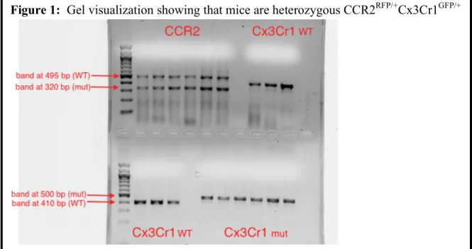
Gel visualization using an ethidium bromide gel was used to confirm that the mice were
heterozygous. Figure 1 shows a sample gel with all three primer sets. CCR2 heterozygotes
display a band at 495 base pairs (bp), indicating that they possess the wild-type CCR2 gene.
They also exhibit a band at 320 bp because they possess a knockout CCR2 gene (replaced with
RFP). The Cx3Cr1 heterozygotes exhibit bands at 410 bp because they have one copy of the
wild-type Cx3Cr1 gene, and bands at 500 bp because they have a Cx3Cr1 knockout (replaced
with GFP). Successfully genotyping these mice was essential in order to further examine the
percentages of CCR2+ and Cx3Cr1+ monocytes in the blood.
Figure 2 shows the levels expression for the various genes examined in this study. IL-1β
is a proinflammatory cytokine produced by macrophages, and is normally found in brain tissues
at low levels but expression increases in the event of disease, injury or inflammation17. In the
exception of the HF infected mice, yet the increase was not significant (Fig. 2A). IL-6 is a
proinflammatory cytokine secreted by adipocytes18. Initially, the infected groups showed
increased levels of IL-6, but levels of IL-6 decreased with age among all groups (Fig. 2B). The
infected LF and HF groups saw a significant decrease in IL-6 expression (LF infected young
mean=3.523E-05 ± 6.747E-06; LF infected old mean= 2.243E-05 ± 6.181E-06. HF infected
young mean=3.215E-05 ± 7.064E-06; HF infected old mean= 1.517E-05 ± 3.976E-06). TNFα is
increased in the serum of obese subjects. TNFα levels increased with age across all groups, but
the most notable increases were seen among the LF uninfected and infected groups (Fig. 2C).
These differences were not statistically significant. MHCII is normally found on antigen
presenting cells, including activated macrophages. The presence of MHCII in the brain is
indicative of microglial activation, as it is not expressed on quiescent (non-activated) microglia.
MHCII levels were similar among the young and old groups, with the exception of the HF
uninfected young mice (no significant difference) (Fig. 2D). CCL2 is a chemokine that recruits
inflammatory CCR2+ monocytes to the brain. Among the young mice, the expression of CCL2
was highest in the HF diet groups. But in the older mice, the LF infected group had the highest
level of CCL2.
As anxiety has been associated with the infiltration of bone-marrow derived monocytes
from the blood to the brain, we examined the blood samples and determined the percentage of
lymphocytes in the blood, as well as the percent of the lymphocytes that express CCR2+ or
Cx3Cr1+ (Figure 3). CCR2+ monocytes are considered pro-inflammatory cytokines that enter
the brain in response to inflammation16. In contrast, Cx3Cr1+ is thought to be anti-inflammatory.
In fact, deletion of Cx3Cr1 results in increased neuroinflammation and has been associated with
decreased neurogenesis and cognitive impairment30. In the young mice, the HF diet groups had
the highest percent lymphocytes in the blood (Fig. 3A). However in the older mice, percentages
were more similar. CCR2 was expressed at the highest level in the lymphocytes of young, LF
uninfected mice (Fig. 3B) (LF uninfected young mean= 7.8 ± 1.7%). In the older mice, CCR2
expression was significantly lower in the HF infected group (HF infected old mean= 1.65 ±
0.4%). Among the young mice, the expression of Cx3Cr1 within lymphocytes was highest
uninfected old mean= 1.03 ± 0.475%). The older mice showed similar expressions of Cx3Cr1 in
Chapter 5: Discussion
Previous studies have shown that elevated levels of the proinflammatory cytokines IL1-β,
IL-6 and TNFα can result in increased anxiety among adult animals14. The statistically
significant decreases in IL-6 expression between the infected young mice and the infected old
mice is contrary to what was expected. This may indicate that while infection provokes an
increase in IL-6 expression initially, as the mice get older and infection progresses, the
inflammatory response is less pronounced and IL-6 expression returns to normal levels. The
finding of high IL-6 levels in infected young mice may have potential clinical implications due
to evidence associating high levels of IL-6 with behavioral and cognitive changes13.
More notable findings, although not statistically significant, include the differences in the
young and old mice in CCL2 and TNF expression. It seems a HF diet may increase CCL2
expression, but with age, this agonistic effect no longer exists. CCL2 expression is most
pronounced in older lean infected mice, indicating that its expression may increase after
prolonged infection. However, the older mice on the HF diet did not have increased levels of
CCL2, indicating that the inflammatory state of obesity may counteract the expression of CCL2
expression in these older mice. This may have serious implications on the inflammatory response
in these mice because CCL2 is the chemokine that attracts the proinflammatory CCR2+
monocytes to the brain. Additionally, higher expression of TNFα in the older lean mice was
contrary to what was expected, as the proinflammatory state of obesity is often accompanied by
increased TNFα expression2. This finding may indicate that another factor may affect the
expression of TNFα in obese mice.
Cx3Cr1 is highly expressed in resident monocytes, whereas CCR2 is highly expressed in
cognitive impairment15. CCR2 is the receptor for CCL215, therefore the high expression of CCR2
in the lymphocytes of the younger lean mice, and low expression in the older HF mice may have
affected the amount of IL-1β, IL-6 and TNFα found in the hippocampus.
An association between a high fat diet and increased neuroinflammation, which in turn
contributes to behavioral changes, has previously been established in male mice in the larger
study. However, in the female mice used in this part of the study, the inflammatory response was
not as pronounced. As a result, if tested, these female mice may not exhibit the behavioral
changes and increases in anxiety that were seen in male mice. The decreased inflammatory
response may be due in part to the female C57BL/6 mice weighing less than the males, or the
males developing metabolic syndrome earlier. Earlier onset of metabolic syndrome would
contribute to an increased inflammatory state in these male mice. It is also possible that estrogen
may have a role in brain inflammation. Women have an increased vulnerability to developing
anxiety, and recent data suggests that female reproductive hormones may have a significant
role31. Additionally, there is evidence suggesting that estrogen plays a role in susceptibility to
inflammation32. One study even found increased expression of IL-1β, IL-6 and TNFα in male
mice upon administration of estrogen32. To adapt our model to examine the role of estrogen in
neuroinflammation due to diet-induced obesity and HSV-1 infection, we could administer
estrogen antagonists to female mice or treat male mice with estrogen. The discovery of a link
between estrogen and inflammatory response may have vast implications for the field of
References:
1. Schwartz, Michal, et al. "How do immune cells support and shape the brain in health,
disease, and aging?" The Journal of Neuroscience 33.45 (2013): 17587-17596.
2. Fantuzzi, Giamila. "Adipose tissue, adipokines, and inflammation." Journal of Allergy and
Clinical Immunology 115.5 (2005): 911-919.
3. Saederup, Noah, et al. "Selective chemokine receptor usage by central nervous system
myeloid cells in CCR2-red fluorescent protein knock-in mice." PloS one 5.10 (2010):
e13693.
4. Karjala, Zuzana, Diane Neal, and James Rohrer. "Association between HSV1 seropositivity
and obesity: data from the National Health and Nutritional Examination Survey,
2007–2008." PloS one 6.5 (2011): e19092.
5. Fernández!Real, José!Manuel, et al. "Burden of Infection and Fat Mass in Healthy Middle!
aged Men." Obesity 15.1 (2007): 245-252.
6. Lumeng, Carey N., Jennifer L. Bodzin, and Alan R. Saltiel. "Obesity induces a phenotypic
switch in adipose tissue macrophage polarization." Journal of Clinical Investigation
117.1 (2007): 175.
7. Maachi, M., et al. "Systemic low-grade inflammation is related to both circulating and
adipose tissue TNFα, leptin and IL-6 levels in obese women." International journal of
obesity 28.8 (2004): 993-997.
8. Hube, F., et al. "Expression pattern of tumour necrosis factor receptors in subcutaneous and
omental human adipose tissue: role of obesity and non-insulin-dependent diabetes
9. Teeling, J. L., and V. H. Perry. "Systemic infection and inflammation in acute CNS injury
and chronic neurodegeneration: underlying mechanisms." Neuroscience 158.3 (2009):
1062-1073.
10.Farr, Susan A., et al. "Obesity and hypertriglyceridemia produce cognitive impairment."
Endocrinology 149.5 (2008): 2628-2636.
11. Morris, Martha Clare, et al. "Dietary fats and the risk of incident Alzheimer disease."
Archives of neurology 60.2 (2003): 194-200.
12.Kronfol, Ziad, and Daniel G. Remick. "Cytokines and the brain: implications for clinical
psychiatry." Cytokines 157.5 (2000).
13. Bale, Tracy L., et al. "Early life programming and neurodevelopmental disorders." Biological
psychiatry 68.4 (2010): 314-319.
14. Janelidze, Shorena, et al. "Low IL!8 is associated with anxiety in suicidal patients: genetic
variation and decreased protein levels." Acta psychiatrica Scandinavica (2014).
15. Gao, Liang, et al. "MCP-1 and CCR2 gene polymorphisms in Parkinson’s disease in a Han
Chinese cohort." Neurological Sciences (2014): 1-6.
16. Morris, David L., et al. "CX3CR1 Deficiency Does Not Influence Trafficking of Adipose
Tissue Macrophages in Mice With Diet!Induced Obesity." Obesity 20.6 (2012):
1189-1199.
17.Rothwell, Nancy J., and Giamal N. Luheshi. "Interleukin 1 in the brain: biology, pathology
and therapeutic target." Trends in neurosciences 23.12 (2000): 618-625.
18.Bastard, Jean-Philippe, et al. "Evidence for a link between adipose tissue interleukin-6
content and serum C-reactive protein concentrations in obese subjects." Circulation
19. McLaughlin, Katie A., et al. "Childhood socio-economic status and the onset, persistence,
and severity of DSM-IV mental disorders in a US national sample." Social Science &
Medicine 73.7 (2011): 1088-1096.
20. Frederick, Carl B., Kaisa Snellman, and Robert D. Putnam. "Increasing socioeconomic
disparities in adolescent obesity." Proceedings of the National Academy of Sciences
111.4 (2014): 1338-1342.
21. Beydoun, Hind A., et al. "Socio-demographic and behavioral correlates of herpes simplex
virus type 1 and 2 infections and co-infections among adults in the USA."
International Journal of Infectious Diseases 14 (2010): e154-e160.
22. Fitzpatrick, Annette L., et al. "Midlife and late-life obesity and the risk of dementia:
cardiovascular health study." Archives of neurology 66.3 (2009): 336-342.
23. Holden, Karen F., et al. "Serum leptin level and cognition in the elderly: Findings from the
Health ABC Study." Neurobiology of aging 30.9 (2009): 1483-1489.
24. Kaczmarczyk, Melissa M., et al. "Methylphenidate prevents high-fat diet (HFD)-induced
learning/memory impairment in juvenile mice." Psychoneuroendocrinology 38.9
(2013): 1553-1564.
25. Kessler, Ronald C., et al. "Prevalence, severity, and comorbidity of 12-month DSM-IV
disorders in the National Comorbidity Survey Replication." Archives of general
psychiatry 62.6 (2005): 617-627.
26. Hettema, John M., Michael C. Neale, and Kenneth S. Kendler. "A review and meta-analysis
of the genetic epidemiology of anxiety disorders." American Journal of Psychiatry
158.10 (2001): 1568-1578.
disease and depressive disorders." Prev Chronic Dis 2.1 (2005): A14.
28.Dowd, Jennifer Beam, Anna Zajacova, and Allison Aiello. "Early origins of health
disparities: burden of infection, health, and socioeconomic status in US children."
Social science & medicine 68.4 (2009): 699-707.
29.Tarter, Kara D., et al. "Persistent viral pathogens and cognitive impairment across the life
course in the third national health and nutrition examination survey." Journal of
Infectious Diseases (2013): jit616.
30.Corona, Angela W., et al. "Indoleamine 2, 3-dioxygenase inhibition attenuates
lipopolysaccharide induced persistent microglial activation and depressive-like
complications in fractalkine receptor (CX 3 CR1)-deficient mice." Brain, behavior,
and immunity 31 (2013): 134-142.
31.Pigott, Teresa A. "Gender differences in the epidemiology and treatment of anxiety
disorders." Journal of Clinical Psychiatry (1999).
32.Harris, Margaret T., et al. "Expression of proinflammatory genes during estrogen!induced
inflammation of the rat prostate." The Prostate 44.1 (2000): 19-25.
33.Wohleb, Eric S., et al. "Re-establishment of anxiety in stress-sensitized mice is caused by
monocyte trafficking from the spleen to the brain." Biological psychiatry 75.12
(2014): 970-981.
Acknowledgements:
This project was supported by the Gump Family Undergraduate Research Fund, administered by