Original Research Article
Spectrum of congenital anomalies of neonates in a tertiary care hospital
in Southern India
Ananya S. L. Tenali
1, Santosh K. Kamalakannan
2*, Kumutha K. Jayaraman
2INTRODUCTION
According to WHO, Congenital anomalies are defined as structural or functional anomalies, including metabolic disorders which are present at the time of birth.1,2
Congenital anomalies account for 11% of neonatal deaths globally and 9% in India.3 The prevalence of birth
anomalies in India is 6-7%.2 Birth defects represent
defective morphogenesis during early fetal life. Maternal risk factors contributing to the occurrence of congenital
anomalies include genetic and environmental factors and their interaction with each other. Well known syndromes like Down’s syndrome and Turner’s syndrome have underlying genetic defects whereas neural tube defects and other musculoskeletal anomalies have a sporadic incidence and are associated with environmental exposures and nutritional deficiencies.1
The most important and common nutritional deficiencies seen in Indian maternal population are iron, folic acid and
1Department of Pediatrics, 2Department of Neonatology, Saveetha Medical College and Hospital, Thandalam,
Chennai, Tamil Nadu, India
Received: 24 December 2017 Accepted: 03 January 2018
*Correspondence:
Dr. Santosh K. Kamalakannan, E-mail: drsantoshkmc03@gmail.com
Copyright: © the author(s), publisher and licensee Medip Academy. This is an open-access article distributed under the terms of the Creative Commons Attribution Non-Commercial License, which permits unrestricted non-commercial use, distribution, and reproduction in any medium, provided the original work is properly cited.
ABSTRACT
Background: Congenital anomalies contribute upto 11% of neonatal deaths globally. Neonates with multiple congenital malformations present a very difficult challenge to the treating physicians. This study was done to know the frequency, pattern of congenital anomalies and various presentations, which may help to develop strategies for patient counseling and management in our setting.
Methods: Retrospective hospital based observational study from the period of June 2015 to June 2017. Neonates born in our hospital during the study period with documented congenital anomalies were included in the study and the data was classified as per European Surveillance of Congenital Anomalies and further analysed.
Results: Total number of neonates with documented congenital anomalies was 40, of which 6 neonates (15%) had multiple anomalies. The anomalies in the study were divided into major and minor anomaly groups. 70% of the anomalies were classified as major anomalies while 30% were classified as minor anomalies. Multiparity and GDM were found to be major risk factors in the mother. Major anomalies identified involved the Musculoskeletal system (21.6%) and cardiovascular system (20%). Minor anomalies included skin disorders (27.7%) followed by Musculoskeletal (16.6%) and genitourinary system (16.6%). Of the 40 anomalous babies five babies expired soon after birth
Conclusions: Antenatal screening is an effective tool to detect Musculoskeletal and CNS anomalies. CVS anomalies may be missed by routine anomaly scan. Early intervention and effective follow up have shown that good outcomes are possible even in while managing some of the major anomalies.
Keywords: Congenital anomalies, EUSCAT classification, Major anomalies, Minor anomalies
zinc deficiencies. Of which, the latter two have been implicated in the occurrence of neural tube defects as established previously from various studies.1-3 In the
recent past, this is being prevented to a large extent by periconceptional folic acid intake which has been a part of National health mission guidelines.2 In India, with
decrease in mortality due to infection and nutritional disorders as a result of the various health programs and schemes conducted by the government, death due to congenital malformation is on the rise.2
Other risk factors include drug exposure, perinatal infection, exposure to radiation, and maternal systemic conditions like diabetes, autoimmune disorders. Congenital anomalies can be classified as major and minor anomalies.1 Major defects are structural
abnormalities that have cosmetic or medical consequences which may require surgical intervention for correction; examples include cleft palate. Minor anomalies are those with no medical or cosmetic significance they are more useful for recognition of specific syndromes though isolated anomalies may occur sporadically.
Both major and minor anomalies may present in various patterns such as being part of a syndrome or along with associations like VACTER association. They may also be a part of developmental field defects, deformations and disruptions. Neonates with multiple congenital malformations present a very difficult challenge to the treating physician. This study was done to know the frequency, pattern of congenital anomalies and various presentations, which may help to develop strategies for patient counselling and management in our setting.
METHODS
This is a retrospective hospital based observational study. The study was done from the period of June 2015 to June 2017. Medical records of all the neonates born in our hospital during the study period were collected and scrutinized. Of which, all the neonates with documented congenital anomalies were included in the study.
Medical records of intra uterine deaths, still births were excluded from the study and were not analyzed. Relevant information regarding maternal age, parity, antenatal anomaly scans, maternal risk factors like gestational diabetes mellitus, pregnancy induced hypertension etc were noted. Neonatal data regarding gestational maturity, birth weight, sex, anomalies present in the neonate and outcomes were documented.
Data entry and data processing were made using a Microsoft Excel (2007) and descriptive statistical analysis was done. All the variables which could result in anomalies were documented and studied. Anomalies in the study population was classified as per European Surveillance of Congenital Anomalies classification guidelines into major and minor anomalies and further,
the spectrum of anomalies were analyzed in system wise manner.
The prevalence of congenital malformations, the proportion of antenatal diagnosis, the and the type and distribution of malformations were analyzed in detail.
RESULTS
Prevalence of congenital malformations
Total number of neonates born during the study period was 950. Total number of neonates with documented congenital anomalies was 40, of which 6 neonates (15%) had multiple anomalies. 18 (45%) of the neonates were preterm while 22 (55%) were of term gestation.
Figure 1: Distribution of neonates based on weight.
Among the babies with anomalies there was a female predominance with 57.5% (23 neonates) being anomalous compared to 17 male neonates. Majority of neonates in the study were within normal weight range (57%) while 25% of the neonates were low birth weight (<2 kg), 10% were very low birth weight (<1.5 kg) and 8% were extreme low birth weight (Figure 1).
Antenatal risk factors associated with congenital anomalies
Among the risk factors studied, multiparity was an important association. With regards to maternal problems, gestational diabetes mellitus was found to be associated with birth of an anomalous baby, with nearly 25% of anomalous neonates being born to diabetic mothers (Table 1).
Regarding the maternal age, a higher proportion of anomalies were noted among teenage mothers and mothers less than 25 years with 37.5% of anomalous babies being born to mothers less than 20 years and another 35% to mothers between 20 to 25 years.
Among older mothers (>35 years) there was only one anomalous baby. Mothers between 26-30 years had 8 anomalous babies (20%) and between 31-35 years had 2 (5%) babies with anomalies.
57.5
25
10 7.5
0 10 20 30 40 50 60 70
Normal LBW VLBW ELBW
Table 1: Maternal risk factors in the study population.
Maternal risk factors
No. of babies
with anomalies Percentage
Consanguinity
Consanguineous
13 in
201consanguinous parents
6.4
Non-consanguineous
27 in 749 non-consanguineous parents
3.4
Antenatal anomaly scan
Anomalies missed 17 42.5 Anomalies detected 23 57.5 Parity
Primi 12 30
Gravida 2 13 32.5 Gravida 3 15 37.5 Antenatal problems
GDM 10 25
PIH 6 15
Anemia complicating
pregnancy 2 5
Varicella infection 1 2.5
UTI 1 2.5
No antenatal
problems 20 50
Antenatal diagnosis of anomalies
The regular antenatal scans done to detect anomalies missed 42.5% of cases.
Majority of which were cardiac and neural tube defects which emphasizes the need for more targeted modalities like fetal echocardiogram and MRI to be a part of antenatal screening in the presence of maternal risk factors, though cost could be a limiting factor.
Table 2: Distribution of major and minor anomalies in each system.
System (n = 60)
Percentage of major anomalies (n = 42) 70%
Percentage of minor anomalies (n = 18) 30%
CNS (11) 18.3% 23.8% (10) 5.5% (1) CVS (12) 20% 26% (11) 5.5% (1) GIT (4) 6.6% 4.7% (2) 11.1% (2) Musculoskeletal
system (13) 21.6% 23.8% (10) 16.6% (3) Skin (6) 10% 2.3% (1) 27.7% (5) Genito urinary
system (8) 13.3% 11.9% (5)
16.6% (3) 16.6 Endocrine (2) 3.3% - 11.1% (2) Respiratory system
(2) 3.3% 2.3% (1) 5.5% (1) Other (2) 3.3% 4.7% (2) -
Classification of anomalies
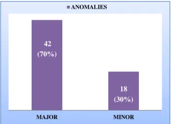
We classified the anomalies as per the European surveillance of congenital anomalies coding system. The anomalies in the study were divided into major and minor anomaly groups. 70% of the anomalies were classified as Major anomalies while 30% were classified as minor anomalies (Figure 2).
Figure 2: EUSCAT classification of anomalies in the study population.
In the present study, major anomalies identified involved the musculoskeletal system (21.6%) and cardiovascular system (20%). Minor anomalies were confined to dermatological disorders (27.7%) followed by musculoskeletal (16.6%) and genitourinary system (16.6%) (Figure 3).
Figure 3: System wise classification of the congenital anomalies.
Outcome of malformed neonates
Of the 40 anomalous babies five babies expired soon after birth. Three babies with neural tube defects were
42
18
MAJOR MINOR
ANOMALIES
(30%) (70%)
18.30%20%
6.60% 21.60%
10%
3% 13.30%
3% 3.30%
0.00% 5.00% 10.00% 15.00% 20.00% 25.00%
SYSTEM WISE DISTRIBUTION
Central Nervous System Cardiovascular System Gastro intestinal System Musculoskeletal System
Skin Respiratory System
operated and were found to be neurologically intact on follow up. Four neonates with cardiac lesions were operated of which 3 have survived. Among the babies with gastro intestinal anomalies three babies had been operated with no post-operative mortality. No mortality was noted in neonates with minor anomalies.
DISCUSSION
With the improvements in healthcare facilities in our country we have been able control infectious diseases and nutritional problems, which used to be major contributing factors to infant mortality.1 If the present trend continues,
congenital malformations and defects at birth would be one of the major determinant of neonatal and infant mortality in future especially in India, similar to the trend prevailing in the western countries at present.2-5
The incidence, pattern and prevalence of congenital anomalies is determined by several factors. Of which; genetic, ethnic and racial background are the key factors. The other factors also include socio economic, cultural and environmental factors and their interaction with the
genetic component which determines the occurrence of an anomaly.
The incidence of anomalies detected in our study was 6.3% which is slightly higher compared to the previous studies done in India by Jehangir et al, Chathurvedi et al and Bhide et al.6-8 But the present study results are
comparable to studies done by Swain et al, Baht BV et al in South India (3.7%), Dolk H et al in Europe (2.39%) and also with the western data from the EUROCAT surveillance.9-11
Several factors like the study population, duration of the study, the period and place where the study is conducted determine the incidence to a great extent therefore comparison of incidence with the present study is relatively difficult. We had a slightly higher incidence compared to previous studies done in the Indian settings. This could be possibly explained by the fact that the study was conducted in a referral hospital which caters high risk pregnancies with higher percentage of consanguinity, low socioeconomic class, nutritional and maternal problems.
Table 3: Overall distribution of congenital defects.
System (Total anomalies) %
with 11 system wise Major anomalies (N) % Minor anomalies (N) %
CNS-11
Hydrocephalus-2 18.1
Spina bifida occulta-1 9 Neural tube defects-6 (4 meningocele,
2 spina bifida anencephaly-1) 54.5 Sacrococcgyeal teratoma -1 9
CVS- 12
Vascular (TGA2, TAPVC1,
Coactation of aorta1)-4 33.3
Single umbilical artery-1 8.3 Septal defects (ASD 3, VSD 2)-5 41.6
Single ventricle physiology-1 8.3 Hypoplastic left heart syndrome -1 8.3
Musculoskeletal system-13
Limb deformities –6 (5 polydactyly,
1 congenital dysplasia of hip)
46.1
Clinodactyly - 1 7.69 Inguinal hernia-1 7.69 (Cleft lip1, cleftlip with cleft palate 2,
isolated cleft palate1)-4 30.7 Umbilical hernia-1 7.69 GIT-4 Vestibulorectal fistula-1 25 Hypertrophic pyloric
stenosis-2 50 Deodenal atresia-1 25
SKIN-6 Congenital icthyosis-1 16.6
Accessory nipples-2 33.3 Preauricular tag-1 16.6 Congenital non-neoplstic naevus-1 16.6 Hemangioma-1 16.6
Genitourinary system-8
Ambiguous genetalia-1 12.5 Cliteromegaly-1 12.5 Hypospadias-2 25 Congenital hydrocele-1 12.5 Bilateral hydronephrosis (>10mm)-2 25 Undescended testes -1 12.5 Respiratory system-2 Congenital lobar emphysema-1 50 Tracheomalacia-1 50
Endocrine-2 - - Congenital adrenal
hyperplasia-2 100
During the study period, a total of 60 anomalies were detected among 40 anomalous babies. Of which, 6 babies had multiple anomalies. 70 % of the total anomalies were major anomalies whereas minor anomalies were 30% (Figure 2).
In the present study, the predominant system involved in the anomalies is musculoskeletal followed by cardiovascular and central nervous system. They represent 59.9% of total defects (Figure 3). These results are similar to those reported by Baht BV et al who found that musculo-skeletal malformations were the commonest.10 Some authors like Verma et al previously
found predominance of central nervous system malformation in India Studies by Swain et al, Datta et al, Khanna et al, Taksande A et al, Anand JS et al have shown that incidence of Central nervous system and musculoskeletal system anomalies are the predominant anomalies noted in accordance with our study findings.12,9,13-16 This change in trend could be possibly
explained by introduction of periconceptional folic acid.
In the current study, Polydactyl is the most common defect of all the isolated birth defects with an incidence rate of 8.33%; which is 38.5% of neonates with musculoskeletal defects. Neural tube defects contributed to 10% of the total anomalies with meningomyelocele being present in three fourth of the cases among the neural tube defects. Other defects being hydrocephalus, Spina bifida and Sacrococcegal teratoma (Table 3).
Multiple defects were present in 15% of the babies (6 of 40 babies with anomalies) with significant associations. VACTER anomaly, Down syndrome, cleft lip and palate, neural tube defects and club foot being commonly found associations. Similar results were found by Charlotte et al where he has shown 18.2% of the neonates with multiple defects in his study.17 This is lower than that observed by
Coulibaly-Zerbo et al who had reported 45.2% in his study.18 However this could be due to the fact that some
of the associations and diseases could not be confirmed because of lack of further work up, early death and logistic reasons.
Prenatal risk factors associated with the occurrence of anomalies is also well established from previous studies. In our study, maternal diabetes is found to be a significantly common association with congenital anomalies.10 anomalous babies were born to diabetic
mothers in the present study. Other risk factors that were identified included maternal obesity, maternal poor socioeconomic status, maternal anaemia, malnutrition, maternal infections which was similar to other studies.12,13,16,19 Present study has also reinforced the
findings that consanguinity is an important risk factor for occurrence of congenital anomalies with 6.4% cases among consanguineous parents (13 among 201 mothers) and 3.4% (27 among 749 mothers) among non-consanguineous parents. Of the anomalous babies included in the present study, 5 died soon after birth. One
baby died post cardiac surgery. The highest death rate was found in neonates with multiple malformations. This is explained by the fact that multiple malformations prevent a harmonious development of the fetus with subsequent multi-organ failure. This finding is also similar to studies like Charlotte et al and Sarkar et al which had similar result.17,20
CONCLUSION
Congenital anomalies are an important cause of mortality and morbidity in developing countries like India. In this study we have demonstrated that multiparity, diabetes mellitus and consanguinity to be major contributing factors. Present study has highlighted that musculoskeletal and cardiovascular anomalies as the commonest major anomaly while dermatological and limb anomalies as most frequent minor anomaly. Antenatal screening is an effective tool to detect musculoskeletal and CNS anomalies. While cardiovascular anomalies may be missed by routine anomaly scan and a second scan may be needed in second trimester, fetal echocardiogram and fetal MRI may be useful in few cases. Early intervention and effective follow up have shown that good outcomes are possible even in while managing some of the major anomalies.
Funding: No funding sources Conflict of interest: None declared
Ethical approval: The study was approved by the Institutional Ethics Committee
REFERENCES
1. World Health Organization. Section on congenital anomalies. Available from: http://www.who.int/mediacentre/factsheets/fs370/en / Accessed October 2012.
2. National health Portal of India. Section on congenital anomalies (birth defects). Available at https://www.nhp.gov.in/disease/gynaecology-and-obstertrics/congenital-anomalies-birth-defects 3. UNICEF. Neonatal Health. Available at
Unicef.in/whatwedo/2/Neonatal-Health
4. Agarwal SS, Singh US, Singh PS, Singh SS, Das VI, Sharma AN, et al. Prevalence and spectrum of congenital malformations in a prospective study at a teaching hospital. Indian J Med Res. 1991;94:413-9. 5. Patel ZM, Adhia RA. Birth defects surveillance
study. Indian J Pediatr. 2005;72:489-91.
6. Jehangir W, Ali F, Jahangir T, Masood MS. Prevalence of gross congenital malformations at birth in the neonates in a tertiary care hospital. In APMC. 2009;3(1):47-50.
7. Chaturvedi P, Banerjee KS. Spectrum of congenital malformations in newborns from rural Maharashtra. Indian J Pediatr. 1989;56:501-7.
prevention, and surveillance implications. PloS One. 2016;11(11):e0166408.
9. Swain S, Agarwal A, Bhatia BD. Congenital malformations at birth. Indian Paediatr. 1994;31:1187-91.
10. Bhat BV, Babu L. Congenital malformations at birth: a prospective study from South India. Indian J Pediatr. 1998;65:873-81.
11. Dolk H, Loane M, Garne E. The prevalence of congenital anomalies in Europe. Advances Exp Med Biol. 2010;686:349-64.
12. Verma M, Chhatwal J, Singh D. Congenital malformations: a retrospective study of 10,000 cases. Indian J Pediatr. 1991;58(2):245-52.
13. Datta V, Chaturvedi P. Congenital malformations in rural Maharashtra. Indian Paediatr. 2000;37:998-1001.
14. Khanna MP, Prasad LS. Congenital malformations in the newborn. Indian J Paediatr. 1967;230:63-71. 15. Taksande A, Vilhekar K, Chaturvedi P, Jain M.
Congenital malformation at birth in central India, Indian J Hum Genetics. 2010;16:159-63.
16. Anand JS, Javadekar BB, Mala B. Congenital malformations in 2000 consecutive births. Indian Pediatr. 1988;25:845-51.
17. Charlotte TN, Aurore ND, Charlotte B, Esther B, Eugene BP. Prenatal diagnosis of congenital malformations in Douala General Hospital. Open J Obstet Gynecol. 2015;5(15):839.
18. Coulibaly-Zerbo F, Amorissani-Folquet M, Kacou-Kakou A, Sylla M, Noua F, Kramo E, et al. Epidemiological study of congenital malformations. Medicine of Black Africa. 1997;44(7):409-14. 19. Ajay K, Kalra K, Sharma V, Singh M, Dayal RS.
Congenital malformations. Indian Paediatr. 1984;21:945-9.
20. Sarkar S, Patra C, Dasgupta MK, Nayek K, Karmakar PR. Prevalence of congenital anomalies in neonates and associated risk factors in a tertiary care hospital in Eastern India. J Clin Neonatol. 2013;2(3):131.