Iran J Allergy Asthma Immunol August 2020; 19(4):437-446. Doi: 10.18502/ijaai.v19i4.4118
Increased Expression of B Lymphocyte Induced Maturation Protein 1
(BLIMP1) in Patients with Common Variable Immunodeficiency (CVID)
Shockrollah Farrokhi1, Faezeh Abbasi-rad2, Nafiseh Esmaeil2, Roya Sherkat3, Reza Yazdani4, Sanaz Afshar-Ghasemlou2, Saba Fekrvand4, and Mazdak Ganjalikhani-Hakemi2,3
1 Department of Immunology, Faculty of Medicine, Boushehr University of Medical Sciences, Boushehr, Iran
2 Department of Immunology, Faculty of Medicine, Isfahan University of Medical Sciences, Isfahan, Iran
3 Acquired Immunodeficiency Research Center, Isfahan University of Medical Sciences, Isfahan, Iran
4 Research Center for Immunodeficiencies, Pediatrics Center of Excellence, Children's Medical Center Hospital,
Tehran University of Medical Sciences, Tehran, Iran
Received: 6 December 2019; Received in revised form: 2 June 2020; Accepted: 11 June 2020
ABSTRACT
Common variable immunodeficiency (CVID) is a primary immune deficiency disorder characterized by a failure in B cell differentiation, impaired immunoglobulin production, and defect in response to vaccines. As a result of defective B cell maturation and differentiation in CVID, the affected patients commonly present with reduced numbers of memory B cell and antibody-secreting plasma cells. B-cell lymphoma 6 protein (BCL6) and B lymphocyte induced maturation protein 1 (BLIMP1) molecules are two important transcription factors that have key roles in the maturation of B cells to plasma cells. Hence, in the current survey, we aimed to evaluate the mRNA and protein expression levels of BCL6 and BLIMP1 in B lymphocytes isolated from peripheral blood in CVID patients.
We collected blood samples from 12 CVID patients and 12 healthy controls. We isolated peripheral blood mononuclear cells (PBMCs) using Ficoll density gradient separation. Then, CD19+ B cells were purified using MACS. The protein expression and transcriptional level of BCL6 and BLIMP1 were respectively measured using flow cytometry and real-time PCR.
Our results showed that the BLIMP1 mRNA expression, as well as BLIMP1 protein expression, were significantly higher in CVID patients compared to control subjects (p=0.009 and p=0.007, respectively). However, we found no significant difference in mRNA and protein expression of BCL6 between patients and healthy controls.
According to our findings, increased mRNA and protein expression levels of BLIMP1 could be involved in defective maturation of B cells in patients with CVID and elucidate mechanistic insights into the pathogenesis of this disorder.
Keywords: B-cell lymphoma 6 protein (BCL6); Common variable immunodeficiency (CVID); Plasma cell
Corresponding Author: Mazdak Ganjalikhani-Hakemi, PhD; Department of Immunology, Faculty of Medicine, Isfahan University
*First two authors contributed equally in this study.
INTRODUCTION
Common variable immunodeficiency (CVID) is a primary immunodeficiency syndrome defined by a decreased serum immunoglobulin level and impaired humoral immune responses associated with increased susceptibility to respiratory infections.1 Its prevalence
is estimated to be 1:50,000 to 1:25,000 cases in Caucasians.2 In addition to respiratory infections,
CVID patients manifest various clinical manifestations consisting of autoimmunity, gastrointestinal problems, lymphoproliferative diseases, malignancies and allergic diseases.3-7 To prevent and reduce infections in CVID
patients, prophylactic antibiotics and intravenous immunoglobulin (IVIG) are commonly used.8
In the past years, a range of abnormalities in innate and adaptive immune systems in CVID patients has been reported such as the reduced number and impaired T-cell stimulatory function of dendritic cells (DCs), decreased T-cell count and proliferative response to mitogens and antigens, defective T-cell signaling, low frequency and reduced suppressive capacity of regulatory T cell (Tregs), uncontrolled T-cell polarization, elevated levels of T-cell activation markers and apoptosis, and abnormality in cytokine production.9-13 The most important immunologic
feature of patients with CVID is a reduced number of the B-lymphocytes, especially the decline of the number of class-switched memory B cells as well as antibody-secreting plasma cells.14,15 Since the defect in
differentiation of B cell into antibody-secreting plasma cells has been confirmed in CVID patients, the assumption of a defect in some transcriptional factors such as B lymphocyte induced maturation protein 1 (BLIMP1) and B-cell lymphoma 6 protein (BCL6) molecules in CVID patients is rational.16-18 BCL6 is a
DNA–binding zinc finger protein and a suppressive nuclear factor16-18 that is found in lymphocytes
especially centroblasts and centrocytes in the germinal centers (GC).16 In the GC, BCL6 binds to a large
number of cell cycle inhibitor genes including Cdkn1 (encodes the P21) and Tp53 (encodes the p53)19 to
suppress them, thus, BCL-6 promotes the proliferation of B cells. BCL-6 is also essential for CD4+ follicular
helper T (TFH) cell differentiation in GC, which itself is
critical for germinal center B cell proliferation.20,21
Accordingly, an increase in B cell proliferation in some patients with CVID who were associated with increased levels of BCL6 has been reported.16-18
Reduction in the expression levels of BCL6 and PAX5 could promote differentiation of memory B cells into antibody-secreting plasma Cells.22 BLIMP1 (B
lymphocyte Induced Maturation Protein 1) is encoded by the PRDM1 gene and is also a suppressor transcription factor that binds to the DNA by a single-zinc finger domain.23,24 BLIMP1 plays a critical role in
the differentiation of B cells into antibody-producing cells and is a required transcription factor for the development and the survival of long-lived plasma cells in the bone marrow.25 The signaling pathway
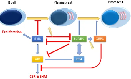
towards differentiation of plasma cells along with the involved proteins and their interaction with upstream and downstream proteins is provided in Figure 1. Moreover, it has been shown that continuous expression of BLIMP1 is essential for the maintenance of plasma cells, however, there is no expression of BLIMP1 in human memory cells.24 Thus, based on the
roles of BCL6 and BLIMP1 in the differentiation and maturation of antibody-secreting cells, in the current study, we evaluated the expression levels of BLIMP1 and BCL6 transcription factors in B cells of the patients with CVID.
MATERIALS AND METHODS
Subjects
The study population included 12 CVID patients who were diagnosed in Alzahra Hospital of Isfahan University of Medical of Sciences and 12 age- and sex-matched healthy controls. CVID patients were diagnosed according to the European Society for
Immunodeficiencies (ESID) criteria26 including
PBMC Isolation and Separation of CD19+ B Cells
Peripheral blood mononuclear cells (PBMCs) were
isolated from fresh-EDTA blood samples by
Lymphodex (BIOZ, Los Altos, CA, USA) density centrifugation. We isolated almost 10×106-15×106
PBMC from 10ml blood of each patient. CD19+ B
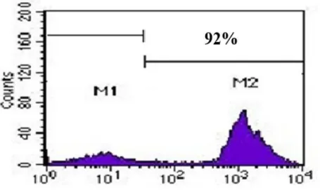
lymphocytes were separated; using positive selection with immunomagnetic microbead columns and B cell isolation kit according to the manufacturer’s protocol (MiltenyiBiotec, Gladbach, Germany). First, the CD19+ cells were magnetically labeled with CD19 MicroBeads. Then, the cell suspension was loaded onto a MACS column which was placed in the magnetic field of a MACS separator. The magnetically labeled CD19+ cells were retained within the column. The unlabeled cells ran through; this cell fraction was thus depleted of CD19+ cells. After removing the column from the magnetic field, the magnetically retained CD19+ cells could be eluted as the positively selected cell fraction. The purity of isolated B-cells was ≥90% as assessed by flow cytometric analysis (anti-CD19 PE) which was repeated 10 times (Figure 2).
Stimulation of B Cells
Purified B cells were washed twice with phosphate-buffered saline (PBS) and the purified B cells were
counted on a hemocytometer slide. Trypanblue exclusion was used to evaluate the viability assessment of the cells. Then, in each well of 24-well plate, 5×10⁵
isolated B cells were cultured in 1 mLRPMI-1640 medium (BIO-IDEA, USA) containing 10% heat-inactivated FBS (Waltham, MA, USA) and 1% penicillin/streptomycin (BioSera, France). Finally, 5 μg/mL of anti-human IgM antibody (Sigma-Aldrich, Germany) and anti-human CD40 antibody (BioLegend, San Diego, CA, USA) were added into the culture medium for B-cell Antigen Receptor (BCR) stimulation and were incubated at 37°C in a 5% CO2 containing humidified atmosphere for 24 hours. After these hours, the B cells were collected for flow cytometry analysis and reverse transcriptase-polymerase chain reaction (RT-PCR).
Flow Cytometry
Flow cytometry was performed to assess the expression of BCL6 and BLIMP1 transcription factors in isolated B cells 24 hours after stimulation with anti-IgM and anti-CD40 antibodies. We fixed and permeabilized the B cells using an Intra Stain kit (EskanTeb Asia, Tehran, Iran). To assay BLIMP1 and BCL6, the CD19+ cells were stained with
PE-labeled-anti-BLIMP1 (Santacruz, Dallas, Texas, USA) and FITC
Table 1. Specific primers designed for RT- q
Amplicon
size TM
Reverse primer (5' → 3') Forward primer (5' → 3')
Gene
213 bp 82.3
TGGCAGACCTGGCATTCA GAAGAAGCAGAACGGCAAGA
BLIMP1
120 bp 79.77
ATCCCTGTGAAATCTGTGGC TGTGACGGAAATGCAGGTTA
BCL6
94 bp 85.29
GTTGTCGACGACGAGCG AGCACAGAGCCTCGCCTTT
β-ACTIN
labeled anti-BCL6 (Santacruz, Dallas, Texas, USA) in the staining buffer. We used normal goat serum as a blocking agent. At least 1×10⁵ cells were scanned using
a Facscalibur FACS (BD Biosciences, San Jose, CA, USA). Finally, data analysis was performed using FlowJo software (Tree Star, Inc, Ashland, OR, USA).
RNA Extraction and cDNA Synthesis
Total RNA was extracted from stimulated B cells using an RNA extraction kit (YektaTajhizAzma, Tehran, Iran), according to the manufacturer's instructions. Subsequently, RNA pellets were dissolved in nuclease-free water and were applied for cDNA synthesis. cDNA strands were generated in a 20-μL reaction mixture by using 500 ng of total RNA as a template, 0.5 μg of oligo (dT)12-18, 0.25 mM deoxyribonucleoside triphosphate mixture, and 200 U of Revert Aid reverse-transcriptase enzyme according to manufacturer instructions (Thermo Fisher Scientific, Waltham, MA, USA), in the presence of a ribonuclease inhibitor.
Quantification of BLIMP1 and BCL6 Transcripts by Real-time PCR
After cDNA synthesis, quantification of the mRNA levels for BLIMP1 and BCL6 was performed by using Maxima SYBER Green/ROX qPCR Master Mix (2X) (ThermoFisher Scientific, Waltham, MA, USA). All primers, which are summarized in Table 1, were designed using Allele ID 7.0 software. PCR reactions were performed in a volume of 10 μL containing 1 μL of cDNA, 0.1 μmol/L of each primer, and 5 μL of Maxima SYBER Green/ROX qPCR Master Mix (2X). Amplification condition was: 95°C for 10 minutes followed by 40 cycles consisting of 15 seconds at 95°C, 60 seconds at 60°C and 60 seconds at 72°C. All PCR assays were performed in duplicate and data analyzed using the comparative threshold cycle method and the 2-ΔΔCt formula, the difference in expression of
these two genes was compared between healthy and patient groups. The expression of β-actin was used as
internal control and RNA samples from the healthy individuals were used as quality control in each RT-PCR reaction.
Statistical Analysis
We used the GraphPad Prism 7.03 software for statistical analyses of the data. Kolmogorov–Smirnov and Shapiro-Wilk tests were used to k data normality. Based on the finding of these tests, we performed parametric and non-parametric analyses (independent sample t-test for data with a normal distribution and Mann– Whitney U test for data with skewed distribution). p-value<0.05 was considered statistically significant.
RESULTS
Clinical Characteristics of Patients
In this study, 12 patients (6 females and 6 males per group) referred to the Al-Zahra Hospital of
Isfahan University of Medical Sciences were
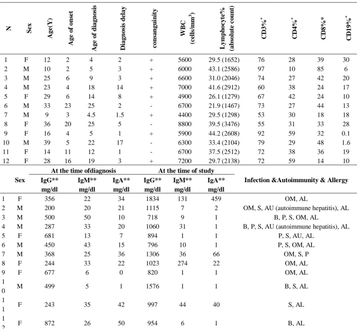
evaluated. The mean age of the patients at the time of this study, at the onset of clinical symptoms and at the time of diagnosis were 22.8 years, 8.49 years, and 13.53 years, respectively. Allergies (11 patients, 91.7%) followed by sinusitis (8 patients, 66.7%) and otitis media (7 patients, 58.3%) were the most prevalent clinical manifestations in our studied patients. Other less common manifestations were pneumonia (5 patients, 41.7%), bronchiectasis (4 patients, 33.3%), and autoimmune disorders (2 patients, 16.7%). Detailed data of demographic and immunological characteristics of the studied CVID patients are provided in Table 2.
Increased Transcript Level of BLIMP1 Gene Expression in B Cells of CVID Patients
We evaluated possible changes in the mRNA level
of BLIMP1 and BCL6 in patients compared with
healthy controls. Our data showed that the expression
CVID patients in comparison with the healthy controls and this was statistically significant (p=0.009). In consistent with our flow cytometry results, the transcript level of BCL6 had no statistical difference between the patients’ and healthy controls’ groups (p=0.45), as is shown in Figure 3.
Increased BLIMP1 and BCL6 Protein Expression in B Cells of CVID Patients
Changes in mRNA expressions disclosed by RT-qPCR are not always a good indicator of changes in protein expression. In this regard, we performed flow
cytometry analysis on isolated CD19+ B cells after 24
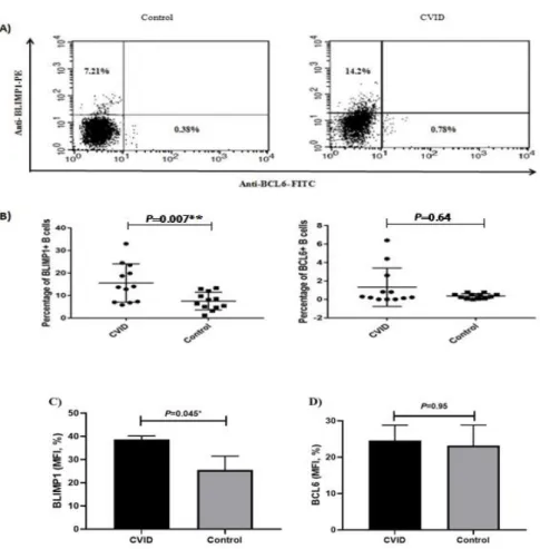
hours of activation and stimulation by anti-IgM and anti-CD40 antibodies. Our result demonstrated that the expression of BLIMP1 protein in patients with CVID was significantly higher compared to that of the healthy controls (p=0.007). Also, our data indicated that the expression of BCL-6 protein was higher in CVID patients, however, this difference was not statistically significant (p=0.64). Figure 4 represents flow cytometry results and MFI data of the BCL-6 and BLIMP1 expression in patients compared to the healthy controls.
Figure 2. The histogram graph demonstrates the purity of isolated B-cells by flow cytometric analysis (anti-CD19PE).
Table 2. Summary of the demographic and immunological characteristics of subjected common variable immunodeficiency (CVID) patients
N Sex
A ge (Y ) A ge o f on se t A ge o f d iagn o si s D iagn o si s d el ay con sa n gu in it y WB C (c el ls /m m 3 ) L ym p h oc y te % (ab sol u te c o u n t) C D 3% * C D 4% * C D 8%* C D 19% *
1 F 12 2 4 2 + 5600 29.5 (1652) 76 28 39 30
2 M 10 2 5 3 + 6000 43.1 (2586) 97 10 85 6
3 M 25 6 9 3 + 6600 31.0 (2046) 74 27 42 20
4 M 23 4 18 14 + 7000 41.6 (2912) 60 38 24 17
5 F 29 6 14 8 + 4900 26.1 (1279) 67 42 24 10
6 M 33 23 25 2 - 6700 21.9 (1467) 73 27 44 13
7 M 9 3 4.5 1.5 + 4400 29.5 (1298) 53 30 18 18
8 F 36 20 25 5 - 8800 39.5 (3476) 55 31 33 28
9 F 16 4 5 1 + 5900 44.2 (2608) 92 59 32 0.1
10 M 39 5 22 17 - 6300 33.4 (2104) 79 29 48 1.6
11 F 14 11 12 1 - 6700 37.5 (2512) 72 38 36 19
12 F 28 16 19 3 + 7200 29.7 (2138) 72 59 14 10
Sex
At the time ofdiagnosis At the time of study
Infection &Autoimmunity & Allergy IgG** mg/dl IgM** mg/dl IgA** mg/dl IgG** mg/dl IgM** mg/dl IgA** mg/dl
1 F 356 22 34 1834 131 459 OM, AL
2 M 200 20 21 1115 7 2 OM, S, AU (autoimmune hepatitis), AL
3 M 500 50 10 718 9 1 B, P, S, OM, AL
4 M 287 33 20 1060 31 1 B, P, S, AU (autoimmune hepatitis), AL
5 F 681 13 7 894 1 1 P, S, AU, AL
6 M 450 43 15 796 10 1 P, S, OM, AL
7 M 368 25 36 1306 36 66 OM, S, P
8 F 244 33 22 1023 274 22 OM, AL
9 F 677 6 0 820 1 1 OM, AL
1
0 M 499 5 1 1576 1 1 B, S, AL
1
1 F 243 35 42 997 44 40 S, AL
1
2 F 872 26 50 954 6 1 B, AL
N: number; M: male; F: female; Y: year; +: positive; -: negative; WBC: white blood cell; Ig: immunoglobulin; OM: otitis media; B: bronchiectasis; P: pneumonia; S: sinusitis; AU: autoimmunity; AL: allergy. * Lymphocyte subset reference ranges (27):
CD3: 16-85% CD4: 28-58% CD8: 19-48% CD19: 7-23%
** Immunoglobulins reference ranges (https://emedicine.medscape.com/article/2157901-overview) Age 0-1 years: IgG (231-1411 mg/dL), IgM (0-145 mg/dL), IgA (0-83 mg/dL)
Figure 4. Flow cytometry analysis and MFI data of B lymphocyte induced maturation protein 1 (BLIMP1) and B-cell lymphoma 6 protein(BCL6) expressing cells in a population of purified CD19+ B cells isolated from patients with common variable immunodeficiency (CVID) and healthy controls. Expression of BLIMP1 and BCL6 transcription factors in B-cell isolated from blood samples of healthy controls and CVID patients. A) Dot plots manifest expression of BLIMP1 and BCL6 in B-cells of representative control and a patient with CVID. The cells were stained with anti-BLIMP1 PE and anti BCL6 FITC after 24 hours of stimulation. The plots are based on the gating of viable cells in the forward and side scatter plots. B) The bar chart demonstrates the mean percentage of BLIMP1 and BCL6 expression in B cells of healthy controls and CVID patients. C) BLIMP1 MFI percentage is significantly elevated in CVID compared to controls while D) the BCL6 MFI percentage is unchanged. Results are presented as Mean±SEM. *p<0.05, **p<0.01 and ***p<0.001
DISCUSSION
BLIMP1 and BCL-6 are two important transcription factors that have important roles in the differentiation and maturation of B cells. Almost 90% of CVID patients have normal numbers of B-cells, along with diminished memory B-cells and plasmablasts that indicates a defect in the final stages of B cell differentiation to antibody-secreting plasma cells.28
Hence, in the present study, we evaluated mRNA and protein expression levels of BLIMP1 and BCL6 in a group of CVID patients to identify underlying mechanisms in the pathogenesis of CVID. Both mRNA and protein expression levels of BLIMP1 were
significantly higher in the CVID group compared to the healthy controls, while there was no significant difference in the mRNA and protein levels of BCL6 between patients and healthy groups.
differentiate into plasma cells (24, 29). In another study on the same set of patients, Ansari et al have shown a significant decrease in the number of plasmablasts in the same CVID patients compared with healthy subjects, which complicates the situation.30 It could be assumed
that this BLIMP1 is probably a protein without proper function in these patients or probably there are other unknown mechanisms that nullify the function and the effects of the high expression level of BLIMP1 in our patients. In other words, a probable loss-of-function mutation or an inhibitory epigenetic effect might suppress the compensatory effect of BLIMP1 expression in these patients. Therefore, it is important to investigate the phosphorylation, dephosphorylation, and histone modification levels of this protein and also its proper interaction with its downstream proteins, such as XBP1 or upstream proteins including signal transducer and activator of transcription 3 (STAT3) and interferon regulatory factor 4 (IRF4). Moreover, there is double-positive feedback between IRF4 and BLIMP1 in B cells. Simultaneously with our study, Afshar et al have investigated XBP1 and IRF4 expression in our subjects and they found a significant increase in the mRNA and protein expression of IRF4 and a slight, but not significant, decrease in the mRNA and protein expression of XBP1 in the B cells.31 Accordingly, high
expression of IRF4 in the patients may be due to the increased BLIMP1 which did not augment the XBP1 expression located in its downstream and consistent with our results, Afshar et al findings also indicate the likelihood of loss-of-function mutation in BLIMP1 and IRF4. It is noteworthy to remind that, increasing IRF4 entails in up-regulation of BLIMP1 and vice versa (Figure 1).
In the present study, we also showed a slight, albeit not significant, increase in the protein level of BCL6 in CVID patients in comparison to the healthy controls. Several transcription factors including Bcl-3, Bcl-6, NF-kB/p52, and IRF-4 are required for the formation of germinal centers (GCs) in the process of B cell differentiation.32 Warnatz et al have shown that GCs are
normal in CVID patients, however, there are different degrees of inappropriate and hyperplastic GCs in most of them.33 High expression of BCL6 in our study and
IRF4 in Afshar et al study in the same CVID patients, is indicative of possible induction of hyperplastic GCs in these patients by stimulation of BCL6 and IRF4 signaling pathways.
In another study, Taubenhelm et al, assessed the
expression level of BLIMP1 and BCL6 molecules in the lymph nodes of CVID patients, but not in peripheral blood.16 In this study, increased expression of BLIMP1
in pre-plasmablast cells in the lymph nodes of CVID patients has been demonstrated. Moreover, consistent with our findings they found the high expression of BCL6 in the early stages of differentiation of B-cell into centrocytes, which had been accompanied by an increase in the proliferation of B cells at this stage.16
The results of this study are consistent with our findings. It has been reported that differentiation of the centrocytes into antibody-secreting plasma cells correlates with high-level expression of BLIMP1, while PAX5, BCL6, and BATCH2 levels are significantly reduced. Indeed, BCL6 is an important transcription factor for the development and propagation of B-cells and directly inhibits BLIMP1.18 This is in contrast to
our findings and we suggest that there may be other less studied molecules or regulatory mechanisms which interfere with the direct action of BCL6 on downregulation of BLIMP1. Another possibility is that, if we assume that BLIMP1 was non- functional in our patients, its high level of expression could be unable to suppress BCL6 expression. As previously mentioned, Ansari et al have indicated that plasmablasts were significantly reduced in our patients, which is consistent with this elevated BCL6 levelBCL6 inhibits differentiation of plasmablasts to plasma cells with repressing BLIMP1, XBP-1, and IRF4.34 Thus, the
non-reduced level of BCL6 in our patients could be interpreted as a probable reason for the low number of their plasmablasts.
We had limitations for isolating B cells from these patients; hence we had to evaluate BLIMP1 expression in stimulated cells. We were not able to compare BLIMP1 expression levels in unstimulated cells from patients and the healthy control group. Therefore, we suggest for assaying this molecule in both unstimulated and stimulated conditions.
CONFLICT OF INTEREST
The authors declare no conflicts of interest.
ACKNOWLEDGEMENTS
This work was financially supported by the Isfahan University of Medical Sciences (394669) and Boushehr University of Medical Sciences (4016).
REFERENCES
1. Yong P, Thaventhiran J, Grimbacher B. “A rose is a rose is a rose,” but CVID is Not CVID common variable immune deficiency (CVID), what do we know in 2011? Adv Immunol. 2011;111:47-107.
2. Hammarström L, Vorechovsky I, Webster D. Selective
IgA deficiency (SIgAD) and common variable
immunodeficiency (CVID). Clin Exp Immunol.
2000;120(2):225-31.
3. Chapel H, Lucas M, Lee M, Bjorkander J, Webster D,
Grimbacher B, et al. Common variable
immunodeficiency disorders: division into distinct clinical phenotypes. Blood. 2008;112(2):277-86. 4. Wehr C, Kivioja T, Schmitt C, Ferry B, Witte T, Eren E,
et al. The EUROclass trial: defining subgroups in
common variable immunodeficiency. Blood.
2008;111(1):77-85.
5. Cunningham-Rundles C, Bodian C. Common variable immunodeficiency: clinical and immunological features of 248 patients. Clin Immunol. 1999;92(1):34-48. 6. Tak Manesh A, Azizi G, Heydari A, Kiaee F, Shaghaghi
M, Hossein-Khannazer N, et al. Epidemiology and pathophysiology of malignancy in common variable
immunodeficiency? Allergol Immunopath.
2017;45(6):602-15.
7. Yazdani R, Heydari A, Azizi G, Abolhassani H, Aghamohammadi A. Asthma and Allergic Diseases in a Selected Group of Patients With Common Variable Immunodeficiency. J Investig Allergol Clin Immunol. 2016;26(3):209-11.
8. Azizi G, Abolhassani H, Asgardoon MH, Rahnavard J, Dizaji MZ, Yazdani R, et al. The use of Immunoglobulin Therapy in Primary Immunodeficiency Diseases. Endocr MetabImmune. 2016;16(2):80-8.
9. Azizi G, Rezaei N, Kiaee F, Tavakolinia N, Yazdani R, Mirshafiey A, et al. T-Cell Abnormalities in Common Variable Immunodeficiency. J Investig Allergol Clin Immunol. 2016;26(4):233-43.
10. Azizi G, Hafezi N, Mohammadi H, Yazdani R, Alinia T, Tavakol M, et al. Abnormality of regulatory T cells in common variable immunodeficiency. Cell Immunol. 2017;315:11-7.
11. Yazdani R, Seify R, Ganjalikhani-Hakemi M,
Abolhassani H, Eskandari N, Golsaz-Shirazi F, et al. Comparison of various classifications for patients with common variable immunodeficiency (CVID) using measurement of B-cell subsets. Allergol Immunopath. 2017;45(2):183-92.
12. Yazdani R, Ganjalikhani-Hakemi M, Esmaeili M, et al. Impaired Akt phosphorylation in B-cells of patients with common variable immunodeficiency. Clin Immunol. 2017;175:124-32.
13. Sharifi L, Tavakolinia N, Kiaee F, Rezaei N, Mohsenzadegan M, Azizi G, et al. A Review on Defects
of Dendritic Cells in Common Variable
Immunodeficiency. ENDOCR METAB IMMUNE.
2017;17(2):100-13.
14. Tam JS, Routes JM. Common variable
immunodeficiency. Am J RhinolAllergy. 2013;27(4):260. 15. Warnatz K, Schlesier M. Flowcytometric phenotyping of common variable immunodeficiency. Cytometry B Clin Cytom. 2008;74(5):261-71.
16. Taubenheim N, von Hornung M, Durandy A, Warnatz K, Corcoran L, Peter H-H, et al. Defined blocks in terminal plasma cell differentiation of common variable
immunodeficiency patients. J Immunol.
2005;175(8):5498-503.
17. Nutt SL, Taubenheim N, Hasbold J, Corcoran LM, Hodgkin PD, editors. The genetic network controlling
plasma cell differentiation. Semin Immunol.
2011;23(5):341-9.
18. Kuo TC, Shaffer AL, Haddad J, Choi YS, Staudt LM, Calame K. Repression of BCL-6 is required for the formation of human memory B cells in vitro. J Exp Med. 2007;204(4):819-30.
19. Phan RT, Dalla-Favera R. The BCL6 proto-oncogene suppresses p53 expression in germinal-centre B cells. Nature. 2004;432(7017):635-9.
20. Nurieva RI, Chung Y, Martinez GJ, Yang XO, Tanaka S, Matskevitch TD, et al. Bcl6 mediates the development of T follicular helper cells. Science. 2009;325(5943):1001-5. 21. Crotty S, Johnston RJ, Schoenberger SP. Effectors and memories: Bcl-6 and Blimp-1 in T and B lymphocyte differentiation. Nat Immunol. 2010;11(2):114.
plasma cells and pre-plasma memory B cells. Immunity. 2003;19(4):607-20.
23. Tsuji S, Cortesão C, Bram RJ, Platt JL, Cascalho M. TACI deficiency impairs sustained Blimp-1 expression in B cells decreasing long-lived plasma cells in the bone marrow. Blood. 2011;118(22):5832-9.
24. Kallies A, Hasbold J, Tarlinton DM, Dietrich W, Corcoran LM, Hodgkin PD, et al. Plasma cell ontogeny defined by quantitative changes in blimp-1 expression. J Exp Med. 2004;200(8):967-77.
25. Turner CA, Mack DH, Davis MM. Blimp-1, a novel zinc finger-containing protein that can drive the maturation of B lymphocytes into immunoglobulin-secreting cells. Cell. 1994;77(2):297-306.
26. Seidel MG, Kindle G, Gathmann B, Quinti I, Buckland M, van Montfrans J, et al. The European Society for
Immunodeficiencies (ESID) Registry Working
Definitions for the Clinical Diagnosis of Inborn Errors of
Immunity. J Allergy CIin Immunol Pract.
2019;7(6):1763-70.
27. Reichert T, DeBruyère M, Deneys V, Tötterman T, Lydyard P, Yuksel F, et al. Lymphocyte subset reference ranges in adult Caucasians. Clin Immunol Immunopathol. 1991;60(2):190-208.
28. Jacquot S, Maçon-Lemaître L, Paris E, Kobata T, Tanaka Y, Morimoto C, et al. B cell co-receptors regulating T cell-dependent antibody production in common variable immunodeficiency: CD27 pathway defects identify subsets of severely immuno-compromised patients. Int Immunol. 2001;13(7):871-6.
29. Martins G, Calame K. Regulation and functions of Blimp-1 in T and B lymphocytes. Annu Rev Immunol. 2008;26:133-69.
30. Ansari M, Yazdani R, Sherkat R, Homayouni V,
Ganjalikhani-Hakemi M, Rezaei A. Decreased
Expression of B cell Maturation Antigen in Patients with Common Variable Immunodeficiency. Pediat Aller Imm Pul. 2017;30:7-13.
31. Afshar-Ghasemlou S, Esmaeil N, Sherkat R, Yazdani R, Abbasi-Rad F, Ganjalikhani hakemi M, et al. Increased IRF4 expression in isolated B cells from Common variable immunodeficiency (CVID) patients. Allergol Immunopath. 2019;47(1):52-9.
32. Reimold AM, Iwakoshi NN, Manis J, Vallabhajosyula P, Szomolanyi-Tsuda E, Gravallese EM, et al. Plasma cell differentiation requires the transcription factor XBP-1. Nature. 2001;412(6844):300.
33. Unger S, Seidl M, Schmitt-Graeff A, Böhm J, Schrenk K, Wehr C, et al. Ill-defined germinal centers and severely reduced plasma cells are histological hallmarks of lymphadenopathy in patients with common variable immunodeficiency. J Clin Immunol. 2014;34(6):615-26. 34. Nutt SL, Fairfax KA, Kallies A. BLIMP1 guides the fate