Plasmon Enhanced Light Harvesting: Multiscale Modeling of the
FMO Protein Coupled with Gold Nanoparticles
Oliviero Andreussi, Stefano Caprasecca, Lorenzo Cupellini, Ingrid Guarnetti-Prandi, Ciro A. Guido,
Sandro Jurinovich, Lucas Viani, and Benedetta Mennucci*
Department of Chemistry, University of Pisa, Via Moruzzi 3, 56124 Pisa, Italy
ABSTRACT: Plasmonic systems, such as metal nanoparticles, are becoming increasingly important in spectroscopies and devices because of their ability to enhance, even by several orders of magnitude, the photophysical properties of neighboring systems. In particular, it has been shown both theoretically and experimentally that combining nanoplasmonic devices with natural light-harvesting proteins substantially increases thefluorescence and absorption properties of the system. This kind of biohybrid device can have important applications in the characterization and design of efficient light-harvesting systems. In the present work, the FMO light-harvesting protein was combined with gold nanoparticles of different sizes, and its photophysical properties were characterized using a multiscale quantum-mechanical classical-polarizable and continuum model (QM/ MMPol/PCM). By optimal tuning of the plasmon resonance of the metal nanoparticles, fluorescence enhancements of up to 2 orders of magnitude were
observed. Orientation effects were found to be crucial: amplifications by factors of up to 300 were observed for the absorption process, while the radiative decay of the emitting state increased at most by a factor of 10, mostly as a result of poor alignment of the emitting state with the considered metal aggregates. Despite being a limiting factor for high-fluorescence-enhancement devices, the strong orientation dependence may represent an important feature of the natural light-harvesting system that could allow selective enhancement of a specific excited state of the complex.
1. INTRODUCTION
Plasmons have gained increasing attention in the manipulation and control of interactions of light with matter, particularly with nanoscale metal systems, doped semiconductors, or gra-phene.1−6 The localized surface plasmon resonance (LSPR) that exists at a surface of a plasmonic nanostructure can intensify light scattering and enhance light concentration around the structure, and this can be exploited to enhance the optical properties of nearby molecular systems. For example, the fluorescence of an emitting system placed in localized“hot spots”can be greatly increased as a result of an enhancement of the excitation field and modifications of the radiative rate that increase the effective quantum efficiency (QE). As a result, afluorophore with a low intrinsic QE can be
transformed into a brighter and more efficient emitter.
Moreover, because of the higher radiative rate, the excited-state lifetime is reduced with a resulting increase in photo-stability. These effects have been largely used to enhance the
fluorescence properties of organic dyes,7−14but more recently it has been shown that the same characteristics can also be exploited to enhance the light-harvesting (LH) properties of natural photosynthetic systems.15−21 In particular, the LH efficiency can be drastically enhanced by tuning the plasmon frequency of the constituent plasmonic particles to coincide with the maximal photon flux of sunlight. These findings suggest that natural LH complexes can serve as molecular building blocks in artificial photosynthetic systems for
high-efficiency sunlight harvesting and solar energy conversion. In parallel, the plasmon-induced enhancements of LH properties can be used for important spectroscopic applications. In fact, the low fluorescence efficiency and limited photostability of natural LH complexes have hampered single-molecule studies to date. This latter instead would be of paramount importance to definitely explain the mechanisms beyond adaptations of these systems to changes in the local electronic structure tofind optimal energy transfer pathways. The relatively recent development of two-dimensional electronic spectroscopy (2DES) has in fact provided a completely new picture of the energy transfer pathways in photosynthetic complexes22−26 since the first application to the photosynthetic Fenna− Matthews−Olson complex (FMO). The coherent oscillations observed for this and other photosynthetic systems in the 2DES data that persist even at room temperature seem to suggest a fundamental role of quantum effects, but an unequivocal proof of this role in photosynthesis or in other biological processes is still missing.27−29
In the present study, we demonstrate via quantum-chemistry-based approaches that plasmonic effects can indeed be used to control the electronic processes in natural LH systems and to
Special Issue: Jacopo Tomasi Festschrift
Received: September 30, 2014
Revised: November 22, 2014
Article
pubs.acs.org/JPCA
selectively enhance their excitation and emission properties. For
this study, we chose the FMO pigment−protein complex
(PPC) of green sulfur bacteria, which has been extensively used as a model system for larger antenna complexes. The main function of FMO is to transfer excitation energy from the chlorosome, the principal LH complex of these anaerobic bacteria, to the reaction center (RC) (see Figure 1a,b).30−35 The whole structure of FMO accounts for three identical
monomer units arranged with C3 symmetry and connected
through ionic bridges between charged residues of adjacent helices. Each monomeric unit contains eight bacteriochlor-ophyll a (BChl) molecules. In the FMO, the excitation energy
flow is achieved by suitable tuning of the excitation energies of the pigments and the pigment−pigment couplings. Since the pigments are equivalent, the energy funnel is mainly determined by the protein environment, which perturbs the excited states through an electrostatic effect due to the bulk and specific pigment−protein interactions and an indirect effect due
to pigment geometry distortions. Experimental22,36 and
theoretical37−42 results indicate that the excitation energy
flows from the pigment facing the outer antenna system to the pigment closer to the RC (see Figure 1c).
In FMO, as in other PPCs, pigments are found at very short distances (less than 10 Å); as a result, strong pigment−pigment interactions determine a delocalization of the excited states over different pigments.43 In this work, we employed a semi-empirical QM approach to describe the excitonic states of FMO by considering a complex made of the eight BChls constituting a monomer unit in combination with gold nanoparticles of different sizes (see Figure 1). The QM description of the pigments was coupled with a polarizable atomistic model (MMPol) to include the protein effects, and a polarizable continuum model (PCM) to describe the metal nanoparticles
(MNPs). The resulting QM/MMPol/PCM multiscale
ap-proach44 was used not only to determine the optimal
composition of the biohybrid device (number and type of metal nanoparticle, hotspot configurations) but also to investigate the plasmonic effects in the different processes that combine to give the resultingfluorescence behavior.
The paper is organized as follows: In section 2 the numerical details of the simulations are summarized. In section 3.1 we present the main properties of the two components of the proposed devices, namely, the FMO protein and the gold metal nanoparticles; in section 3.2 the results obtained for different setups of the devices are presented and discussed. Section 4 provides concluding remarks.
2. METHODOLOGY
Simulations were performed with a development version of the
Gaussian 09 simulation package45 using the ZINDO
semi-empirical method with Zerner’s spectroscopic parametriza-tion46 to describe the excited states of the eight interacting BChls contained in the monomer unit. The remaining parts of the FMO system, comprising the protein matrix and the BChls of the second and third monomers, were modeled with an atomistic classical polarizable approach based onfixed charges and induced dipoles.47 In the present calculations, protein amino acids were described using the recent charge and polarizability parameters derived by Wang and co-workers in the context of the Amber forcefield.48,49BChl parameters were derived as described in ref 42. In this work we used the crystal structure ofProsthecochloris aestuariiFMO protein determined at 1.3 Å resolution (PDB code 3EOJ).50Since the occupancy of
the BChl 8 site is 0.341 molecules/site, two different
conformations of FMO (holo and apo) can be found, and
Figure 1.(a) Schematic representation of the biohybrid system. (b) Close-up view of the biometal junction highlighting the sections of the FMO trimer considered in the QM, MM, and MMPol layers. (c) Relative positioning of chlorophylls in the FMO monomer. (d) Fragment of the bacteriochlorophyll considered in the QM calculations. Thefigure was rendered with the help of MView, www.mview-tools.com.
they are related to the structures in which the BChl 8 site is occupied or not occupied by the pigment, respectively. In our simulation, we used the holo structure considering eight BChls per monomer. Thefirst six residues are missing in the crystal structure, and the residue Asp7 contains only two atoms. Thus, we replaced the Asp group with a methyl acetate (Ace) capping group for the C-terminal side. We also manually added the missing residue Glu214 and adjusted the conformation of Lys215, which is present only in the apo form. For the protein missing atoms, the apo coordinates were used and a standard protonation pattern for titrable residues was assumed. All of the
histidine (His) residues were considered to be in the ε
configuration except for His110, -145, -290, and -298, which were considered with hydrogen at theδposition to allow axial coordination with the Mg atom of BChls. After addition of the hydrogen atoms, two minimization steps were performed using the Amber 12 suite of programs.51In thefirst step we relaxed all of the hydrogen atoms, all of the atoms of the terminal Ace group, and residues 213−215. Then the FMO trimer was built using the crystallographic symmetry transformation of the monomer unit, and a second minimization step of the trimer structure was performed to relax the hydrogen atoms in order to avoid close contacts at the interface of the different monomers; the heavy atoms of the tail of BChl 8 that were missing in the structure were also relaxed.
In the QM analysis we considered only one monomer unit: BChls 1 to 7 were selected from monomer unit A, whereas BChl 8 was selected from unit B because of the smaller distances between this pigment and the other selected ones. The system is shown in Figure 1c.
The nanoplasmonic devices were modeled as continuum dielectric bodies exploiting a modified version of the PCM,52as described in refs 53−55. As the absorption/emission bands of BChls in FMO end up in the red region of the visible spectrum, we decided to consider gold nanoparticles as the plasmonic system, since gold shows resonances at lower energies with respect to other metals used in plasmon-enhancing devices (e.g., silver). The dielectric constant of bulk gold at optical frequencies was taken from experimental data as reported by Palik.56Single aggregates placed along the symmetry axis of the
FMO trimer on the RC side (“down”) were considered,
together with pairs of aggregates placed symmetrically along the axis in the so-called hotspot configuration (see Figure 1a). Single aggregates placed on the chlorosome side (“up”) of the protein were also considered, but the results showed similar qualitative features as the results for the“down”configurations and will not be discussed in the following. The aggregates were modeled as the union of interlocking spheres with a center− center distance equal to the sphere radius (see Figure 1a). For simplicity, only linear aggregates were considered, and the number of spheres in the aggregate was optimized so as to tune the plasmon resonances in the spectral region of absorption/ emission of the FMO complex, i.e., around 770−830 nm (see Figure 3).
3. RESULTS AND DISCUSSION
3.1. The Isolated Components: FMO and MNPs. 3.1.1. FMO Complex. We computed the low-energy excited states of the coupled system using the ZINDO/MMPol method and selected the first eight bright states (S0−S8), which result from mixing of the Qytransitions localized on the
BChls. The transition energies and electric transition dipole moments are reported in Table 1.
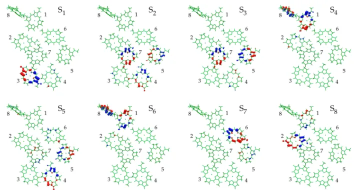
From the analysis of the transition densities reported in Figure 2, we assigned the nature and the composition of the
first eight excitonic states, which correspond to locally excited Qy states and do not show charge transfer character. The
excitonic picture shows that the lowest-energy states are localized on BChls 3, 4, 5, and 7, which face the RC side. In particular, the lowest state, S1, which is responsible for the redmost peak in the spectrum, is almost entirely localized on BChl 3, in agreement with the literature.40,41,57−59 The high-energy states are localized on BChls 1, 2, and 6, which are located on the baseplate side. BChl 8, which seems to be the linker between the baseplate and the FMO complex, has been identified in previous works as the blue-shifted pigment. From our analysis, BChl 8 contributes to middle-energy states S4and
S6. This may be due to the fact that BChl 8 is the most external pigment in the FMO and therefore the most exposed to the solvent, which we did not include in our simulation.
Finally, we simulated the low-energy part of the absorption spectrum using a convolution of Gaussian line shape functions with variableσin order to betterfit the experimental data. The spectrum is reported in Figure 3, and compared with the low-temperature experimental one.60All of the main features of the experimental spectrum are well-reproduced by the theoretical simulation, showing that the QM/MMPol approach used is
able to describe the interpigment and pigment−protein
interactions. Indeed, when we switched off the electrostatic environmental effects, the resulting in vacuo simulated spectrum lost the characteristic band shape. In a more detailed comparison, it can be seen that the simulated peaks are spread over a wider range of wavelength with respect to the experimental ones, indicating that the semiempirical model seems to overestimate the effects of geometry distortion and polarization on the excitation energies.
3.1.2. Metal Nanoparticles. In order to have large enhancements of the FMO properties, the metal aggregates need to be chosen in such a way that their plasmon resonances end up in the region of BChl absorption and emission. While spherical gold aggregates show resonances at about 500 nm, elongated aggregates show localized plasmons at longer wavelengths and thus represent the main targets of the considered application. The plasmonic properties of the metal aggregates can be characterized with the exploited methodology by looking at the different components of the polarizabilities computed using the PCM operators as described previously.54 While the intensity of the plasmonic resonance depends significantly on the size (radius) of the spheres used to model the MNP, the position of the resonance depends only on the aspect ratio of the aggregate (the longest dimension divided by Table 1. Excitonic Energies, Electric Transition Dipole Moments, and Oscillator Strengths of the First Eight Bright States Computed at the ZINDO/MMPol Level of Theory along with the Main Contributions from the Eight BChls
state E(eV) λ(nm) μ2(D2) f contrib.
S1 1.285 965 155 0.755 3
S2 1.361 911 271 1.401 4, 5, 7
S3 1.389 893 148 0.779 5, 7
S4 1.401 885 171 0.909 1, 8
S5 1.421 872 170 0.914 4, 5
S6 1.438 862 201 1.095 1, 8
S7 1.507 823 111 0.635 6
S8 1.532 809 90 0.522 1, 2
The Journal of Physical Chemistry A Article
the diameter). Moreover, multiple aggregates can interact strongly among each other and at short interaggregate separations can present plasmonic resonances at lower energies with respect to isolated aggregates. For the considered applications, single elongated aggregates and aligned pairs of elongated ones were considered, with aspect ratios between 2.5 and 4.5, which correspond to linear systems composed of approximately four to eight interlocking spheres. In order to keep the overall dimensions of the system within the limits imposed by the electrostatic assumption intrinsic in our model (i.e., smaller than the wavelength of the incident/emitted radiation), a sphere radius of 125 Å was used for all of the simulated metal particles. Although the largest systems treated in this work were at the limit of validity of the quasistatic approximation, the results for these systems can still serve as guidance.
In Figure 4 we report the real and imaginary components of the metal polarizability along the main axis of the different aggregates. From the calculations it appears that the single aggregates composed of six spheres and the pairs of aggregates composed of five spheres present resonances in the required region of the visible spectrum. The coupling between the aggregates is responsible for a shift of about 50 nm in the plasmon peaks at the shortest interaggregate separation considered. In order to be of use in experimental applications, the plasmonic properties of the MNPs were tuned in the experimental spectral operation region of FMO. Therefore, from the reported polarizability trends, the single aggregates of
five to seven spheres and the pairs of aggregates offive and six spheres were selected as the best candidates to enhance the spectroscopic properties of the FMO-based device. Throughout the rest of this article, single aggregates are labeled SN, whereN indicates the number of spheres forming the aggregate, while the notation DN is used for double systems, where pairs of aggregates surround the protein system in the so-called hotspot configuration.
3.2. The Biohybrid System. It is usually assumed that under natural light-harvesting conditions, the highest excited states of the FMO complex are responsible for collecting the energy incoming from the chlorosomes. BChls in the center of the protein are instead responsible for the transmission of the excitation energy toward the trapping site, which corresponds to BChl 3. In an artificial device, however, the wavelength of the excitation light can be arbitrarily tuned and different excited states and BChls can be arbitrarily selected.
To take advantage of the different possibilities offered by such an artificial design, we considered two different cases:first, we analyzed the process that more closely reproduces the natural conditions, i.e., using an excitation wavelength in the high-energy tail of the FMO absorption band (770−780 nm); second, we characterized the behavior of the device under conditions of optimal absorption by considering excitation in one of the high-absorption regions of the complex (around 800 nm). The two different cases correspond to mediation of the
Figure 2.Transition density isosurfaces for thefirst eight excitonic states of the complex.
Figure 3. Absorption spectrum of the whole system of eight BChls computed with the semiempirical ZINDO approach in the presence of the protein environment treated at the QM/MMPol/MM level of theory. The experimental absorption spectrum of FMO is reported in the inset. The computed spectrum was shifted so as to align the energy of the most intense peak with the corresponding experimental one.
absorption of the system by completely different excited states and, consequently, by different BChls. In particular, while the process at 776 nm would mostly excite the highest-energy states S7and S8localized on BChls 1, 2, and 6 at the top of the FMO complex, the radiation at 800 nm would be absorbed mostly by the central BChls of the aggregate and would mainly involve the S3, S4, S5, and S6states.
While two different processes were considered for the absorption of the system, experimental evidence25allows us to consider only the lowest excited state, S1localized on BChl 3, as
the emitting state, with an emission peak at about 830 nm. The present choices of absorbing and emitting states are based on the comparison of experimental and simulated absorption spectra of isolated FMO, as reported in Figure 3. Indeed, the effect of the metal particles on the excitation energies and molecular transition dipole moments is expected to be negligible and will not be considered in the following discussion. Moreover, only electrostatic interactions between the two systems were explicitly taken into account in the model. This implies that no chemical interactions (e.g., formation of chemical bonds or electron transfer processes) or structural deformations were included.
3.2.1. Kinetic Model for Fluorescence Enhancement. The
fluorescence enhancement (FE) induced by the metal aggregate is due to a series of concurring effects that involve both the absorption and emission properties of the system. With the assumption of a stationary flow of energy in the system, the
metal-induced fluorescence enhancement can be computed
with a simple kinetic model provided that the following assumptions hold true: (i) selective irradiation of the absorber states; (ii) direct energy transfer from the absorber state to the emitting state; (iii) absence of energy back-transfer; (iv) negligible population of the excited states with respect to the ground state.
According to these assumptions, the expression for the overallfluorescence enhancement is
= = ∑ Γ
∑ Γ
I I
P
P
FE i i i
i i i (0)
em rad
em (0) rad(0)
(1)
whereIis the emission intensity,Piis the population of theith
state,Γiradis its radiative rate, and the summation runs over the emissive states only. Quantities with the (0) superscript are
those without the metal aggregate. Withe the assumption that the only emissive state is the lowest one, eq 1 reduces to
= Γ
Γ P
P
FE 1 1
rad 1 (0) 1 rad(0) (2)
The population P1 can be obtained by solving the following
system of equations under stationary conditions:
∑
= = − Γ + Γ
= = − + Γ + Γ
→ → ⎧ ⎨ ⎪ ⎪ ⎩ ⎪ ⎪ P
t k P P
P
t A k P
0 d
d ( )
0 d
d ( )
j j j
j
j j j j j
1 abs
1 1rad 1nrad 1
abs 1 nrad rad (3) This gives
∑
=Γ + Γ → → + Γ + Γ
⎛
⎝
⎜⎜ ⎞⎠⎟⎟
P k A
k 1
j j
j
j j j
1 1 rad 1 nrad abs 1 abs 1 rad nrad (4)
where the indexjruns over the absorbing states,kj→1is the rate of energy transfer from state j to state 1, Γinrad is the nonradiative rate of state i, and Aiabs the corresponding
absorption coefficient.
The radiative and nonradiative rates as well as the absorption coefficients were calculated using the method in ref 54. Even though we do not have direct access to the different transfer rateskj→1, experimental spectroscopic data suggest that they are all higher than the rates of most of the competing decay processes, as the overall energy relaxation time is as short as 1.5 ps. Thus, under the assumption of a fast internal relaxation regime, i.e.,kj→1≫Γjrad,Γjnrad, the dependence on the relaxation
rates disappears, and the above equations simplify to
= ∑
Γ + Γ
P
A j j 1
abs abs
1rad 1nrad (5)
and
= ∑
∑
Γ
Γ + Γ = ·
A
A
FE j j AE QY
j j abs abs abs Abs(0) 1 rad 1 rad 1 nrad (6)
where the overall enhancement has been divided into one absorption-specific factor, the absorption enhancement (AE),
Figure 4.Real (dotted lines) and imaginary (continuous lines) components of the polarizability of the whole metal system along its main axis for (left) single aggregates and (right) pairs of aggregates with a separation of 100 Å. Each aggregate was built as a combination of a variable number of aligned spheres ranging from four to eight.
The Journal of Physical Chemistry A Article
= ∑
∑
A
A
AE j j
j j abs abs
abs Abs(0)
(7)
and one emission-specific factor, the quantum yield (QY),
= Γ
Γ + Γ
QY 1
rad
1rad 1nrad (8)
In order to give a qualitative analysis of the trends computed for the above quantities, we recall here that higher enhance-ments are usually obtained because of the presence of an intense plasmon resonance and/or constructive interference between the excited-state transition dipole and the induced dipole on the metallic system. The magnitude and sign of the latter are strongly related to the relative orientation of the molecular and metallic systems as well as to the signs of the real components of the polarizability. In particular, from the analysis of the transition dipoles of the eight excited states, the states with the largest components along the main axis of the system
are S2, S3, S4, and S5, while S1, S6, S7, and S8are mostly oriented perpendicular to the metal aggregates.
3.2.2. Absorption Enhancement. We first analyze the behavior of the AEs (see eq 7) for the different aggregates and for the two processes considered, as reported in Figure 5. As in both excitation processes the absorption is mediated by more than one excited state (two for the excitation at 776 nm and four for the one at 800 nm), an implicit average of the relative orientations of the BChls is obtained, with the states oriented along the main metal axis contributing more significantly to the final absorption. From the graphs reported in Figure 5, the enhancements appear to be significant for all of the aggregates considered and in both absorption processes, with only one system (the S7 aggregate) showing quenching of absorption at intermediate distances.
Considering the process at 776 nm, the trends for the different aggregates well reflect the differences in plasmonic resonances, with the D5 aggregate showing the highest enhancement, followed by the S6 aggregate; in fact, both show an intense resonance very close to the considered
Figure 5.Absorption enhancements for the two different excitation processes considered. (left) Excitation at 776 nm, for which the absorption is mediated by the highest excited states of the BChls complex, S7and S8, involving the pigments in the top (chlorosome) side of the protein. (right)
Excitation at 800 nm, for which the absorption is mediated by the intermediate excited states S3, S4, S5, and S6of the BChl complex, involving mainly
the central pigments of the system. Theyaxes are on a logarithmic scale.
Figure 6.(left) Radiative and (center) nonradiative decay rates of the lowest excited states of the BChl complex close to the different metal aggregates as functions of the protein−metal separation. Theyaxes are on logarithmic scales. (right) Fluorescence quantum yields of the lowest excited states of the different metal aggregates as functions of the protein−metal separation.
absorption wavelength, with a strong imaginary component of the polarizability (see Figure 4). On the other hand, the D6 aggregate also shows an important enhancement at short distances and a significantly slower decay with increasing metal−protein separation. This can be explained by the shift of the plasmon peak due to weakening of the interaggregate coupling as the two metal particles are moved away from each other: as the distance increases, the plasmon resonance moves from∼830 nm toward higher energy and thus is closer to the incoming radiation wavelength. As for the two last aggregates, while their imaginary polarizabilities are of similar magnitude, the absorption enhancements show a marked difference, with the S7 aggregate presenting lower enhancements and quenching at intermediate distances. This can be rationalized in terms of the real components of the polarizabilities of the two aggregates, which have different signs and thus contribute constructively or destructively to the total transition dipole of the system. Such an effect, which is also present for the other aggregates, is more evident in this case because the plasmon resonance is far from the considered wavelength.
In the second process, at 800 nm, significantly stronger absorption enhancements are obtained for all of the aggregates, with up to a 300-fold increase with respect to the isolated protein, in good agreement with a recent experimental measurement on the LH2 complex of purple bacteriabacteria.15 Such a strong enhancement is due to the higher number of states involved in the process and to the more optimal orientation of these states with respect to the metal aggregates. Similar to the above results, in this case the aggregates showing the largest enhancements are the ones with stronger resonances at the considered wavelength, namely, the D5, D6, and S6 aggregates. Also in this case, the D6 aggregate shows a slower decay of the enhancement with increasing metal−protein separation due to the shift of its plasmon peak, thus becoming the best-performing aggregate at most distances. Of the remaining aggregates, contrary to what was found for the process at 776 nm, the S7 aggregate, which shows a plasmon resonance at lower energy, displays higher enhancements than the S5 aggregate at shorter distances. Nonetheless, also in this setup destructive interference takes place for the S7 aggregate at intermediate distances, and nonmonotonic behavior is observed.
3.2.3. Emission Quantum Yield.We now analyze the second factor contributing to the overallfluorescence enhancement of the proposed devices, namely, the quantum yield of the emitting state (see eq 6). In Figure 6 we report the radiative
and nonradiative decay rates of the lowest-energy state of the BChls complex together with their combination in the form of the QY (eq 8).
As discussed above, the considered emissive state is mostly localized on BChl 3, whose transition dipole moment is oriented perpendicular to the main axis of the metal particles. For this reason, enhancements of the radiative decay rates are limited to 1 order of magnitude in the best case, while many setups show negligible enhancements or quenching of radiative emission. The two best-performing aggregates, D6 and S7, are the ones showing the plasmon resonance at the same wavelength as the emission process (see Figure 4). The S5 aggregate, presenting the plasmon resonance at much higher energies, is the worst-performing one, with radiative decay quenching at all metal−protein distances. The S6 and D5 aggregates present similar values of the real and imaginary components of the polarizabilities at the considered wave-length. Therefore, they show the same order of magnitude of enhancement at shorter distances. Nonetheless, as the interaggregate separation is increased, the resonance of the D5 aggregate shifts toward higher energies, thus reducing the amplification of the radiative decay rate and eventually resulting in radiative quenching.
The nonradiative decay rates present more regular behaviors, decreasing monotonically for all of the different aggregates. This excitation energy decay channel becomes negligible with respect to the radiative process at protein−metal separations larger than 300 Å. At such distances, the different aggregates show nonradiative decay rates that are consistent with the positions of the specific plasmon resonances, with the S7 aggregate presenting the largest values, followed by the S6 and D6 aggregates and then the S5 and D5 ones. At shorter distances, the pairs of aggregates show higher nonradiative decay rates with respect to the single aggregates as a result of the shift in the corresponding plasmon peaks at lower energies. The quantum yield, i.e., the ratio of the radiative decay rates to the total decay rate of the system, can be easily computed from the data discussed above. For distances as large as 300 Å, all of the computed QYs approach unity, reflecting the fact that the nonradiative channel becomes negligible. At shorter distances the different aggregates show similar QY values, with differences of up to a factor of 3 at most. In particular, the highest QY is obtained for the D6 aggregate at short distances, while the D5 aggregate shows the lowest QY. Such a difference is a direct consequence of the different behaviors of the radiative decay rates, since the nonradiative rates show very
Figure 7.Totalfluorescence enhancements as functions of metal−protein separation for the different proposed devices and for the two processes considered: (left) absorption at 776 nm and (right) absorption at 800 nm. Emission from the lowest excited state at 830 nm is assumed in both cases. Theyaxes are on a logarithmic scale.
The Journal of Physical Chemistry A Article
similar trends and intensities. The S7 aggregate is the one showing the lowest values of QY at intermediate and large separations, reflecting the slower decay of the nonradiative channel compared with the other systems.
3.2.4. Fluorescence Enhancement. Last, we analyze the
overall fluorescence enhancement (FE) induced by the
different devices, as reported in Figure 7. As noted above, the aggregates show significant differences in AE, while the QYs are more homogeneous. Thus, the computed FEs mostly reflect the behaviors described for the absorption process. In particular, the systems that show higher performances at the two different absorption wavelengths are the D5 aggregate at 776 nm and the D6 aggregate at 800 nm. As a matter of fact, while the D5 aggregate is able to maximally amplify only the absorption process, the D6 aggregate is able to combine a large amplification of the radiative decay at short distances with a large amplification of the absorption process at larger distances, as the increasing interaggregate separation shifts the plasmon resonance at higher energies. In the systems composed of pairs of aggregates, less regular behavior is observed at both absorption wavelengths as the protein−metal separation increases. As discussed above, this is the due to the combined effect of increasing the separation between the two systems while at the same time shifting the plasmon peak of the metal in or out of resonance with the molecular excitation.
Among the single aggregates, the best-performing device is the S6 aggregate, which consistently has a better match of its plasmon resonance with the two absorption wavelengths. The S5 aggregate shows nonmonotonic behavior for both processes, with a maximum at the optimal separation of 80 Å and overall 2-fold and 4-fold enhancements for absorption at 776 and 800 nm, respectively. The S7 aggregate also shows nonmonotonic behavior, but with a minimum at larger distances:fluorescence quenching by up to a factor of 2 is observed at 120 Å for the process at 776 nm, for which the plasmonic properties of the aggregate are less in resonance with the absorption step. Fluorescence quenching is also observed for the process at 800 nm, but at larger distances (∼350 Å) and of minor intensity (up to a factor of 1.1).
Although no assumptions were made concerning the polarization of the incident light, the anisotropy of the MNPs considered implies that maximum enhancement will be obtained under illumination with light polarized along the direction of the largest elongation of the aggregates. Similarly, as the emission is dipolelike, with the emitting dipole also oriented along the major axis of the NP, the placement of the hypothetical system receiving the FMO energy should take into account such anisotropy.
4. CONCLUSIONS
A multiscale approach combining a QM description of the pigment complex, a polarizable MM model for the protein matrix, and a PCM formulation for the metal nanoparticles has been used to study the plasmonic effects on the light-harvesting properties of natural photosynthetic systems. In the FMO− gold NP devices analyzed here, high fluorescence enhance-ments were observed for different setups. Enhancements of up to 2 orders of magnitude were obtained upon irradiation of the intermediate excited states of the protein, which show stronger absorption enhancements because of their better alignment with the metal aggregates. Orientation effects seem to be crucial: amplifications of up to a factor of 300 were observed for the absorption process, while the radiative decay of the emitting
state increased at most by a factor of 10, mostly as a result of poor alignment of the emitting state with the considered metal aggregates. As a result, for all of the simulated devices, absorption enhancement seems to play the crucial role in
determining the overall fluorescence enhancement of the
device.
Despite being a limiting factor for high-fl uorescence-enhancement devices, the strong orientation dependence of the energy transfer found in the considered processes may represent an important feature of the natural light-harvesting system. Moreover, the reported differences in orientation could allow selective enhancement of a specific excited state of the complex: alternative setups with respect to the ones suggested in this work could be devised in which the metal aggregates are in the plane of the protein and aligned along the transition dipole of the lowest excited state, thus amplifying the emission process more specifically.
■
AUTHOR INFORMATIONCorresponding Author
*E-mail: [email protected]. Notes
The authors declare no competingfinancial interest.
■
ACKNOWLEDGMENTSWe gratefully acknowledge Prof. Jacopo Tomasi for his huge impact as an inspirator and mentor, especially for all the advices that he has given us in many years. We thank him in particular for having introduced most of us to the world of theoretical chemistry, with passion and enthusiasm. The authors
acknowl-edge the European Research Council (ERC) for financial
support in the framework of the Starting Grant (EnLight-277755).
■
REFERENCES(1) Moskovits, M. Surface-Enhanced Spectroscopy.Rev. Mod. Phys.
1985,57, 783−826.
(2) Camden, J. P.; Dieringer, J. A.; Zhao, J.; Van Duyne, R. P. Controlled Plasmonic Nanostructures for Surface-Enhanced Spectros-copy and Sensing.Acc. Chem. Res.2008,41, 1653−1661.
(3) Willets, K. A.; Van Duyne, R. P. Localized Surface Plasmon Resonance Spectroscopy and Sensing.Annu. Rev. Phys. Chem. 2007,
58, 267−297.
(4) Saha, K.; Agasti, S. S.; Kim, C.; Li, X.; Rotello, V. M. Gold Nanoparticles in Chemical and Biological Sensing.Chem. Rev. 2012,
112, 2739−2779.
(5) García de Abajo, F. J. Graphene Plasmonics: Challenges and Opportunities.ACS Photonics2014,1, 135−152.
(6) Aroca, R. F. Plasmon Enhanced Spectroscopy.Phys. Chem. Chem. Phys.2013,15, 5355−5363.
(7) Cosa, G. Single-Molecule Fluorescence: Assembling Nano-antennas.Nat. Chem.2013,5, 159−160.
(8) Ming, T.; Chen, H.; Jiang, R.; Li, Q.; Wang, J. Plasmon-Controlled Fluorescence: Beyond the Intensity Enhancement.J. Phys. Chem. Lett.2012,3, 191−202.
(9) Dragan, A. I.; Bishop, E. S.; Casas-Finet, J. R.; Strouse, R. J.; McGivney, J.; Schenerman, M. A.; Geddes, C. D. Distance Dependence of Metal-Enhanced Fluorescence. Plasmonics 2012, 7, 739−744.
(10) Nabika, H.; Takase, M.; Nagasawa, F.; Murakoshi, K. Toward Plasmon-Induced Photoexcitation of Molecules. J. Phys. Chem. Lett.
2010,1, 2470−2487.
(11) Lakowicz, J. R.; Ray, K.; Chowdhury, M.; Szmacinski, H.; Fu, Y.; Zhang, J.; Nowaczyk, K. Plasmon-Controlled Fluorescence: A New Paradigm in Fluorescence Spectroscopy.Analyst2008,133, 1308.
(12) Tam, F.; Goodrich, G. P.; Johnson, B. R.; Halas, N. J. Plasmonic Enhancement of Molecular Fluorescence. Nano Lett. 2007,7, 496− 501.
(13) Zhang, J.; Fu, Y.; Chowdhury, M. H.; Lakowicz, J. R. Metal-Enhanced Single-Molecule Fluorescence on Silver Particle Monomer and Dimer: Coupling Effect between Metal Particles.Nano Lett.2007,
7, 2101−2107.
(14) Anger, P.; Bharadwaj, P.; Novotny, L. Enhancement and Quenching of Single-Molecule Fluorescence.Phys. Rev. Lett.2006,96, No. 113002.
(15) Wientjes, E.; Renger, J.; Curto, A. G.; Cogdell, R.; van Hulst, N. F. Strong Antenna-Enhanced Fluorescence of a Single Light-Harvesting Complex Shows Photon Antibunching. Nat. Commun.
2014,5, No. 4236.
(16) Goliney, I. Y.; Sugakov, V. I.; Valkunas, L.; Vertsimakha, G. V. Effect of Metal Nanoparticles on Energy Spectra and Optical Properties of Peripheral Light-Harvesting LH2 Complexes from Photosynthetic Bacteria.Chem. Phys.2012,404, 116−122.
(17) Beyer, S. R.; Ullrich, S.; Kudera, S.; Gardiner, A. T.; Cogdell, R. J.; Köhler, J. Hybrid Nanostructures for Enhanced Light-Harvesting: Plasmon Induced Increase in Fluorescence from Individual Photo-synthetic Pigment−Protein Complexes.Nano Lett. 2011,11, 4897− 4901.
(18) Kim, I.; Bender, S. L.; Hranisavljevic, J.; Utschig, L. M.; Huang, L.; Wiederrecht, G. P.; Tiede, D. M. Metal Nanoparticle Plasmon-Enhanced Light-Harvesting in a Photosystem I Thin Film.Nano Lett.
2011,11, 3091−3098.
(19) Nieder, J. B.; Bittl, R.; Brecht, M. Fluorescence Studies into the Effect of Plasmonic Interactions on Protein Function.Angew. Chem., Int. Ed.2010,49, 10217−10220.
(20) Carmeli, I.; Lieberman, I.; Kraversky, L.; Fan, Z.; Govorov, A. O.; Markovich, G.; Richter, S. Broad Band Enhancement of Light Absorption in Photosystem I by Metal Nanoparticle Antennas.Nano Lett.2010,10, 2069−2074.
(21) Mackowski, S.; Wormke, S.; Maier, A. J.; Brotosudarmo, T. H. P.; Harutyunyan, H.; Hartschuh, A.; Govorov, A. O.; Scheer, H.; Brauchle, C. Metal-Enhanced Fluorescence of Chlorophylls in Single Light-Harvesting Complexes.Nano Lett.2008,8, 558−564.
(22) Brixner, T.; Stenger, J.; Vaswani, H.; Cho, M.; Blankenship, R.; Fleming, G. Two-Dimensional Spectroscopy of Electronic Couplings in Photosynthesis.Nature2005,434, 625−628.
(23) Engel, G. S.; Calhoun, T. R.; Read, E. L.; Ahn, T. K.; Mančal, T.; Cheng, Y. C.; Blankenship, R. E.; Fleming, G. R. Evidence for Wavelike Energy Transfer through Quantum Coherence in Photo-synthetic Systems.Nature2007,446, 782−786.
(24) Collini, E.; Wong, C. Y.; Wilk, K. E.; Curmi, P. M. G.; Brumer, P.; Scholes, G. D. Coherently Wired Light-Harvesting in Photo-synthetic Marine Algae at Ambient Temperature.Nature 2010,463, 644−647.
(25) Panitchayangkoon, G.; Hayes, D.; Fransted, K. A.; Caram, J. R.; Harel, E.; Wen, J.; Blankenship, R. E.; Engel, G. S. Long-Lived Quantum Coherence in Photosynthetic Complexes at Physiological Temperature.Proc. Natl. Acad. Sci. U.S.A.2010,107, 12766−12770.
(26) Fuller, F. D.; Pan, J.; Gelzinis, A.; Butkus, V.; Senlik, S. S.; Wilcox, D. E.; Yocum, C. F.; Valkunas, L.; Abramavicius, D.; Ogilvie, J. P. Vibronic Coherence in Oxygenic Photosynthesis.Nat. Chem.2014,
6, 706−711.
(27) Scholes, G. D.; Fleming, G. R.; Olaya-Castro, A.; van Grondelle, R. Lessons from Nature about Solar Light Harvesting. Nat. Chem.
2011,3, 763−774.
(28) Ball, P. The Dawn of Quantum Biology. Nature 2011, 474, 272−274.
(29) Lambert, N.; Chen, Y. N.; Cheng, Y. C.; Li, C. M.; Chen, G. Y.; Nori, F. Quantum Biology.Nat. Phys.2012,9, 10−18.
(30) Blankenship, R. E.Molecular Mechanisms of Photosynthesis, 2nd ed.; Wiley-Blackwell: Chichester, U.K., 2014.
(31) Oostergetel, G. T.; van Amerongen, H.; Boekema, E. J. The Chlorosome: A Prototype for Efficient Light Harvesting in Photosyn-thesis.Photosynth. Res.2010,104, 245−255.
(32) Matthews, B. W.; Fenna, R. E.; Bolognesi, M. C.; Schmid, M. F.; Olson, J. M. Structure of a Bacteriochlorophyll a-Protein from the Green Photosynthetic BacteriumProsthecochloris aestuarii.J. Mol. Biol.
1979,131, 259−285.
(33) Fenna, R. E.; Matthews, B. W. Chlorophyll Arrangement in a Bacteriochlorophyll Protein from Chlorobium limicola.Nature 1975,
258, 573−577.
(34) Dostál, J.; Vácha, F.; Pšenčík, J.; Zigmantas, D. 2D Electronic Spectroscopy Reveals Excitonic Structure in the Baseplate of a Chlorosome.J. Phys. Chem. Lett.2014,5, 1743−1747.
(35) Borisov, A. Y. On the Structure and Function of“Chlorosomes” of Green Bacteria.Biophysics (Oxford)2012,57, 562−564.
(36) Hayes, D.; Engel, G. S. Extracting the Excitonic Hamiltonian of the Fenna−Matthews−Olson Complex Using Three-dimensional Third-Order Electronic Spectroscopy. Biophys. J. 2011, 100, 2043− 2052.
(37) Schmidt am Busch, M.; Müh, F.; El-Amine Madjet, M.; Renger, T. The Eighth Bacteriochlorophyll Completes the Excitation Energy Funnel in the FMO Protein.J. Phys. Chem. Lett.2011,2, 93−98.
(38) König, C.; Neugebauer, J. Protein Effects on the Optical Spectrum of the Fenna−Matthews−Olson Complex from Fully Quantum Chemical Calculations. J. Chem. Theory Comput.2013, 9, 1808−1820.
(39) Olbrich, C.; Jansen, T. L. C.; Liebers, J.; Aghtar, M.; Strümpfer, J.; Schulten, K.; Knoester, J.; Kleinekathöfer, U. From Atomistic Modeling to Excitation Transfer and Two-Dimensional Spectra of the FMO Light-Harvesting Complex.J. Phys. Chem. B2011,115, 8609− 8621.
(40) Cole, D. J.; Chin, A. W.; Hine, N. D. M.; Haynes, P. D.; Payne, M. C. Toward ab Initio Optical Spectroscopy of the Fenna− Matthews−Olson Complex.J. Phys. Chem. Lett.2013,4, 4206−4212. (41) Gao, J.; Shi, W.; Ye, J.; Wang, X.; Hirao, H.; Zhao, Y. QM/MM Modeling of Environmental Effects on Electronic Transitions of the FMO Complex.J. Phys. Chem. B2013,117, 3488−3495.
(42) Jurinovich, S.; Curutchet, C.; Mennucci, B. The Fenna− Matthews−Olson Protein Revisited: A Fully Polarizable (TD)DFT/ MM Description.ChemPhysChem2014,15, 3194−3204.
(43) Renger, T.; Müh, F. Understanding Photosynthetic Light-Harvesting: A Bottom Up Theoretical Approach.Phys. Chem. Chem. Phys.2013,15, 3348−3371.
(44) Andreussi, O.; Biancardi, A.; Corni, S.; Mennucci, B. Plasmon-Controlled Light-Harvesting: Design Rules for Biohybrid Devices via Multiscale Modeling.Nano Lett.2013,13, 4475−4484.
(45) Frisch, M. J.; Trucks, G. W.; Schlegel, H. B.; Scuseria, G. E.; Robb, M. A.; Cheeseman, J. R.; Scalmani, G.; Barone, V.; Mennucci, B.; Petersson, G. A.; et al.Gaussian 09, revision D.01; Gaussian, Inc.: Wallingford, CT, 2009.
(46) Zerner, M. C. Semiempirical Molecular Orbital Methods.Rev. Comput. Chem.1991,2, 313−365.
(47) Curutchet, C.; Muñoz-Losa, A.; Monti, S.; Kongsted, J.; Scholes, G. D.; Mennucci, B. Electronic Energy Transfer in Condensed Phase Studied by a Polarizable QM/MM Model.J. Chem. Theory Comput.
2009,5, 1838−1848.
(48) Wang, J.; Cieplak, P.; Li, J.; Hou, T.; Luo, R.; Duan, Y. Development of Polarizable Models for Molecular Mechanical Calculations I: Parameterization of Atomic Polarizability. J. Phys. Chem. B2011,115, 3091−3099.
(49) Wang, J.; Cieplak, P.; Li, J.; Wang, J.; Cai, Q.; Hsieh, M.; Lei, H.; Luo, R.; Duan, Y. Development of Polarizable Models for Molecular Mechanical Calculations II: Induced Dipole Models Significantly Improve Accuracy of Intermolecular Interaction Energies. J. Phys. Chem. B2011,115, 3100−3111.
(50) Tronrud, D. E.; Wen, J.; Gay, L.; Blankenship, R. E. The Structural Basis for the Difference in Absorbance Spectra for the FMO Antenna Protein from Various Green Sulfur Bacteria.Photosynth. Res.
2009,100, 79−87.
(51) Case, D. A.; Darden, T. A.; Cheatham, T. E., III; Simmerling, C. L.; Wang, J.; Duke, R. E.; Luo, R.; Walker, R. C.; Zhang, W.; Merz, K. M.; et al.Amber 12; University of California: San Francisco, 2012.
The Journal of Physical Chemistry A Article
(52) Tomasi, J.; Mennucci, B.; Cammi, R. Quantum Mechanical Continuum Solvation Models.Chem. Rev.2005,105, 2999−3093.
(53) Corni, S.; Tomasi, J. Excitation Energies of a Molecule Close to a Metal Surface.J. Chem. Phys.2002,117, 7266−7278.
(54) Andreussi, O.; Corni, S.; Mennucci, B.; Tomasi, J. Radiative and Nonradiative Decay Rates of a Molecule Close to a Metal Particle of Complex Shape.J. Chem. Phys.2004,121, 10190−10202.
(55) Vukovic, S.; Corni, S.; Mennucci, B. Fluorescence Enhancement of Chromophores Close to Metal Nanoparticles. Optimal Setup Revealed by the Polarizable Continuum Model.J. Phys. Chem. C2009,
113, 121−133.
(56) Palik, E. D.Handbook of Optical Constants of Solids; Academic Press: Orlando, FL, 1985; Vol.1.
(57) Müh, F.; Madjet, M. E. A.; Adolphs, J.; Abdurahman, A.; Rabenstein, B.; Ishikita, H.; Knapp, E. W.; Renger, T.α-Helices Direct Excitation Energy Flow in the Fenna−Matthews−Olson Protein.Proc. Natl. Acad. Sci. U.S.A.2007,104, 16862−16867.
(58) Olbrich, C.; Strümpfer, J.; Schulten, K.; Kleinekathöfer, U. Theory and Simulation of the Environmental Effects on FMO Electronic Transitions.J. Phys. Chem. Lett.2011,2, 1771−1776.
(59) Shim, S.; Rebentrost, P.; Valleau, S.; Aspuru-Guzik, A. Atomistic Study of the Long-Lived Quantum Coherences in the Fenna− Matthews−Olson Complex.Biophys. J.2012,102, 649−660.
(60) Wendling, M.; Przyjalgowski, M. A.; Gülen, D.; Vulto, S. I. E.; Aartsma, T. J.; van Grondelle, R.; van Amerongen, H. The Quantitative Relationship between Structure and Polarized Spectros-copy in the FMO Complex of Prosthecochloris aestuarii: Refining Experiments and Simulations.Photosynth. Res.2002,71, 99−123.