Address for correspondence
Dr. Farhana Quyum, Assistant Professor, Department of Dermatology & Venereology, Ashiyan Medical College Hospital,
Dhaka, Bangladesh
E-mail: [email protected]
Original Article
Epidemiological indicators and clinical profile of
leprosy cases in Dhaka
Introduction
Leprosy is a chronic granulomatous slowly progressive infection caused by the obligate
intracellular organism Mycobacterium leprae affecting mainly the peripheral nerves, skin and certain other tissues and results in disabling deformities.1-3 It leads to considerable physical
and psychological disabilities. This has made leprosy among the most feared and stigmatizing of all diseases.4 The disease occurs worldwide,
with most cases in the tropical areas of Asia and Africa.5 South-East Asia contributes 72% cases
worldwide.6
Farhana-Quyum*, Mashfiqul-Hasan**, Wahida Khan Chowdhury***, M A Wahab
Department of Dermatology and Venereology, Ashiyan Medical College Hospital, Dhaka, Bangladesh
** Department of Endocrinology, Bangabandhu Sheikh Mujib Medical University, Dhaka, Bangladesh
*** Department of Dermatology and Venereology, Shahabuddin Medical College Hospital, Dhaka, Bangladesh
¶ Department of Dermatology and Venereology, Bangabandhu Sheikh Mujib Medical University, Dhaka, Bangladesh
Abstract Objective To evaluate epidemiological indicators and clinical profile of leprosy patients in Dhaka city.
Methods In this cross-sectional observational study 722 new leprosy patients registered in six different clinics of The Leprosy Mission International – Bangladesh, Dhaka program over two and half years (January 2011 to June 2013) were included. Demographic details and clinical characteristics during diagnosis were recorded.
Results Out of 722 patients, 390 (54%) were males and 332 (46%) were females. Proportion of cases under 15 year age was 8.7%. Borderline tuberculoid was the most common form of the disease (81.0%) followed by tuberculoid (9.3%), lepromatous (4.3%), borderline lepromatous (3.5%), borderline (1.8%) and pure neural (0.1%). Proportion of multibacillary leprosy was 22.4%. Most of the patients had duration of symptoms from 6 months to 1 year (53.0%). 12% of patients had history of contact with leprosy patients. Type 1 reaction was more prevalent than type 2 (7.6% and 2.9%, respectively). Proportion of cases with grade 2 disability was 5.9%. Most common presentation was with hypopigmented macule with obvious margin, marked anesthesia and mild infiltration. Ulnar nerve was the most common nerve to be involved (15.8%).
Conclusion Leprosy cases are being frequently diagnosed in Dhaka city. Epidemiological
indicators reflect that there may be ongoing disease transmission and relative delay in diagnosis despite a strong surveillance program.
Keywords
According to World Health Organization (WHO) in Bangladesh there were 3970, 3688 and 3141 new case detections respectively in 2011, 2012 and 2013. In 2013, among the new cases number of multibacillary (MB) leprosy was 1380 (43.9%), number of females was 1237 (39.4%), number of children was 166 (5.3%) and number of cases with grade 2 disabilities was 341 (10.8%).6 Although Bangladesh has
achieved the leprosy elimination goal by the end of December 1998, 2 year ahead the target set by WHO, leprosy is still not an uncommon disease in Bangladesh and it is endemic in Bandarban, Nilphamari, Khagrachari, Rangamati, Gaibandha, Rangpur, Lalmanirhat, Dianajpur districts and Dhaka and Chittagong Metro.7
Prompt recognition and treatment are essential to limit morbidity and loss of quality of life in leprosy patients.8 As a result of increasing
surveillance the disease pattern is continuously changing. So, this study was conducted to evaluate the current epidemiological indicators and clinical patterns of leprosy patients attending six clinics of The Leprosy Mission International – Bangladesh (TLMI-B), Dhaka program.
Methods
It was a cross-sectional observational study carried out over two and half year period from January 2011 to June 2013. All new leprosy cases registered over this period at 6 different clinics of TLMI-B, Dhaka program were included in the study. TLMI-B is working in conjunction with National Leprosy Elimination Program in Dhaka and other areas of Bangladesh. Among their 15 clinics in Dhaka, most are based in government and non-government medical colleges covering the major portion of Dhaka city. The 6 clinics (clinics in Dhaka Medical College Hospital, Sohrawardi Medical College, Bangladesh Medical College,
Mansur Ali Medical College, Al-Falah clinic, Maniknagar clinic) were selected as they cover wider areas and have comparatively higher case detection rate. The patients were detected in the field level by experienced field workers, appointed to visit the clinic at a particular day and verified by registered physician. The clinical and epidemiological details of the patient were recorded including age, sex, occupation, duration of disease (as described by the patient as time interval between first noticing the lesion and diagnosis), contact history, peripheral nerve involvement (thickening of nerve with or without palsy), disability, presence of reaction at diagnosis, eyebrow hair loss, involvement of nose and pinna, types of disease and laboratory diagnosis of bacterial index (BI) for acid-fast bacilli (AFB). The type of leprosy according to Ridley-Jopling classification was defined clinically, supplemented by slit skin smear when available. The disease was labelled as paucibacillary (PB) when the number of skin lesion was upto 5 and multicabacillary (MB) when the number of lesion exceeded 5 or the smear was positive.7 Diagnosis was based on clinical ground by the cardinal signs of leprosy and supported by demonstration of AFB in slit-skin smear, which was performed in cases who gave consent to undergo the procedure. Skin biopsy for histopathological evaluation was done in a few cases. The skin lesions were tested for anaesthesia and peripheral nerves were palpated for thickening and tenderness. Type I reaction was diagnosed when the lesions were raised, warm and erythematous with enlarged, tender peripheral nerve adjacent to the lesion. On the other hand, type II reaction was diagnosed by the presence of multiple tender nodules with systemic features.7 Isolated nerve tenderness was
labelled separately as neuritis. Disability grading was recorded as suggested by WHO.9 For
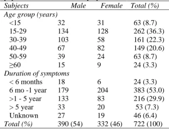
Table 1 Age and sex distribution of the study population and duration of symptoms.
Subjects Male Female Total (%) Age group (years)
<15 32 31 63 (8.7)
15-29 134 128 262 (36.3)
30-39 103 58 161 (22.3)
40-49 67 82 149 (20.6)
50-59 39 24 63 (8.7)
≥60 15 9 24 (3.3)
Duration of symptoms
< 6 months 18 6 24 (3.3)
6 mo -1 year 179 204 383 (53.0) >1 - 5 year 133 83 216 (29.9)
> 5 year 33 20 53 (7.3)
Unknown 27 19 46 (6.4)
Total (%) 390 (54) 332 (46) 722 (100) Table 2 Clinical manifestations of leprosy (n=722). Characters of lesions N (%)
Macule 494 (68.4%)
Plaque 214 (29.6%)
Nodule 26 (3.6%)
Ulcer 22 (3.0%)
Hypopigmented 681 (94.3%)
Erythematous 64 (8.9%)
Obvious margin 701 (97.1%)
Unclear margin 40 (5.5%)
Marked anesthesia 637 (88.2%)
Some anesthesia 92 (12.7%)
No anesthesia 12 (1.7%)
Heavy infiltration 69 (9.5%) Moderate infiltration 140 (19.4%)
Mild infiltration 462 (64.0%)
Madarosis 22 (3.0%)
Ear lobe enlargement 32 (4.4%)
Claw hands or feet 20 (2.8%)
Shortening of finger 8 (1.1%)
Foot drop 5 (0.7%)
Wrist drop 1 (0.1%)
Saddle nose 7 (1.0%)
Lagophthalmos 1 (0.1%)
Enlargement of nipple 1 (0.1%) Photophobia with lacrimation 1 (0.1%) Paralysis of the finger 3 (0.4%)
subjected to any additional visit or procedure for the study other than that needed for clinical management. Data were recorded during registration of the leprosy patients with informed consent.
Results
The total number of patients was 722 (304 in 2011, 337 in 2012 and 82 in first 6 months of 2013). The age and sex distribution of the patients are shown in Table 1. Most of the patients (26.9%) belonged to 15-29 year age group with a male-female ratio=5:4. The youngest patient was 2½-year-old and the eldest patient was 70-year-old. The number of child (<15 years) cases was 63 (8.7%) and female cases was 332 (46%). Duration of symptoms varied from1 month to 15 years, but the majority (53%) had symptom duration from 6 months to 1 year (Table 1). Eighty seven (12%) patients had history of contact with leprosy patients. Intra-familial contact was more than extra-familial contact (71 versus 16). Most of the affected females were homemakers and most of the affected males were physical labourers. The characteristics of skin lesions and frequencies of other presenting features are shown in Table 2.
Borderline tuberculoid was the most common form of the disease (81.0%) followed by tuberculoid (9.3%), lepromatous (4.3%), borderline lepromatous (3.5%), borderline (1.8%) and pure neural (0.1%). PB presentation was more common than MB presentation (77.6% and 22.4%, respectively. Type 1 lepra reaction was more prevalent than type 2 (7.6% vs 2.9%). 9.7% patients had disabilities at time of diagnosis. The rate was significantly higher in MB cases (40.6%) in comparison to PB cases (0.9%) (χ2
=233, p<0.001). Disabilities affecting the feet were more frequently observed than hands and eyes. Both the reactions as well as disabilities occurred more commonly in MB patients (Table 3). Number of cases with grade 2 disability was 43 (5.9%).
Table 3 Reactions and disability in leprosy patients.
PB (n=562) MB (n=160) Total (n=722) Reaction
Type 1 13 (2.3%) 42 (26.2%) 55 (7.6%)
Type 2 0 (0.0%) 21 (13.1%) 21 (2.9%)
Neuritis 6 (1.1%) 29 (18.1%) 35 (4.8%)
No reaction 543 (96.6%) 79 (49.4%) 622 (86.1%)
Disability Hand
Grade 1 2 (0.4%) 9 (5.6%) 11 (1.5%)
Grade 2 0 (0.0) 27 (16.9%) 27 (3.7%)
Feet
Grade 1 1 (0.2%) 26 (16.2%) 27 (3.7%)
Grade 2 3 (0.5%) 26 (16.2%) 29 (4.0%)
Eyes
Grade 2 0 (0.0%) 3 (1.9%) 3 (0.4%)
Disability affecting at least one or more sites
5 (0.9%) 65 (40.6%) 70 (9.7%)
No disability 557 (99.1%) 95 (59.4%) 652 (90.3%)
Table 4 Nerve involvement in leprosy patients (n=722).
Thickened nerve Non tender Tender Total
Great auricular 65 3 68 (9.4%)
Ulnar 89 25 114 (15.8%)
Radial 57 10 67 (9.3%)
Median 6 1 7 (1.0%)
Common peroneal 69 14 83 (11.5%)
Posterior tibial 47 12 59 (8.2%)
Superficial cutaneous 24 1 25 (3.5%)
Facial 7 0 7 (1.0%)
Supratrochlear and sural 14 0 14 (1.9%)
At least one or more nerve involvement - - 143 (19.8%)
No nerve involvement - - 579 (80.2%)
Table 5 Slit skin smear and skin biopsy results in leprosy patients (n=722).
Investigation N (%)
Slit-skin smear
BI 0 71 (9.8%)
BI 1+ 2 (0.3%)
BI 2+ 5 (0.7%)
BI 3+ 4 (0.5%)
BI 4+ 13 (1.8%)
BI 5+ 27 (3.7%)
Not done 600 (83.1%)
Skin biopsy
Tuberculoid 4 (0.5%)
Borderline tuberculoid 1 (0.1%)
Lepromatous 1 (0.1%)
Not done 716 (99.2%)
4). Slit-skin smears were studied in 122 cases. Among them 51 were found positive and 71 were negative (Table 5). Only 6 patients
underwent skin biopsy for histopathological examination. In all of those cases leprosy was confirmed (tuberculoid 4, borderline tuberculoid 1 and lepromatous 1).
Discussion
In this study, out of 722 newly detected cases, borderline tuberculoid was far more prevalent than other types of leprosy: a result similar to different studies done in different countries.4,10-15
increasing awareness and relatively strong surveillance in this capital city.
Majority of affected patients were in their productive phase of life with a peak in the 15-29 year age group. As we know if the transmission of leprosy is being reduced in an area, it is expected that the number of children affected will decrease.16 Proportion of child cases (under
15 years of age) in our study was 8.7%. The proportion is higher in comparison to the country as a whole (5.3%), but lower than many other regions of south Asia6 indicating an
ongoing transmission. Though in many studies sex ratio was strongly in favour of male.4,12,14,17-19
We found a male-female ratio = 5:4. As there is concern that women may have less access to health care specially in developing areas, a higher ratio often reflects that the women are not getting adequate facility.16 The ratio we get was
reassuring in this regard.
The lag period in between onset of symptoms and the diagnosis is less than one year in most of the cases, reflecting the strong surveillance by both government and non-government sectors. Familial clustering was quite common as 9.8% patients had history of intra-familial contact. Affected people were predominantly from lower socioeconomic group. The most common presentation was with hypopigmented macule with obvious margin, marked anesthesia and mild infiltration. Overall prevalence of reaction was much higher in multibacillary in relation to paucibacillary patients, which is similar to that reported by Teixeira et al.20
The proportion of new cases with grade 2 disabilities often indicates the quality of case-detection activities and when high reflects underdetection due to various reasons.6 In the
present study proportion of such cases (5.9%) was lower than many areas but still significant. The reason behind such prevalence of
disabilities may be social stigma and misconception regarding the disease resulting in delay in health care seeking. Grade 2 disability of feet was more common and disability rate was higher in multibacillary patients.
Slit-skin smear is an important tool for confirming leprosy. But as a substantial bacillary load is required for the skin smear result to be positive, the test is invariably negative in paucibacillary leprosy.8 So in our study
population, the most of the paucibacillary patients did not undergo slit-skin smear examination. Even among 122 patients who underwent this examination BI was zero in most of them (n=71). But this negative result does not exclude the clinical diagnosis of leprosy. Skin biopsy for histopathology can be done in difficult cases to exclude the differentials.
Though nerve enlargement is one of the cardinal signs of leprosy, in our study only 19.8% had one or more nerve enlargement. This reflected the fact that nerve enlargement is common but not universal in leprosy. Among the enlarged nerve ulnar nerve was most commonly affected followed by common peroneal nerve. This result is similar to other studies.4,17,18,22
Conclusion
Leprosy cases are being frequently diagnosed in Dhaka city. Epidemiological indicators reflect that despite a strong surveillance program there may be ongoing disease transmission and relative delay in diagnosis.
References
1. Lockwood DNJ. Leprosy. In: Burns T, Breathnach S, Cox N, Griffths C, editors. Rook’s Textbook of Dermatology, 8th ed. Oxford: Blackwell Publishing; 2010. p. 32.1-32.20.
2. Edward C, Klatt EC, Robbins SL, Cotran RS, editors. Robbins Pathologic Basis of Disease, 6th ed. Philadelphia: Saunder Elsevier; 2010. p. 385-7.
3. Jopling WH, McDougall AC, editors. Handbook of Leprosy. Oxford: Heinemann professional publishing; 1988. p. 114-5. 4. Khan I, Khan AR, Khan MS.
Clinicopathological study of 50 cases of leprosy in Northern Pakistan. J Pak Assoc Dermatol. 2012;22:200-6.
5. Levinson W, Jqwetz E, editors. Medical Microbiology and Immunology, 6th ed. New York: McGraw-Hill; 2000. p. 140-1. 6. World Health Organization. Weekly
Epidemiological Report. 2014;89:389-400. 7. National guidelines and technical manual
on leprosy. Ministry of Health and Family Welfare, Govt. of Bangladesh and World Health Organization 2005. 3rd ed. p. 4, 55. 8. Soomro FR, Pathan GM. Leprosy
awareness among doctors during survey of District Larkana. Infect Dis J. 2005;14:98-9.
9. James WD, Berger TG, Elston DM. editors. Andrew’s Diseases of the skin Clinical Dermatology, 11th ed. Philadelphia: Saunders Elsevier; 2011. p. 334-44.
10. Haque MA, Sharmin S, Ekram ARMS, Mahmood I. Epidemilogical trends of Leprosy in Rajshahi district. J Teach Assoc RMC Rajshahi. 2009;22:88-92.
11. Soomro FR, Pathan GM, Bajaj DR, Bhatti NS. Leprosy in Larkano region; an analysis of 102 cases from 2001-2011 at leprosy centre Larkano, Sindh, Pakistan. J Pak Assoc Dermatol. 2012;22:126-9.
12. Jindal N, Shanker V, Taqta GR et al. Clinico-epidemiological trends of leprosy in Himachal Pradesh; a five-year study. Indian J Lepr. 2009;81:173-9.
13. Toweir AA, Chaudhary RC. Review of leprosy cases in Benghagi, Libyan Arab Jamahiriya, 1994-98. East Mediterr Health J. 2000;6:1098-1102.
14. Boggild AK, Correia JD, Keystone JS, Kain KC. Leprosy in Toronto: an analysis of 184 imported cases. Can Med Assoc J. 2004;170:55-9.
15. Barbosa-junior AA, Jambeiro J, Jonelia SO, Cirqueira, Silva TC. Retrospective histopathological classification of 1,108 skin biopsies from patients clinically suspected of having leprosy from Bahia, Northeast Brazil. Revista da Sociedade Brasiteira de Medicina Tropical. 1998;31:533-7.
16. World Health Organization. Enhanced global strategy for further reducing the disease burden due to leprosy (2011-2015) Operational guidelines (Updated) 2009. SEA-GLP-2009.4.
17. Golfurshan F, Sadaghi M, Goldust M. Leprosy in Iran: an analysis of 195 cases from 1994-2009. J Pak Med Assoc. 2011;16:558-61.
18. Hussein A, Mohammed H, Eltahir A et al. Frequency of neurological deficits in Sudanese lepromatic patients. Sudan J Med Sci. 2010;5:17-24.
19. Tiwary PK, Kar HK, Sharma PK et al. Epidemiological trends of leprosy in an urban leprosy center of Delhi: a retrospective study of 16 years. Indian J Lepr. 2011;83:207-8.
20. Teixeira MA, Silveira VM, Franca ER. Characteristics of leprosy reaction in paucibacillary and multibacillary individual attended at two reference centers in Recife, Pernambuco. Rev Soc Bras Med Trop. 2010;43:287-92.
21. Bongorno MR, Pistone G, Note S. Tuberculoid leprosy and type 1 lepra reaction. Travel Med Infect Dis. 2008;6:311-4.