Address for correspondence Prof. Zahra Rahnama
Department of Dermatology, University of Medical Sciences, Kerman, Iran
Email: [email protected]
Original Article
Prevalence of thyroid disorders in patients with
alopecia areata
Zahra Rahnama*, Saeedeh Farajzadeh**, Saman Mohamamdi*, Mohammad Ali Masoudi*
* Department of Dermatology, University of Medical Sciences, Kerman, Iran
** Department of Pediatric Dermatology, Dermatology and Leishmania Research Center, University of Medical Sciences, Kerman, Iran
Abstract Objective To study the prevalence of autoimmune thyroid disorders – thyroid auto-antibodies in patients with alopecia areata (AA) in Kerman, a city in South-East part of Iran.
Methods 52 patients with AA from those attending the dermatology ward of Afzalipour hospital in Kerman were enrolled. An equal number of age- and sex-matched controls (n=52) was included. Physical examination of thyroid was done for all patients and controls. The rate of positivity of anti-thyroid peroxidase (TPO) antibodies and abnormal thyroid hormone levels in both groups were measured and compared.
Results In both cases and controls, 48.1% and 51.9 % were males and females, respectively. The mean age in group of cases and control groups were 30.55 and 31.80 years, respectively. The number of lesions ranged from 1 to 10, and the duration of disease ranged from 0.6 to 96 months. No meaningful statistical difference was seen between prevalence of thyroid disorders in patients of AA and controls.
Conclusion In this study no correlation between AA and thyroid disorders was noted.
Key words
Alopecia areata, thyroid disorders, autoimmune disorders.
Introduction
Alopecia areata (AA) is a common hair disorder that manifests as sudden appearance of patches of nonscarring alopecia. Around 1-2% of communities are affected and it usually
commences in childhood-juvenile period.1
Potentially, every hair-bearing area of skin can be involved; however, AA usually affects scalp. The disease may progress to involve the whole scalp (alopecia totalis) or total body (alopecia
universalis). AA, may be associated with specific signs in nails, as well.2 Hair loss affects the quality of life and may lead to social
loneliness especially in children and
adolescents.3
Although many theories have been propounded, its etiology and pathogenesis have not been elucidated yet, but many evidences support the theory that AA is an autoimmune disease
affected by genetic, psychological and
shown. In histopathology examination of AA a lymphocytic infiltration around follicular root of
anagen and early catagen hairs was found.8
In many cases AA is associated with other
autoimmune diseases such as atopy,6
Hashimoto’s thyroiditis,9 diabetes mellitus and vitiligo. There are studies regarding increased risk of autoimmune disorders in first degree relatives of children with AA, especially thyroid
disorders.10 A few reports regarding probable
association of AA with celiac disease (gluten sensitive enteropathy) exist.11 There are reports of abnormal thyroid function tests in patients with AA9,10,12 as 8-10% cases of alopecia totalis and alopecia universalis are reported with
increased prevalence of thyroid dysfunction.13
Despite many reports regarding association of AA with thyroid disease, no complete consensus exists about prevalence of thyroid disease and
thyroid dysfunction in AA.14 As few studies exist
in this field, we decided to study the prevalence of thyroid disease and thyroid autoantibodies in patients with AA in Kerman.
Methods
This was a case-control study comprising of 52 cases of AA who visited dermatology ward in Afzalipour hospital and private clinics in Kerman, a city located in South-East of Iran. These cases were enrolled with simple sampling method. Inclusion criteria for the cases in this study consisted of clinical diagnosis of AA by a dermatologist. Exclusion criteria for the study consisted of history of thyroid disease, pregnancy and other autoimmune diseases.
The second group (control group) had also 52 cases from those who visited in dermatological clinics and had no history of AA and other autoimmune or thyroid diseases. Written consent form was obtained from patients or their parents. Demographic data including age, sex, involved area, number of patches, size of the lesions, duration and severity of disease and type of the lesions were recorded. For all the cases in both groups physical examination for thyroid was undertaken and blood samples for studying of dysfunction and diseases of thyroid were
collected. Serum levels of anti-thyroid
peroxidase (TPO) antibodies, TSH, free T3 and free T4 with chemiluminescent method were done. All the laboratory tests were carried out in the same conditions.
Finally the rates of positivity of anti-TPO and serum level of thyroid hormones were compared in the 2 groups.
Demographic data were recorded in a
predesigned form. The relationship between dysfunction of thyroid gland and AA was assessed by χ2 (Chi square), and Fischer bicaudal exact test and T test used for analysis of data. P value of less than 0.05 was statistically considered meaningful. Data analysis was done by SPSS software. The process of the study was explained for cases in both groups and consent form was obtained, as well.
Results
In general 48.1% of the cases were male and 51.9% were female and no meaningful
difference was seen (Table 1). The average age
Table 1 Age and sex distribution in two groups.
Cases Controls Total P value
Male 29 (55.8) 21 (40.4) 50(48.1) 0.169
Female 23 (44.2) 31 (59.6) 54(51.9)
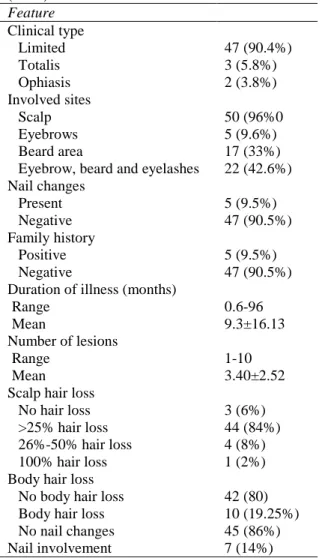
Table 2 Disease characteristic in the study population (n=52).
Feature Clinical type
Limited 47 (90.4%)
Totalis 3 (5.8%)
Ophiasis 2 (3.8%)
Involved sites
Scalp 50 (96%0
Eyebrows 5 (9.6%)
Beard area 17 (33%)
Eyebrow, beard and eyelashes 22 (42.6%) Nail changes
Present 5 (9.5%)
Negative 47 (90.5%)
Family history
Positive 5 (9.5%)
Negative 47 (90.5%)
Duration of illness (months)
Range 0.6-96
Mean 9.3±16.13
Number of lesions
Range 1-10
Mean 3.40±2.52
Scalp hair loss
No hair loss 3 (6%)
>25% hair loss 44 (84%)
26%-50% hair loss 4 (8%)
100% hair loss 1 (2%)
Body hair loss
No body hair loss 42 (80)
Body hair loss 10 (19.25%)
No nail changes 45 (86%)
Nail involvement 7 (14%)
of the cases was 30.55 and 31.80 in case and
control groups, respectively (p>0.05). The
duration of lesions ranged from 0.6 up to 96 months, and the number of lesions ranged from 1 up to 10. Table 4 shows these results.
Table 2 shows the different involved areas by AA. Eighty percent of the cases with AA had no involvement of body hairs, and 86% had no nail involvement.
The seropositivity rate of anti-TPO in case group and control group were 11.5% and 5.8%, respectively (Table 3).
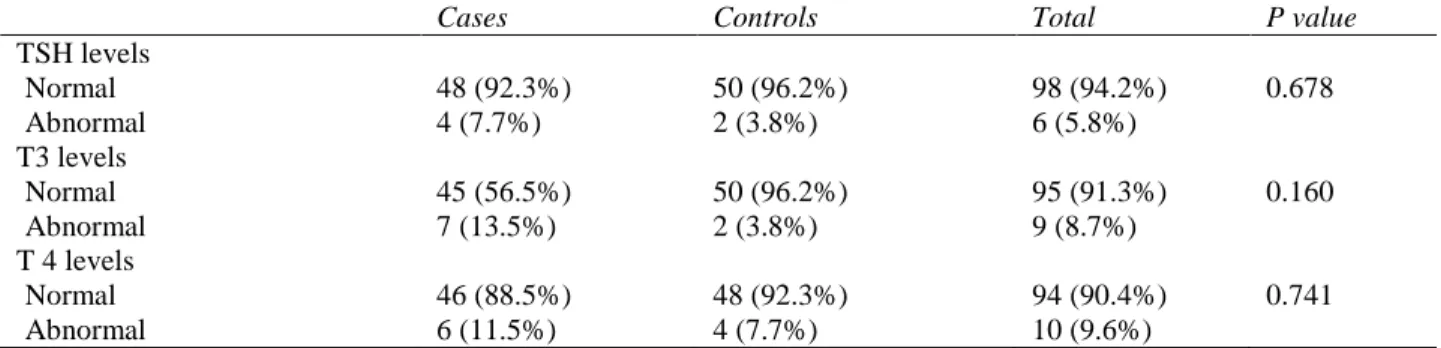
Frequency of TSH dysfunction in thyroid gland in case group and control group were 7.7% and 3.8%, respectively (p>0.05), (Table 4). T3 dysfunction in case group and control group were 7 and 2 individuals, respectively (p>0.05). Similarly, there was no difference in the rate of dysfunction of T4 in the two groups (p>0.05).
Discussion
In this case-control study, in the case group there were 52 patients with AA, and in control group 52 individuals with other skin diseases were enrolled. We tried to consider all confounder factors. In our study, there was no gender difference. Alopecia in juvenile group has the same prevalence in both sexes.1-3 The age of onset in our study was in accordance with previous studies1,2,14 with the onset of first episode in the first 4 decades of life. Based on the previous studies alopecia in juvenile period is the most sever type with the worst probable
outcome.15 The duration of lesions ranged
between 0.6 and 96 months and the number of lesions from 1-10. Eighty percent of the cases with AA had no involvement of body hairs, and 86% were free from nail involvement.
The positivity rate of thyroid dysfunction and dysfunction of TSH, T3 and T4 in the case group and control group showed no meaningful statistical difference (p>0.05). Also there was no meaningful statistical difference between thyroid
diseases and severity of AA (p>0.05). Our
results are in agreement with many studies.12,13,16
Table 3 Frequency of anti-TPO between two groups.
Cases (n=52) Controls (n=52) Total P value
Normal 46 (88.5%) 49 (94.2%) 95 (91.3%) 0.488
Abnormal 6 (11.5%) 3 (5.8%) 9 (8.7%)
*Based on Chi-square, Data are shown as abundance (%)
Table 4 Abundance rate of dysfunction in thyroid gland in the study.
Cases Controls Total P value
TSH levels
Normal 48 (92.3%) 50 (96.2%) 98 (94.2%) 0.678
Abnormal 4 (7.7%) 2 (3.8%) 6 (5.8%)
T3 levels
Normal 45 (56.5%) 50 (96.2%) 95 (91.3%) 0.160
Abnormal 7 (13.5%) 2 (3.8%) 9 (8.7%)
T 4 levels
Normal 46 (88.5%) 48 (92.3%) 94 (90.4%) 0.741
Abnormal 6 (11.5%) 4 (7.7%) 10 (9.6%)
*Based on Chi-square, Data are shown as abundance (%)
well.13 These results are in concordance with our
study.
In another study done by Kasumagic-Halilovic
et al.12 thyroid gland disorders were seen in 11.4% of the patients and no correlation between these diseases and the severity or duration of AA were seen. These results are very similar to our study.
In still another study accomplished by
Ghalamkar et al.16 in BooAli hospital in Tehran
(2001), 70 cases were examined in 2 groups. Each group consisted of 18 female cases (51%) and 17 male cases (49%) at age 23±10 years. None of the cases in control group had thyroid disease, whereas 11.4% of cases in case group had thyroid disorders and the statistical
difference was meaningful (P>0.06). The results
of this study might be doubtful due to the small sample size, whereas our study has larger sample size.
Conclusion
Despite a small arithmetical difference in the frequency of thyroid dysfunctions in patients with AA and control, the results of our study
rejected any association of thyroid disorders with AA.
References
1. Wasserman D, Guzman-Sanchez DA, Scott D, McMichael A. Alopecia areata. Int J Dermatol. 2007;46:121-31.
2. Kavak A, Yesildal N, Parlak A et al. Alopecia areata in Turkey: demographic and clinical features. J Eur Acad Dermatol Venereol. 2008;22:977 81.
3. Gulec AT, Tanriverdi N, Duru C et al. The role of psychological factors in alopecia areata and the impact of the disease on the quality of life. Int J Dermatol. 2004;43:352-6.
4. Goh C, Finkel M, Christos PJ, Sinha AA. Profile of 513 patients with alopecia areata: associations of disease subtypes with atopy, autoimmune disease and positive family history. J Eur Acad Dermatol Venereol. 2006;20:1055-60.
5. Martinez-Mir A, Zlotogorski A, Ott J et al. Genetic linkage studies in alopecia areata. J Invest Dermatol Symp Proc. 2003;8:199-203.
6. Hordinsky M, Ericson M. Autoimmunity: alopecia areata. J Invest Dermatol Symp Proc. 2004;9:73-8.
8. Kavak A, Yesildal N, Parlak A et al. Alopecia areata in Turkey: demographic and clinical features. J Eur Acad Dermatol Venereol. 2008;22:977 81.
9. Kasumagić-Halilović E. Thyroid autoimmunity in patients with alopecia areata. Acta Dermatovenerol Croat. 2008;16:123-5.
10. Puavilai S, Puavilai G, Charuwichitratana S et al. Prevalence of thyroid diseases in patients with alopecia areata. Int J Dermatol. 1994;33:632-3.
11. Corazza GR, Andreani ML, Venturo N et al. Celiac disease and alopecia areata: report of a new association. Gastroenterology. 1995;109:1333-7.
12. Kasumagic-Haliovic E. Thyroid autoimmunity in alopecia areata. Acta Dermatovenereol Croat. 2008;16:123-5.
13. Seyrafi H, Akhiani M, Abbasi H et al. Evaluation of the profile of alopecia areata and the prevalence of thyroid function test abnormalities and serum autoantibodies in Iranian patients. BMC Dermatol. 2005;5:11. 14. Sharma VK, Dawn G, Kumar B. Profile of
alopecia areata in Northern India. Int J Dermatol. 1996;35:22-7.
15. De Waard-van der Spek FB, Oranje AP, De Raeymaecker DM, Peereboom-Wynia JD. Juvenile versus maturity-onset alopecia areata--a comparative retrospective clinical study. Clin Exp Dermatol. 1989;14:429. 16. Ghalamkar F, Mohsenzadeh M, Velai N.