Address for correspondence Dr. Abdul Malik Tareen, PhD Assistant Professor,
CASVAB,
University of Baluchistan, Quetta, Pakistan Email: [email protected]
Original Article
An effective combined therapy of meglumine
antimoniate and co-trimoxazole for cutaneous
leishmaniasis: A study in Quetta City
Bilal Ahmed*, Muhammad Iqbal Tareen*, Ashok Kumar**, Saira Baloch†, Hazir Rehman‡, Zahid Mustafa‡, Abdul Malik Tareen‡
*Department of Dermatology, Bolan Medical College, Quetta ** Department of Pathology, Isra University, Hyderabad, Pakistan
† Medical Research Centre, Liaquat University of Medical and Health Sciences, Jamshoro, Pakistan
‡ CASVAB, University of Baluchistan, Quetta, Pakistan.
Abstract
Objective To evaluate the efficacy of combined therapy of meglumine antimoniate and co-trimoxazole in cutaneous leishmaniasis (CL).Methods 180 patients with active lesions of CL, confirmed by fine needle aspiration cytology (FNAC) were treated with combined therapy of meglumine antimoniate (Glucantime) and co-trimoxazole (Septran) for 20 days.
Results Out of 180 patients, 160 (88.9%) were cured. A few patients reported nausea, vomiting, glossitis, skin rashes and folate deficiency.
Conclusion The combined therapy of meglumine antimoniate (Glucantime) and co-trimoxazole (Septran) is more effective than the previously reported combined therapies.
Key words
Cutaneous leishmaniasis, meglumine antimoniate, co-trimoxazole, combined therapy.
Introduction
Leishmaniasis is a protozoal infection, transmitted by the bite of a female sandfly, which transmits the causative organism
Leishmania. Leishmaniasis is broadly divided
into three categories: cutaneous,
mucocutaneous and visceral leishmaniasis.1
Cutaneous leishmaniasis (CL) is considered as an important health problem in many parts of the world, especially the Mediterranean regions of Africa and almost all countries of the Middle East. The disease has emerged as an important public problem in many tropical countries, including Pakistan where the disease
is prevalent and cases have been reported from many parts of the country.2,3,4
Leishmaniasis has affected 1 to 1.5 million people globally and is a major health concern in Pakistan.5,6 The disease is caused by more
than 20 different species of the Leishmania.
Among them L. tropica is the commonest
form, recorded in Pakistan. CL has been reported in the Northern part of the country as well as in South Waziristan, Pakistan. The CL causes skin lesions on face, arms and legs. Small lesions are usually self-healing, but when multiple and large, they result in disfiguring and disability of the involved part of the body.7,8
with an extreme hot and cold climate
(minimum -12C in January and maximum
42C in June/July.9 Quetta, the capital of
Balochistan is the central city and highly exposed to the refugees and travelers from Afghanistan and Iran. This has been major factor in the increase of this infestation in this area, as disease is highly endemic in Iran and Afghanistan.10 CL, which was reported to be
prevalent in only a couple of districts of Balochistan, has now spread to the entire province.19
Uptill now, no effective vaccines are available
against Leishmaniasis and the treatment
depends exclusively on chemotherapy. In several parts of the world, disease still has a therapeutic problem.6,11 Despite all efforts there
is no safe, cheap, simple and effective treatment for CL. In majority of the cases pentavalent antimony compounds are still the drug of choice for treatment. Using this compound 40% of cases in certain regions have the disadvantage of both clinical resistance and toxicity. In some studies, drugs such as rifampicin, allopurinol, dapsone and nifurtimox have good response.12,13,14
Two classes of drugs are currently being used in our study area, which include meglumine antimoniate and co-trimoxazole. The mode of action of meglumine antimoniate has not been determined yet; but the evidence for inhibition
of glycolysis in the parasite at the
phosphofructokinase reaction has been found.
Because it is not absorbed on oral
administration, it must be administered parenterally. For co-trimoxazole drug, we still do not know its mode of action against CL.
The present study assessed the in vivo efficacy
of combined therapy of meglumine
antimoniate and co-trimoxazole against
cutaneous leishmaniasis. Findings from the current study will be helpful to control
cutaneous leishmaniasis in the high prevalent area of Pakistan.
Methods
Study area and selection of cases
The study was conducted at the Department of
Dermatology, Bolan Medical Complex
Hospital, Quetta, from January 2009 to October 2009. A total of 200 cases of CL were enrolled with typical clinical lesions, involving both the upper and the lower extremities. Inclusion criteria were the patients of any age, gender, race, occupation and residence. After taking a detailed history, complete physical examination was done.
Diagnostic techniques
Fine needle aspiration cytology (FNAC) and slit-skin smear were performed to collect the samples and diagnose the registered patients with active cutaneous lesions, diagnosed as CL patients by our hospital dermatologist.
Fine needle aspiration cytology (FNAC)
A needle aspirate was obtained by using 10 ml syringe with a 20-gauge needle. The needle was inserted through intact skin into the edge of the lesion. The needle was then moved back and forth, and rotated; aspiration was applied at the same time to get the small cut pieces of infected tissues in the syringe. The aspirate was used to prepare smears, which were then dried and stained with Giemsa stain.
Slit-smear method
tissue pulp was smeared on a slide and allowed to air dry. The slides were stained with Leishman stain for microscopic examination.
Microscopic analysis
All the prepared slides were examined by performing microscopy. A light microscope (Nikon, Tokyo, Japan) with 4, 20 and 40 lenses was used. Many fields were visualized to confirm Leishman-Donovan (LD) bodies.
Treatment regimen and follow-up
All the patients were treated with meglumine
antimoniate 600mg/day (adults) and
15mg/kg/day (children) intramuscularly and co-trimoxazole orally in dose of 320 mg once daily for adults and 160 mg for children for 20 days. The patients were followed up for 3 weeks. On 21st day patients were evaluated clinically and the FNAC was performed for further confirmation. These patients were also followed up for another 4 months for relapse.
A patient was considered cured if there was clinical healing of the target lesion and a negative smear for LD bodies at the end of treatment.
Results
Of 200 patients enrolled, 128 were males and 72 females with different age groups (15 to 65 years). Microscopic examination of the smears of all the patients showed presence of many LD bodies. All the patients were given a combined therapy of meglumine antimoniate and co-trimoxazole for 20 days. 20 out of 200 patients did not complete the treatment and therefore were excluded from the study. Out of the remaining 180 patients, 160 (88.9%) showed complete healing response and
negative smear (Table 1). In the remaining, 20
(11.1%) patients, there was partial healing of the lesions and FNAC reports were positive for LD bodies.
Table 1 Treatment response at the end of treatment (n=180).
Treatment response N (%)
Cured 160 (88.9)
Failure 20 (11.1)
The patients, who showed complete healing and negative reports for LD bodies were followed up for a mean of 120 days after completion of the treatment. None of the patients had a relapse during the follow up period. The results show that the combination therapy has a cure rate of 88.8%.
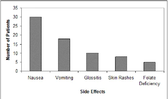
During treatment, some patients noticed side effects (Figure 1). These were nausea in 30 (16.7%) patients, vomiting in 18 (10%), glossitis in 10 (5.6%), skin rashes in 8 (4.4%) and folate deficiency in 5 (2.8%). The patients were given symptomatic treatment. The side effects subsided after completion of the treatment.
Discussion
The treatment of CL has been a major problem for the patients and health authorities. Currently, antimonials are the sole available pharmaceutical drug for its treatment, which needs multiple injections.9,15 The resulting drug
efficacy against the causative organism is low. In the present study, a combined therapy of antimonial and co-trimoxazole was given to the CL patients. 88.9% of the treated patients were cured. Further follow-up of 4 months post-treatment was also done and showed no relapse of the disease which was confirmed by negative report for LD bodies in the FNAC of the healed areas.
Previously, a randomized controlled study was performed in Pakistan to observe the efficacy of meglumine antimoniate alone to treat CL. It
was reported that the cure rate for
intramuscular administered meglumine
Figure 1 Side effects observed during the treatment.
There have been a number of studies done on combined therapy of antimonials and the other drug groups to cure CL, the results still remain short of expectation. A randomized, controlled study in southern Pacific coast of Colombia was performed for single drug therapy of meglumine antimoniate (Glucantime) and in combination with allopurinol to treat CL patients. The reported cure rate for the patients treated with meglumine antimoniate alone was 36%, while those, who received a combined
therapy was 74%.17 In Peru, a randomized
clinical trial with combined therapy of pentavalent antimony and imiquimod was done, which showed 75% cure rate.18 In the
presented work, we treated CL patients with the combined therapy of antimonials and co-trimoxazole, which showed the higher cure rate of 88.9%. To our knowledge, this combined therapy appears to be more effective than the other combinations tried before.
In conclusion, the present study reports that a new combination of meglumine antimoniate and co-trimoxazole for CL patients is more effective and well tolerated in our region. This combination should be tested in other areas where the disease is endemic. This might increase the cure rate of the patients and
References
1. Reithinger R, Dujardin JC, Louzir H, Pirmez C, Alexander B, Brooker S. Cutaneous leishmaniasis. Lancet Infectious Diseases. 2007;7:581-6. 2. Bhutto AM, Soomro RA, Nonaka S,
Hashiguchi Y. Detection of new endemic areas of cutaneous leishmaniasis in Pakistan: a 6-year study. Int J Dermatol. 2003;42:543-8.
3. Burney MI, Lari FA. Status of Cutaneous Leishmaniasis in Pakistan. J Pak Med Assoc. 1986;25:101-8.
4. Pearson RD, Querez AS. Leishmania species: visceral (kala-azar), cutaneous, and mucosal leishmaniasis. In: Mandell GL, Bennett JE, Dolin R, eds. Principles and Practice of Infectious Diseases. New York: Churchill Livingstone; 1995. pp. 2428-42.
5. Alvar J, Velez ID, Bern C et al. Leishmaniasis worldwide and global estimates of its incidence. PloS One. 2012;7:35671–2
6. Herwaldt BL. Leishmaniasis. Lancet. 1999;354:1191-9.
7. Ghazi RR, Ali R. Cutaneous leishmaniasis in Uthal, Balochistan with a note on its status in Pakistan. Proc Parasitol. 1998;5:40-5.
9. Juma KK. Present situation of cutaneous leishmaniasis in Balochistan, Pakistan. Pak J Biol Sci. 2004;7;698-702
10. Fereidoun A, Mohsen J, Hossein H. Epidemiology and Control of Common Disorders in Iran, 3rd Edi. Tehran: Khosravi Pub; 2009.
11. Murray HW, epin JP, TB, Nutman SL et al. Recent advances: tropical medicine. Br Med J. 2000;320:490-4.
12. Ayub S, Gramiccia M, Khalid M et al. Cutaneous leishmaniasis in Multan: species identification. J Pak Med Assoc. 2003;53:445-7.
13. Jaffernay M, Nighat R. Cutaneous leishmaniasis in Pakistan. Int J Dermatol. 2001;40:159.
14. Kolaczinski J, Brooker S, Reyburn H, Rowland M. Epidemiology of anthroponotic cutaneous leishmaniasis in Afghan refugee camps in northwest Pakistan. Trans R Soc Trop Med Hyg. 2004;98:373-8.
15. Juma KK. Kaal Daana (Cutaneous Leishmaniasis) in South-West Pakistan: A preliminary study. Türkiye Parazitoloji Dergisi. 2004;28:5-11.
16. Munir A, Janjua SA, Hussain I. Clinical efficacy of intramuscular meglumine antimoniate alone and in combination with intralesional meglumine antimoniate in the treatment of old world cutaneous leishmaniasis. Acta Dermatovenerol Croat. 2008;16:60-4.
17. Samuel M, Joseph MJ. Allopurinol in the treatment of American cutaneous leishmaniasis. N Engl J Med. 1992;326:741-4.
18. Miranda-Verastegui C, Tulliano G, Gyorkos TW et al. First-line therapy for human cutaneous leishmaniasis in Peru using the TLR7 agonist imiquimod in combination with pentavalent antimony. PLoS Negl Trop Dis. 2009;3:e491. 19. WHO Control of Leishmaniasis. WHO