SINGLE NUCLEOTIDE POLYMORPHISMS IN VITAMIN A, FOLATE AND CHOLINE RELATED GENES AND INTERACTION WITH MATERNAL VITAMIN INTAKE AND
NEUROBLASTOMA
Angela Liu Mazul
A dissertation submitted to the faculty at the University of North Carolina at Chapel Hill in partial fulfillment of the requirements for the degree of Doctor of Philosophy in the Department
of Epidemiology in the Gillings School of Global Public Health.
Chapel Hill 2016
ABSTRACT
Angela Liu Mazul: Single Nucleotide Polymorphisms In Vitamin A, Folate And Choline Related Genes And Interaction With Maternal Vitamin Intake And Neuroblastoma
(Under the direction of Andrew Olshan)
Previous epidemiologic studies suggest maternal vitamin supplementation during pregnancy reduces the risk of neuroblastoma. We hypothesize offspring and maternal genetic variants in vitamin A, folate and choline-related genes are associated with neuroblastoma and are modified by maternal intake of vitamin A, folate, and choline
The Neuroblastoma Epidemiology in North America (NENA) study recruited 563 affected child-parent sets through the Children’s Oncology Group’s (COG) Childhood Cancer Research Network. We ascertained pre-pregnancy supplementation and estimated usual maternal dietary intake with questionnaires and genotyped genetic variants related to folate, choline and vitamin A pathways from DNA extracted from saliva. A log-linear model was employed to estimate additive offspring and maternal risk ratios and stratum-specific risk ratios by COG prognostic risk-classification and age at diagnosis and for gene-environment interactions. For replication for the offspring main effects, we used a genome-wide offspring case-control study from Children’s Hospital of Philadelphia (CHOP).
We found nine SNPs in/near 4 folate-related genes that were FDR-corrected significantly associated with intermediate-risk neuroblastoma but none replicated in the CHOP replication. FDR-corrected significant maternal results were found within the high-risk neuroblastoma strata and offspring age of diagnosis < 1 year with rs6776706 and rs11103603, respectively. No
significant gene-environment interaction was found for pre-pregnancy vitamin supplementation. However from diet, we found a maternal rs729147-vitamin A interaction when vitamin A was dichotomized at the Recommended Dietary Allowance. Gene-choline interactions were found for offspring SNPs located in MTHFD1L and TYMS.
Our results suggest that some genetic variants involved in vitamin A and choline may be associated with neuroblastoma. The significant maternal variants and their joint effects with maternal vitamin A intake suggest a relationship between neuroblastoma and vitamin A. We also found variants related to one-carbon metabolism are not strongly associated with
ACKNOWLDGEMENTS
I would like to express my sincerest gratitude to my advisor and mentor, Dr. Andrew Olshan. I very much appreciate his guidance, expertise, patience, and support throughout my studies at UNC. I would also like to thank my committee members, Drs. Stephanie Engel, Clarice Weinberg, Anna Maria Seiga-Riz and Fei Zou, for their expertise and suggestions throughout this dissertation and the manuscripts.
I would also like to thank the UNC Lineberger's Cancer Control Education Program and the PIs Drs Kurt Ribsil and Jo Anne Earp for the support and opportunity for collaboration across training programs. A very special thanks to Dr. Michael O’Malley who was great at bringing people together and provided a great example of what cancer researchers should strive to be. Special thanks go to all the student services staff, Nancy Colvin, Carmen Woody, Jennifer Moore, and Valerie Huddock for all their help throughout these 5 years.
I would like to thank all the NENA staff, Kathryn Carrier, Elyssa Trani, Kim Ludwig, Kathy Wisniewski, and Jean Strelitz, for all their hard work. Also for all the NENA families who took the time to participate in this study at such a difficult time in their lives.
TABLE OF CONTENTS
LIST OF TABLES ... xiii
LIST OF FIGURES ... xiv
LIST OF ABBREVIATIONS ... xv
CHAPTER 1. BACKGROUND AND LITERATURE REVIEW ... 1
1.1 Dissertation Aims ... 1
1.2 Neuroblastoma Overview ... 4
1.2.1 Biologic Characteristics ... 4
1.2.2 Clinical Characteristics ... 5
1.2.3 Molecular Characteristics ... 8
1.2.4 Neuroblastoma Risk-Classifications ... 9
1.3 Neuroblastoma Descriptive Epidemiology ... 11
1.3.1 Incidence and Mortality in the United States ... 11
1.3.2 Time Trends in the United States ... 13
1.3.3 International Incidence and Time Trends ... 14
1.4 Neuroblastoma Risk Factors ... 15
1.4.1 Genetic Basis for Neuroblastoma ... 15
1.4.1.1 Familial Neuroblastoma... 16
1.4.1.2 Spontaneous Neuroblastoma ... 17
Summary. ... 22
1.4.2.1 Vitamin supplementation ... 24
Summary ... 27
1.4.2.2 Other possible risk factors ... 27
1.5 Literature on Vitamin Pathways ... 29
1.5.1 Vitamin A ... 29
1.5.1.1 Biologic literature ... 29
1.5.1.2 Epidemiologic literature ... 34
1.5.2 Folate ... 35
1.5.2.1 Biologic literature ... 35
1.5.2.2 Epidemiologic literature ... 40
1.5.3 Choline ... 44
1.5.3.1 Biologic literature ... 44
1.5.3.2 Epidemiologic literature ... 47
1.5.4 Summary of Literature on Vitamin A, Folate and Choline Pathways ... 48
1.6 Summary of Literature Review ... 49
CHAPTER 2. AIMS AND METHODS ... 51
2.1 Study population ... 51
2.1.1 COG and CCRN ... 51
2.1.2 Neuroblastoma Epidemiology in North America (NENA) ... 52
2.1.3 Recruitment ... 52
2.1.3.1 Institutional phase ... 52
2.1.3.2 Case Status ... 54
2.1.3.3 Phase I ... 55
2.1.3.4 Phase II ... 56
2.1.4 Study population ... 58
2.2 Measurements ... 60
2.2.1 Clinical and Biologic outcomes ... 60
2.2.2 Environmental Exposures ... 60
2.2.2.1 Maternal Dietary Questionnaire ... 60
2.2.2.2 Maternal Prenatal Vitamin Supplementation... 62
2.2.2.3 Nutrients ... 62
2.2.2.4 Maternal Questionnaire ... 65
2.2.3 Genetic Exposure ... 65
2.2.3.1 DNA collection and extraction ... 65
2.2.3.2 Genotyping ... 66
2.2.3.3 Candidate genes ... 66
2.2.4 Covariates ... 70
2.2.4.1 Genetic Ancestry ... 71
2.2.4.2 Covariate description ... 72
2.3 Analysis ... 72
2.3.1 Genotyping Quality Control ... 72
2.3.2 Offspring Genetic effect ... 73
2.3.3 Maternal Effect ... 75
2.3.4 Gene-Environment Interaction ... 77
2.3.5 Missing Paternal Genotype ... 78
2.3.6 Stratifying by Risk-Classification and Offspring Age at Diagnosis ... 79
2.3.7 Definition of Genetic Model and Environment ... 79
2.3.8 Replication ... 80
2.3.9.1 Correction for Multiple Testing ... 82
2.3.9.2 Bias in measuring pregnancy diet and exposures ... 82
2.3.9.3 Selection ... 83
2.3.9.4 Assumptions for a case-parent triads ... 84
2.4 Statistical Power ... 84
2.4.1 Genetic effect ... 84
2.4.2 Stratification by Risk-Classification and Age ... 85
2.4.3 Gene Environment Interaction ... 86
2.5 Strengths and Limitations ... 87
CHAPTER 3: AIM 1 RESULTS ... 89
3.1 Overview ... 89
3.2 Introduction ... 90
3.3 Methods ... 91
3.3.1 Study Sample ... 91
3.3.2 Candidate Genes and SNP selection ... 93
3.3.3 DNA collection and Genotyping ... 94
3.3.4 Genetic Quality Control ... 94
3.3.5 Biological and Clinical Variables ... 95
3.3.6 Maternal Vitamin Use ... 96
3.3.7 Diet and Nutrient Classification ... 97
3.3.8 Statistical Analysis ... 97
3.3.9 Replication Study ... 99
3.3.10 Sensitivity Analysis ... 100
3.4 Results ... 100
3.4.2 Sensitivity Analysis ... 107
3.5 Discussion ... 107
CHAPTER 4: AIM 2 RESULTS ... 112
4.1 Overview ... 112
4.2 Background ... 113
4.3 Methods ... 114
4.3.1 Study Sample ... 114
4.3.2 Candidate Genes and SNP selection ... 115
4.3.3 DNA collection and Genotyping ... 115
4.3.4 Genotyping Quality Control ... 116
4.3.5 Biological and Clinical Variables ... 117
4.3.6 Maternal Vitamin Use ... 117
4.3.7 Diet and Nutrient classification ... 118
4.3.8 Statistical Analysis ... 119
4.3.9 Replication Study ... 120
4.3.10 Sensitivity Analysis ... 121
4.4 Results ... 122
4.4.1 Descriptive Statistics ... 122
4.4.2 Folate ... 123
4.4.3 Choline ... 123
4.4.5 Replication Study ... 127
4.4.6 Sensitivity Analysis ... 127
4.5 Discussion ... 129
CHAPTER 5: DISCUSSION AND CONCLUSIONS ... 133
5.2 Summary of Results ... 134
5.2.1 Aim 1 ... 134
5.2.2 Aim 2 ... 135
5.3 Strengths and Limitations ... 136
5.3.1 Strengths ... 136
5.3.2 Limitations ... 137
5.4 Implications and Conclusions ... 141
5.4.1 SNP Main effects ... 141
5.4.1.1 Previously studied SNPs ... 141
5.4.1.2 Offspring and Maternal SNP Main effects ... 142
5.4.1.3 Gene-environment interaction ... 144
5.4.2 Consideration for Future Studies ... 145
5.4.3 Public Health Implications ... 147
5.5 Summary ... 148
APPENDIX 1. RESULTS FROM OFFSPRING VITAMIN A-RELATED SNPS IN NENA AND CHOP REPLICATION STUDY ... 150
APPENDIX 2. RESULTS FROM MATERNAL VITAMIN A-RELATED SNPS... 163
APPENDIX 3. RESULTS FROM OFFSPRING FOLATE AND CHOLINE-RELATED SNPS IN NENA AND CHOP REPLICATION STUDY... 173
APPENDIX 4. RESULTS FROM MATERNAL FOLATE AND CHOLINE-RELATED SNPS ... 199
APPENDIX 5. QQ PLOTS ... 216
LIST OF TABLES
Table 1. International Neuroblastoma Pathologic Classification ... 10
Table 2. Children Oncology Group risk-classification ... 10
Table 3. A summary of genes related to neuroblastoma predisposition from Familial and GWA Studies ... 23
Table 4. Summary of possible risk factors of neuroblastoma ... 28
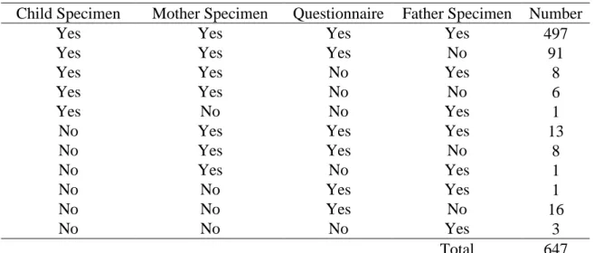
Table 5. Number of returned materials ... 60
Table 6. Descriptive statistics of NENA. Continuous variables are represented as mean ± standard deviation and categorical variables are N (%) ... 64
Table 7. Candidate gene list ... 68
Table 8. Mating types and frequencies in case-parent triad ... 75
Table 9. Mating types and frequencies in case-parent triad for maternal effects ... 76
Table 10. Power for offspring genetic effect ... 85
Table 11. Power for risk-classification and < 1 year and greater than or equal to 1 year of age ... 86
Table 12. Power for gene-environment interaction ... 87
Table 13. Descriptive statistics for triads with genetic data ... 101
Table 14. Offspring FDR-corrected significant SNPs results for intermediate risk group ... 102
Table 15. Maternal FDR-corrected significant SNPs results ... 103
Table 16. Descriptive statistics of maternal usual dietary nutrient levels and supplemental pre-pregnancy vitamin consumption ... 123
LIST OF FIGURES
Figure 1. Distribution of the Location of Primary Tumor by Age of Diagnosis ... 6
Figure 2. Neuroblastoma survival curves stratified by risk type ... 12
Figure 3. Incidence rate in millions of person-years of neuroblastoma from 1975 to 2006 directly standardized to the 2000 population ... 14
Figure 4. Vitamin A transport and metabolism ... 33
Figure 5. Folate metabolism and one-carbon pathway within a cell ... 39
Figure 6. Choline metabolism and relationship with one-carbon pathway ... 46
Figure 7. Flowchart of NENA recruitment ... 59
Figure 8. Causal diagram. Blue is the “environmental” variable and orange represents the exposure and outcome ... 71
Figure 9. Flowchart of DNA collection, genotyping and genetic quality control for mothers, fathers and children in NENA. ... 93
Figure 10. Flowchart for genetic and questionnaire quality control for triads and dyads. ... 95
Figure 11. A) Offspring and B) Maternal interaction with co-dominant rs729147 with vitamin A dichotomized at the RDA (700 µg RAE) ... 105
Figure 12. A) Offspring and B) Maternal interaction with co-dominant rs729147 “total” maternal vitamin A exposure ... 106
Figure 13. A) Offspring and B) maternal interaction with codominant rs173857 and maternal choline dichotomized at the 25th percentile ... 125
LIST OF ABBREVIATIONS
5-MeTHF 5-methyltetrahydrofolate ADH Alcohol dehydrogenases AdoHcy S-adenosoylhomocysteine AdoMet S-adenosylmethionine ALK Anaplastic lymphoma kinase
ATIC 5-aminoimidazole-4-carboxamide ribonucleotide formyltransferase/IMP cyclohydrolase
BARD1 BRCA1-associated RING domain-1 BCMO1 Beta-carotene 15,15'-monooxygenase BHMT Betaine homocysteine methyltransferase
C Child
CASC15 Cancer susceptibility candidate 15 CBS Cystathionine-β-synthase
CCRN Childhood cancer research network CDP-choline Cytidine diphosphocholine
CEL Carboxyl ester lipase
CEPH Centre de l'Étude du Polymorphisme
CES Carboxylesterase
CHKA Choline kinase A
CHOP Children Hospital of Philadelphia CHPT1 Choline phosphotransferase
CI Confidence interval
CNV Copy number variant
CRABPI Cellular retinoic acid-binding protein CRalBP Cellular retinaldehyde–binding protein CRBP Cellular retinol-binding proteins
DDX4 DEAD (Asp-Glu-Ala-Asp) box polypeptide 4 isoform DFE Dietary folate equivalence
DHFR Dihydrofolate reductase DHQ Dietary history questionnaire dTMP Deoxythymidine monophosphate dUMP Deoxyuridine monophosphate
DUSP12 Dual-specificity phosphatase 12 gene
F Father
FDR False discovery rate
FFQ Food frequency questionnaire FOLH1 Folate hydrolase 1
FPGS Folylpolyglutamate synthase FTHFS 10-formylthf synthethase
GART Phosphoribosylglycinamide formyltransferase, phosphoribosylglycinamide synthetase, phosphoribosylaminoimidazole synthetase
GWA Genome-wide Association
HACE1 Encoding HECT domain–and ankyrin HSD17B12 Hydroxysteroid (17-beta) dehydrogenase 12 HWE Hardy Weinberg equilibrium
ICCC International Classification of Childhood Cancer IL31RA Interleukin-31 receptor A precursor
INSS International neuroblastoma staging system LCRA Lead clinical research associate
LD Linkage disequilibrium
LIN28B Lin28 homolog B repeat–containing E3 ubiquitin protein ligase 1 LRAT Lecithin retinol acyltransferase
M Mother
MAF Minor allele frequency MKI Mitosis-karyorrhexis index
MTHFD1L Methylenetetrahydrofolate dehydrogenase 1-
MTHFD2 Methylenetetrahydrofolate dehydrogenase 2, methenyltetrahydrofolate cyclohydrolase
MTHFR Methylenetetrahydrofolate reductase (NAD(P)H)
MTR 5-methyltetrahydrofolate-homocysteine methyltransferase
MTRR 5-methyltetrahydrofolate-homocysteine methyltransferase reductase NBPF17P Neuroblastoma breakpoint family member 17, pseudogene
NENA Neuroblastoma Epidemiology in North America NHANES National Health and Nutrition Examination Surveys
OR Odds ratio
OR Odds ratio
PCYT1A Phosphate cytidylyltransferase 1
PEMT Phosphatidylethanolamine N-methyltransferase PHOX2B Paired-like homeobox 2b
PI Principle investigator PLD2 Phospholipase D2
RA Retinoic acid
RAE Retinol activity equivalent RALDH Retinaldehyde dehydrogenases RAR Retinoic acid receptor
RARE Retinoic acid response elements RBP Retinol binding protein
RDA Recommended dietary allowance RDH Retinol dehydrogenases
REHs Retinyl ester hydrolases
RFMMB Risk Factor Monitoring and Methods Branch
RR Risk ratio
RXR Retinoid X receptor
SEER Surveillance, Epidemiology, and End Results SHMT Serine hydroxymethyltransferase
SLC22A3 Solute carrier family 22, member 3 SLC22A4 Solute carrier family 22
SNAP SNP Annotation and Proxy Search SNP Single nucleotide polymorphism STRA6 Stimulated by retinoic acid 6 TDT Transmission disequilibrium test
THF Tetrahydrofolate
TYMS Thymidylate synthetase
UNC University of North Carolina at Chapel Hill
CHAPTER 1. BACKGROUND AND LITERATURE REVIEW
1.1 Dissertation Aims
Neuroblastoma is an embryonic tumor arising from a malignancy within cells of the neural crest.1,2 While 7.2% of all childhood cancers are neuroblastomas, it disproportionately accounts for 15% of all childhood cancer-related deaths.3,4 It is the most common cancer in infancy and is thought to occur by either environmental or genetic disruption of normal embryonic development.5 Familial cases of neuroblastoma have been associated with specific mutations in the PHOX2B and ALK genes. Among non-familial cases, recent genome-wide association (GWA) studies have identified several common variants of interest.6-9
Previous epidemiologic studies have found evidence of an inverse association between maternal prenatal vitamin use and neuroblastoma,10,11 suggesting that maternal pregnancy vitamin status may play a role in neuroblastoma development. Thus, for this study we focused on three vitamins with biologic plausibility: vitamin A, folate and choline.
Vitamin A is required for many growth and developmental processes including
Since maternal pre-pregnancy vitamin use has been previously associated with
neuroblastoma and the biologic plausibility of these vitamins,10 we are interested in common single nucleotide polymorphism in genes involved in vitamin A, folate and choline metabolism and transport pathways as well as interactions with maternal pregnancy vitamin intake from diet and vitamin supplementation.
Neuroblastoma Epidemiology in North America (NENA) is a case-parent triad study. NENA recruited families with cases of neuroblastoma under 6 years of age from the Childhood Cancer Research Network (CCRN), a registry of childhood cancer treated in Children’s
Oncology Group’s (COG) hospitals in North America. Buccal DNA was collected from the child and both biologic parents. If the child was deceased, then banked samples were requested from COG. A self-administered questionnaire was mailed to the biologic mother to assess vitamin intake through diet and supplements pre-pregnancy and during pregnancy. It also asked for demographic data and other lifestyle factors including tobacco and alcohol use, medication use and family history. NENA recruited a total 626 parent-child trios or dyads.
The specific aims of this project are:
Aim 1. Evaluate the association between maternal and offspring single nucleotide polymorphisms (SNPs) in genes involved in vitamin A related pathways with the risk of neuroblastoma
Aim 1a. Evaluate effects of offspring variants and maternal variants on the risk of
neuroblastoma stratified by offspring age at diagnosis and neuroblastoma Children’s Oncology group (COG) risk-classification.
Aim 1b. Describe the gene-environment interactions of maternal vitamin A intake during pregnancy with the offspring genotype for SNPs in the vitamin A pathway on the risk of neuroblastoma.
Aim 1c. Describe the gene-environment interactions of maternal vitamin A intake during pregnancy with the maternal genotype for SNPs in the vitamin A pathway on the risk of neuroblastoma in the offspring.
Aim 2. Evaluate the association between maternal and offspring SNPs in genes involved in folate and choline related pathways with the risk of neuroblastoma.
Aim 2a. Evaluate these offspring and maternal variants on the risk of neuroblastoma markers stratified by age at diagnosis and neuroblastoma risk-classification as defined by COG
guidelines.
Aim 2b. Describe the gene-environment interactions of maternal folate and choline intake during pregnancy with the offspring genotype in folate and choline related pathway on the risk of neuroblastoma.
during pregnancy with the maternal genotype in folate and choline related pathway on the risk of neuroblastoma in the offspring.
1.2 Neuroblastoma Overview
1.2.1 Biologic Characteristics
Neuroblastoma is an embryonic tumor of the sympathetic nervous system arising in the neural crest with embryonic origins.1,2 Neurulation is a complicated folding process during embryogensis that transforms the neural plate into the neural tube. As the plate folds, the neural plate borders join and become the neural crest. As the neural tube closes, the neural crest is disconnected from the ectoderm. A neural tube closes, the neural crest cells migrate.22 As these neural crest cells migrate, they further differentiate into the sympathetic nervous system.
Neuroblastoma tumors are thought to derive from stem cells in the sympathetic nervous system that did not properly differentiate.
Neuroblastoma is a heterogeneous malignancy with variable site of origin, clinical presentation and cellular composition.1 These tumors have been categorized into four basic morphologic categories:
1. Neuroblastoma (Schwannian stroma-poor)
2. Ganglioneuroblastoma, intermixed (Schwannian stroma-rich)
3. Ganglioneuroblastoma, nodular (composite Schwannian stroma-rich/stroma-dominant and stroma-poor)
4. Ganglioneuroma (Schwannian stroma-dominant)23
recruited by the malignant neuroblasts.24 Schwann cells in the tumors produce anti-proliferative and differentiation-inducing factors, thus indicating less aggressive disease.23
Neuroblastoma tumors are less differentiated than are ganglioneuroblastoma tumors. Ganglioneuromas arise spontaneously from maturation of neuroblastic tumors (i.e. all
Ganglioneuromas were once neuroblastomas in an earlier phase). Ganglioneuroblastoma falls in between neuroblastoma and ganglioneuromas in terms of differentiation.23 In addition to
spontaneous differentiation, neuroblastoma undergoes spontaneous regression more than any other cancer type, which most likely related to apoptosis of undifferentiated cells.4 Although most clinically diagnosed neuroblastic tumors do not undergo spontaneous maturation or
spontaneous regression after detection,23 it is estimated that over 10% of cases of neuroblastoma are missed due to spontaneous regression.25
1.2.2 Clinical Characteristics
Figure 1. Distribution of the Location of Primary Tumor by Age of Diagnosis
Reproduced from “Sympathetic Nervous System Tumors: SEER Pediatric Monograph” by M. Goodman et al., 2008.26
Symptoms vary depending on the location of the tumor. Approximately 50% of patients have localized or regional disease, 35% have regional lymph node spread at the time of diagnosis and the rest have widespread disease.3 Patients with localized disease are typically
asymptomatic and are often diagnosed when testing for unassociated conditions. Some
The current staging for neuroblastoma was defined by the International Neuroblastoma Staging System (INSS) and criteria are based on clinical features.29
Stage 1 tumors are localized, do not involve vital structures, are confined to one body compartment and lymph nodes do not contain tumor cells.
Stage 2A tumors are also localized and confined to one side of the body, but cannot be completely removed. Lymph nodes do not contain tumor cells.
Stage 2B tumors are localized, but nearby lymph nodes show tumor cells. Lymph nodes on the other side of the body can be enlarged but do not contain cancer cells. Stage 3 tumors can fall into two categories. Either the tumor crosses the midline of the body and cannot be surgically removed, or the tumor is restricted to one side of the body, but there are enlarged lymph nodes on the opposite side of the body that contain cancer cells.
Stage 4 tumors have spread further than stage 3 to distant lymph nodes, bone, bone marrow, liver, skin, and/or other organs.
Stage 4S tumors tend to regress without any treatment. The criteria for these tumors are: the child is younger than 1 year of age and a localized primary tumor has only spread to the skin, lymph nodes or liver, although very small amounts may be seen in the bone marrow.
1.2.3 Molecular Characteristics
There are many genetic aberrations commonly found in neuroblastoma tumors that are highly correlated with survival and prognosis. The genetic aberration most commonly associated with poor neuroblastoma outcomes is the amplification of the proto-oncogene MYCN. 31 MYCN amplification of 50 to 100-fold occurs in about 20% of primary tumors and is strongly correlated with advanced disease.32,33 Somatic DNA sequence mutations have not been found in MYCN, suggesting that the wild-type protein is contributing to tumorigenesis or to pathogenicity of the tumor.34 Additionally, transgenic mice that are genetically engineered to overexpress MYCN in the neural crest develop neuroblastoma several months after birth, suggesting MYCN can initiate tumorigenesis.35
The number of copies of chromosomes in a tumor cell, or ploidy, can be an important prognostic factor in children under the age of 2.36,37 Patients with lower grade of disease tend to be hyperdiploid or near-triploid (three sets of chromosomes), while patients with a higher grade of disease are nearly diploid. 38 This is likely because whole chromosome gains and losses are associated with a defect in mitosis, leading to tumor cell death and more favorable outcomes, while more malignant tumors have a defect in chromosomal stability, resulting in chromosomal rearrangements.
Recently with DNA sequencing, additional somatic changes have been identified. In one study, somatic mutations were identified in ARID1A and ARID1B in 11% of the samples and were associated with early treatment failure and decreased survival.42 In a sample of 240 “high-risk” cases, ALK, PTPN11, ATRX, MYCN and NRAS were found to be somatically altered.7 These studies revealed that high-risk neuroblastoma has markedly fewer somatic mutations than adult solid tumors, which has a stronger environmental contribution than childhood tumors. This suggests germline variants, copy number variants and epigenetic modifications drive high-risk neuroblastoma.7,42
1.2.4 Neuroblastoma Risk-Classifications
The Children’s Oncology Group (COG) separated neuroblastoma into three prognostic risk-classifications defined by International Neuroblastoma Staging System (INSS), age at diagnosis, MYCN oncogene status, International Neuroblastoma Pathologic Classification
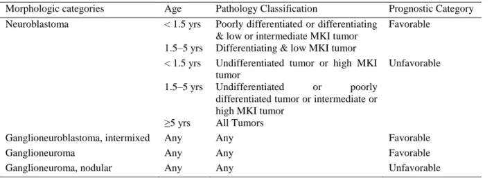
(INPC), and DNA ploidy index.43 The INPC risk-classification is based on tumor classifications, grade of neuroblastic differentiation and mitosis-karyorrhexis index (MKI) (Table 1). There are three COG prognostic risk-classifications: low-risk, intermediate-risk and high-risk (Table 2). Although these categories are prognostic, there is little evidence that favorable tumors progress to unfavorable tumors, suggesting they may be etiologically distinct.44 Brodeur et al.
Table 1. International Neuroblastoma Pathologic Classification
Morphologic categories Age Pathology Classification Prognostic Category
Neuroblastoma < 1.5 yrs Poorly differentiated or differentiating
& low or intermediate MKI tumor
Favorable
1.5–5 yrs Differentiating & low MKI tumor
< 1.5 yrs Undifferentiated tumor or high MKI
tumor
Unfavorable
1.5–5 yrs Undifferentiated or poorly
differentiated tumor or intermediate or high MKI tumor
≥5 yrs All Tumors
Ganglioneuroblastoma, intermixed Any Any Favorable
Ganglioneuroma Any Any Favorable
Ganglioneuroma, nodular Any Any Unfavorable
Yrs: Years; MKI: Mitosis-karyorrhexis index
Table 2. Children Oncology Group risk-classification
Risk INSS Stage Age MYCN INPC
Classification
DNA ploidy
Low risk 1 Any Any Any Any
2A/2B <12 mos Any Any Any
≥12 mos Non-Amplified Any -
>12 mos Amplified Favorable -
4S < 12 mos Non-Amplified Favorable >1
Intermediate Risk
3 < 12 mos Non-Amplified Any Any
≥12 mos Non-Amplified Favorable -
4 < 18 mos Non-Amplified Any Any
4S < 12 mos Non-Amplified Any =1
< 12 mos Non-Amplified Unfavorable Any
High Risk 2A/2B ≥12 mos Amplified Unfavorable -
3 < 12 mos Amplified Any Any
≥12 mos Non-Amplified Unfavorable -
≥12 mos Amplified Any -
4 <12 mos Amplified Any Any
≥18 mos Any Any -
4S <12 mos Amplified Any Any
INSS: International Neuroblastoma Staging System; INPC: International Neuroblastoma Pathological Classification; Mos: Months; -: Not Applicable
treatment of surgery, radiation and chemotherapy followed preventative medication (usually 13-cis-retinoic acid) for a year.
1.3 Neuroblastoma Descriptive Epidemiology
1.3.1 Incidence and Mortality in the United States
Each year approximately 1,500 cases in Europe, 700 cases in the United States (U.S.), and 70 cases in Canada are diagnosed with neuroblastoma.2,46,47 The overall age-standardized incidence rate according to Surveillance, Epidemiology, and End Results (SEER) from 2006 to 2010 is 7.83 per million. However, the neuroblastoma incidence rate is higher among younger children. The average annual age-standardized incidence rate of neuroblastoma is 54.1 per million person-years for children less than 1 year old, 18.8 per million person-years for children 1 to 4 years old and 3.0 per million person-years for children 5 to 9 years old. Incidence of neuroblastoma is slightly higher in males than in females (7.7 per million vs 6.9 per million).48 The difference in incidence by gender is greatest in infants under 1 year of age.26 There are also racial/ethnic trends in incidence. European Americans have a higher rate of infant
neuroblastoma than African Americans, but this trend does not persist in older children aged 1 to 14 years old and could be due to differences in detection.26,48
Although the five-year survival rate for all neuroblastoma is 69%, this is highly variable by COG risk-classification. The five-year survival for high-risk neuroblastoma is about 20%.1,50 Both low-risk and intermediate-risk neuroblastoma have a good survival rate of about 90% to 95%.4 Because of the difference in proportion of high-risk neuroblastoma by race, 5 year overall survival and 5 year event-free survival is highly correlated with race.49 Figure 2 shows the survival curves stratified by risk-group over enrollment in COG from 1986 to 2001. This figure shows that high-risk neuroblastoma has very poor survival that plateaus around 5 years after enrollment in COG.
Figure 2. Neuroblastoma survival curves stratified by risk type
Produced from “Neuroblastoma” by J. Maris, M. et al., 2007, Lancet, 369: 2111.4
organs.51-53 Neuroblastoma and its sequelae have been shown to cause strain on the family unit and contribute to learning and psychological distress. The 20-year incidence of chronic health conditions in survivors of neuroblastoma is 41%.2 These lasting effects, along with the high mortality, emphasize the need to improve prevention of neuroblastoma.
1.3.2 Time Trends in the United States
The incidence of neuroblastoma in the United States has not changed in recent years. In a study from SEER, the annual percent change from 1994 to 2004 was not statistically
Figure 3. Incidence rate in millions of person-years of neuroblastoma from 1975 to 2006 directly standardized to the 2000 population
Adapted from “Incidence, Survival, and Prevalence of Neuroendocrine Tumors Versus Neuroblastoma in Children
and Young Adults: Nine Standard SEER Registries, 1975-2006” by Navalkele et al., 2007, Pediatrics Blood &
Cancer, 56. 54
Overall survival has been improving for neuroblastoma in the United States.56 From 1975 to 2006, mortality over all ages has declined from 75% to 40%. Since infants have more
favorable outcomes, survival for infants with neuroblastoma has been relatively stable since the mid-1970’s with five year survival ranging from 87% to 95%. Although older children tend to have less favorable outcomes, 5-year survival rates have improved from 35% in the 1970s to 65% in 2002 possibly due to better treatment options.57
1.3.3 International Incidence and Time Trends
standards of medical care and technology are less likely to incidentally diagnose neuroblastoma that does not present clinically. Similarly, survival in neuroblastoma has seen dramatic
improvement from the 1980’s in higher-income countries, while lower income countries have improved at a slower rate. 60,61
Incidence rates have been increasing in Europe, but this trend is most likely due to better diagnostic tools and improving ability to differentiate neuroblastoma from other types of
cancer.62 Neuroblastoma screening has been implemented city-wide or country-wide in many countries including Japan, Germany and Canada.63,64 As expected, these regions have
experienced an increased incidence of neuroblastoma.65 However, these programs did not lower the number of high-risk tumors or deaths related to neuroblastoma and were all abandoned.63,64 These screening programs were most likely detecting low risk cases that would not have been previously clinically detected and regressed without treatment.
1.4 Neuroblastoma Risk Factors
1.4.1 Genetic Basis for Neuroblastoma
Neuroblastoma is both genetically and clinically heterogeneous. Cases of neuroblastoma can present with conditions associated with the sympathetic nervous system such as congenital central hypoventilation syndrome, Hirschsprung disease, pheochromocytoma, and
the development of disease and malignant transformation is modified by environmental
exposures.1 Common germline mutations contribute to this minimum genetic threshold, but other events must also occur for malignant transformation.
To further support this theory, the fetal environment for the tumor is very different from the infant environment. During development, humans create more cells than necessary.69,70 As the embryo grows, the cells will go through stages of differentiation and apoptosis. In order for neuroblastoma to be clinically detected, the tumors that arise prenatally must maintain the ability for uninhibited replication in both the fetal environment and the environment after birth. To maintain this unabated replication, somatic mutations must occur early in development and again after birth.71 In autopsies of infants whose cause of death was not cancer, the incidence of neuroblast pre-cancer is higher than the incidence of neuroblastoma.25 These tumors that regress after birth likely did not acquire the necessary hits to lose the ability to respond to apoptotic signals after birth.
1.4.1.1 Familial Neuroblastoma
In a small subset of patients neuroblastoma also present with other sympathetic nervous systems conditions.9 Genes involved in these comorbid conditions have been studied in relation to neuroblastoma. A loss of function in paired-like homeobox 2b (PHOX2B), a gene related to congenital central hypoventilation syndrome,74 has been observed in 6.4% of familial
neuroblastoma cases and almost exclusively in cases of neuroblastoma with associated conditions of the neural crest.75
Given the rarity and the incomplete penetrance of familial neuroblastoma, identifying underlying genetic causes based on multi-case families has been difficult. Linkage analysis found a significant peak at 16p12–13 in seven families, but subsequent association analysis did not map a gene to this region.76 Mossé et al. identified anaplastic lymphoma kinase (ALK) as a major familial neuroblastoma gene in a significant linkage peak on the short arm of chromosome 2 (2p23–p24) in 20 neuroblastoma families. Resequencing of coding exons revealed three distinct germline mutations (Table 3). Families that did not have an ALK mutation harbored a PHOX2B mutation, suggesting that either mutations in ALK or PHOX2B causes familial neuroblastoma.77
1.4.1.2 Spontaneous Neuroblastoma
Common polymorphisms
In a GWA study of 1,032 European American cases of neuroblastoma registered in COG and 2,043 European American disease-free control subjects from the Children’s Hospital of Philadelphia Health Care Network, Maris and colleagues identified three common SNPs (rs6939340, rs4712653, and rs9295536) at 6p22 within the predicted gene cancer susceptibility candidate 15 (CASC15).6,78 These three SNPs are in high linkage disequilibrium (LD; r2 = 0.731-0.873) and yield allelic odds ratios that range from 1.39 to 1.40. These three SNPs were also significant in two replication series, one of high-risk cases in COG and another from the United Kingdom. When stratified by risk type, these SNPs were overrepresented in high-risk cases and among cases with aggressive disease. Two SNPs at chromosome 20p11 (rs3790171 and
rs7272481 within SLC24A3) were also genome wide significant, but did not retain significance after adjustment for population substructure.6
A second GWA study was conducted limiting the cases to the 397 high-risk cases and the same 2,043 controls. In this subset, the previously identified SNPs remained significant and an additional six common intronic SNPs (rs3768716, rs17487792, rs7587476, rs6712055,
rs6435862, and rs6715570) in BARD1 (BRCA1-associated RING domain-1) were also
significant in both the discovery and replication sets.79 These six SNPs, located in the 2q35 locus and are in relatively high LD (r2 =0.47–0.96). The odds ratios for these SNPs ranged from 1.59 to 1.63 in the discovery set. Genome wide significant associations were not seen between these SNPs in BARD1 and low-risk or intermediate-risk neuroblastoma. BARD1 has also been
suppression function of BRCA1. There is no interaction observed between the most significant SNPs in the 6p22 locus and the 2q35 locus in this study.
Researchers have also identified a common germline copy number variant (CNV) associated with neuroblastoma in the same case-control study.80 The deletion polymorphism spans less than 145 kb at 1q21.1 located within neuroblastoma breakpoint family member 17, pseudogene (NBPF17P). Expression of this transcript is associated with the underlying CNV genotype in neuroblastoma tumors and with expression in fetal brain and sympathetic nervous systems in normal tissue. There were no significant interactions of this CNV with previously associated 6p22 risk alleles.
Nguyen and colleagues developed a gene-centric method to analyze the association of 15,885 genes annotated in UCSC Genome Browser with neuroblastoma in the expanded GWA study of 1,627 cases and 3,254 controls.83 In addition to identifying previously significant genes, the dual-specificity phosphatase 12 gene (DUSP12) at chromosome band 1q23.3 was also associated. When the sample was restricted to a subset of 574 low-risk cases and 1,722 matched control subjects, DUSP12 along with three genes in two chromosome bands (5q11.2 and
11p11.2) were significant. DDX4 (DEAD (Asp-Glu-Ala-Asp) box polypeptide 4 isoform) and IL31RA (interleukin-31 receptor A precursor) are located in 5q11.2. HSD17B12 (hydroxysteroid (17-beta) dehydrogenase 12) is located at chromosome band 11p11.2. DUSP12 contains 1 SNP and HSD17B12 contains 3 SNPs that were genome-wide significant, while IL31RA and DDX4 did not include any SNPs that were genome-wide significant. There was no significant
interaction among these three loci (p-value ranges from 0.45–0.91).
Another candidate gene analysis based on imputed genotypes was conducted with the TP53 locus with the same 2,101 cases and 4,202 controls of European ancestry. Two imputed rare variants rs35850753 and rs78378222 (minor allele frequency= 3.0% and 1.0%, respectively) were significant at a genome-wide level. In 176 case patients, the imputed SNPs were genotyped and there was 96% concordance between the measured and imputed genotypes at those loci. Additionally, these results were replicated in an African ancestry cohort with 365 cases and 2491 controls through imputation. PCR genotyping was performed on 351 neuroblastoma case
patients and 780 control subjects in an Italian cohort. The effect estimate was in the same direction and statistically significant. When pooled across the replication sets the estimated OR for rs35850753 was 2.7 (95% CI: 2.0,3.6) and for rs78378222 was 2.3 (95% CI: 1.8,2.9).84
Sequencing results
Recently whole genome and exome sequencing completed on tumors and whole blood from neuroblastoma patients to investigated germline variants associated with neuroblastoma. Genes that harbored clinically annotated variants from the ClinVar database and loss-of-function variants in cancer genes were identified in the 222 cases compared to the 1,974 adult European American controls from the Exome Sequencing Project,.7 Five candidate genes were nominated as having putative germline pathogenic variants: ALK, CHEK2, PINK1, TP53, and BARD1.7 Two genes, BARD1 and ALK, were previously identified in GWA studies.77,79 CHEK2 is has been previously linked with breast and prostate cancer.87,88 TP53 is associated with Li-Fraumeni syndrome, which greatly increases the risk of cancer and has been reported in neuroblastoma families.89 PINK1 has been previously associated with early-onset Parkinson’s disease90.
Summary.
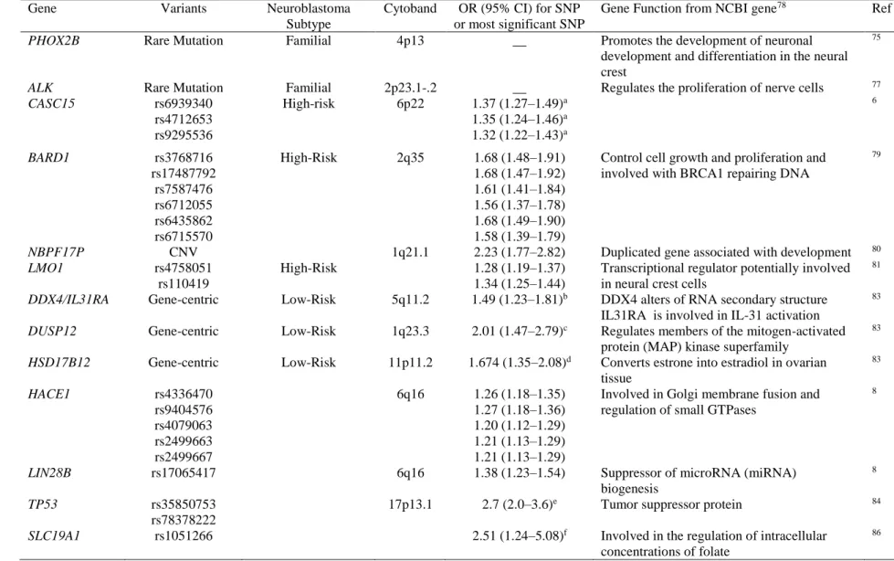
Knudson and Strong proposed that early life cancers have a genetic basis and that familial cases present earlier and with multiple primary sites, as seen in neuroblastoma. Numerous studies suggest that there are common variants that are associated with neuroblastoma. Because of the changing fetal environment, there is evidence that neuroblastoma has an underlying genetic basis that is modified by the environment. Table 3 provides a summary of all the studies and the variants that have been associated with neuroblastoma. Although these studies did not find an association between variants within vitamin pathways and neuroblastoma, these studies are genome-wide and may not be adequately powered to find small effects in a few genes due to correction for multiple testing. In addition to genetic factors, neuroblastoma can be influenced by environmental factors, such as the fetal environment.2 Current studies have not looked at
23
Table 3. A summary of genes related to neuroblastoma predisposition from Familial and GWA Studies
Gene Variants Neuroblastoma
Subtype
Cytoband OR (95% CI) for SNP
or most significant SNP
Gene Function from NCBI gene78 Ref
PHOX2B Rare Mutation Familial 4p13 __ Promotes the development of neuronal
development and differentiation in the neural crest
75
ALK Rare Mutation Familial 2p23.1-.2 __ Regulates the proliferation of nerve cells 77
CASC15 rs6939340
rs4712653 rs9295536
High-risk 6p22 1.37 (1.27–1.49)a
1.35 (1.24–1.46)a
1.32 (1.22–1.43)a
6
BARD1 rs3768716
rs17487792 rs7587476 rs6712055 rs6435862 rs6715570
High-Risk 2q35 1.68 (1.48–1.91)
1.68 (1.47–1.92) 1.61 (1.41–1.84) 1.56 (1.37–1.78) 1.68 (1.49–1.90) 1.58 (1.39–1.79)
Control cell growth and proliferation and involved with BRCA1 repairing DNA
79
NBPF17P CNV 1q21.1 2.23 (1.77–2.82) Duplicated gene associated with development 80
LMO1 rs4758051
rs110419
High-Risk 1.28 (1.19–1.37)
1.34 (1.25–1.44)
Transcriptional regulator potentially involved in neural crest cells
81
DDX4/IL31RA Gene-centric Low-Risk 5q11.2 1.49 (1.23–1.81)b DDX4 alters of RNA secondary structure
IL31RA is involved in IL-31 activation
83
DUSP12 Gene-centric Low-Risk 1q23.3 2.01 (1.47–2.79)c Regulates members of the mitogen-activated
protein (MAP) kinase superfamily
83
HSD17B12 Gene-centric Low-Risk 11p11.2 1.674 (1.35–2.08)d Converts estrone into estradiol in ovarian
tissue
83
HACE1 rs4336470
rs9404576 rs4079063 rs2499663 rs2499667
6q16 1.26 (1.18–1.35)
1.27 (1.18–1.36) 1.20 (1.12–1.29) 1.21 (1.13–1.29) 1.21 (1.13–1.29)
Involved in Golgi membrane fusion and regulation of small GTPases
8
LIN28B rs17065417 6q16 1.38 (1.23–1.54) Suppressor of microRNA (miRNA)
biogenesis
8
TP53 rs35850753
rs78378222
17p13.1 2.7 (2.0–3.6)e Tumor suppressor protein 84
SLC19A1 rs1051266 2.51 (1.24–5.08)f Involved in the regulation of intracellular
concentrations of folate
86
1.4.2 Environmental Exposures
1.4.2.1 Vitamin supplementation
Studies have shown that folic acid supplementation during the preconception period lowers the risk of neural tube defects as well as several childhood cancers including
neuroblastoma.48,91-93 Neural tube defects occur when the neural tube does not close fully. Since neural tube defects occur within close proximity to the neural crest, it is possible that both can arise from related errors in signaling.71 Although the United States food supply was fortified with folic acid at the beginning of 1998,94 women of reproductive age from 2003 to 2006 in NHANES still are estimated to have daily folic acid intake levels lower than the recommended level of 400 μg for women of childbearing age.95,96 From 1999-2006 NHANES, 74% of women reported taking folic acid containing multivitamin/multimineral supplements at one point in pregnancy. The percentage of women taking supplements also differs by trimester. Only 63% of mothers reported taking vitamins in the 1st trimester, 80% in the 2nd trimester and 90% in the 3rd
trimester.97 Since the neural crest migration and differentiation usually begins at around 5 weeks, this usage pattern suggests that many women may not be taking supplements during the most crucial time of fetal neuronal development. In addition to lower folic acid intake, less than 3% of the US population has folic acid consumption above the tolerable upper intake level
(1000µg/day), above which there may be adverse health events as set by the Institute of Medicine.98
response rate for both cases and controls were very high (85% and 87%, respectively). Since the purpose of the study was to describe the role of prenatal medication usage in neuroblastoma, no specific question about prenatal vitamin use. The prenatal vitamin data was collected from mothers who answered an open-ended question about other medications prescribed by doctors during the pregnancy. The reported unadjusted odds ratio was 0.5 (95% CI: 0.3, 0.7) for self-reported vitamin use versus no vitamin use. Due to the open-ended question used, these results may not be an accurate reflection of vitamin use.11 In a study where neuroblastoma cases were recruited at St. Jude in the same time period, about 90% of the mothers took prescription vitamins while 3.7% of the mothers took non-prescription vitamins,100 suggesting that most of the women taking vitamins were by prescription.
These results were replicated in the largest case-control study (530 cases and 500
controls) to date with maternal vitamin supplementation information. Cases were enrolled from COG from 1992–1994 and 73% provided interviews. Controls were recruited with random digit dialing (72% were interviewed) and matched on date of birth with the cases. Mothers were specifically asked whether vitamin or mineral supplements were used during the pregnancy with neuroblastoma by trimester. The odds ratio for daily vitamin use during the pregnancy or 1 month before pregnancy versus no vitamin use during the pregnancy or 1 month before
this study clearly points to an inverse association between prenatal vitamins during pregnancy and neuroblastoma.
A small German study reported a positive association between maternal vitamin use and neuroblastoma. It was conducted from 1992 to 1994 in West Germany with cases from the German Childhood Registry (N=158). Controls were randomly sampled from the local resident registration offices and matched on community and age. This study looked at multiple childhood cancers and the authors used all controls for this analysis (2,057 controls). A questionnaire assessed whether the mother took vitamin, folate, or iron supplements during pregnancy. The results were adjusted for the matching factors and sex, age, year of birth, degree of urbanization, and socioeconomic status. Mothers who took vitamin, folate, or iron supplements were 1.5 (95% CI: 1.06, 2.13) times as likely to have a child with neuroblastoma as mothers who did not take supplements. However, the proportion of vitamin supplementation among controls in this study are much lower than in other studies in the US10,11 and Germany in 1998.101 Additionally, this study recruited cases from West Germany, while the other studies are North American, which could explain the different vitamin supplementation pattern.
excluded cases that occurred from 1995 to 1999, while the Canadian study includes these years, allowing for potential misclassification of the exposure. Additionally, there were few cases of neuroblastoma post-fortification in the Canadian study. As with all ecologic studies the results are affected by other changing factors such as variation in patterns of personal vitamin
supplementation during pregnancy.
Summary
Lowering the incidence of neural tube defects has been attributed to folic acid
supplementation in food and is considered one of public health’s biggest successes. Although an effect of maternal prenatal vitamins and dietary vitamin intake on neuroblastoma has not been well established, there is clear suggestive evidence for a protective association. The studies that have been done are small, but the largest suggest that there is a negative association. The
inconsistent results could be due to gene-environment interactions and different environmental exposure patterns. However, case-control could be biased due to selection bias, since it would be difficult for the cases, who are usually recruited from a large registry, and the controls to arise from the same population. The controls could also fail to be representative of the sample population by either self-selection in sampling, or differential recall of the exposure variable.
1.4.2.2 Other possible risk factors
28
Table 4. Summary of possible risk factors of neuroblastoma
Exposure Comments References
Maternal Alcohol Use Most studies report a positive association with daily or binge drinking pre-pregnancy or pre-pregnancy. Two studies
reported a null association
100,102-104
Electromagnetic Field Studies have found an association or elevated odds ratio with paternal occupations that have exposures to
electromagnetic fields such as those involved with power plants. One study found a null association.
105-110
Pesticides Studies of associations with paternal or maternal occupations that work with pesticides pesticide use have been
mixed with both positive and null results. A meta-analysis also found null result as well.
103,105-107,109,111-116
Other occupational exposures
Maternal exposures to hair dye or maternal occupation of hairdresser or barber either before pregnancy or during pregnancy was associated with neuroblastoma. Maternal exposures to acetone, lead, petroleum, occupation in service retail and paternal exposures creosote, dioxin, lead, petroleum, occupation materials handling have also been associated with neuroblastoma in one study.
102,106,112,117
Use of Diuretics Three studies have identified an imprecise, but positive association with diuretics. Another study found a positive
association with diuretics and antihypertensive drugs.
100,102,118,119
Use of Pain Medications or Codeine
Three studies have found a positive association with non-prescription pain relievers and codeine during pregnancy. No association was found with drugs taken for fever during pregnancy New York State study and any type of pain medication in a German study.
11,100,118,120
Birth weight
Most studies have found a suggestive positive association with low and high birth weight. However, only a few studies have adjusted for gestational age, but there is a suggestive relationship with small for gestational age babies. Studies suggest a U-shape curve in which both low birth weight and high birth weight at associated. Additionally a meta-analysis found associations with both low birth weight and high birth weight.
103,121-132
History of Asthma or Allergies
Studies have identified an inverse association between childhood allergies and later development of neuroblastoma. In one study, family history of asthma has also been associated, but in another maternal history of asthma is not.
103,133
Parental Demographics
No clear association has been seen in maternal age. There is suggestive evidence of low or high maternal age associated with neuroblastoma. However, there are many studies showing null effects. Fewer studies have looked at paternal age, but there is one study that found an association with higher paternal age.
103,106,121,122,124-126,128-131,133,134
Tobacco Use
Most studies did not find an association with maternal tobacco use. One reported a weak positive association with maternal smoking pre-conception and during pregnancy, while a couple reported non-significant elevated odds ratios. Paternal smoking has been less studied and yielded mostly null results.
100,102-104,122,127,134-136
Maternal
Recreational Drug Use
A positive association with a broadly defined recreational drug use was seen in two studies. In one study, marijuana use in the first trimester had the strongest association. Another study did not find an association, but other cancer cases served as the controls.
127,137
Sex Hormones Two studies identified a positive association, especially in stage 1 or 2 cases. However, one study with subjects
reporting exposure was very small. These results failed to replicate in 3 other studies.
1.5 Literature on Vitamin Pathways
Epidemiologic studies have suggested that the prenatal environment is important for the risk of neuroblastoma. Maternal vitamin intake has been consistently associated with decreased risk of neuroblastoma and likely modifies the risk of mutational “hits” occurring. There are 3 vitamins that could potentially be of importance with neuroblastoma. Vitamin A is essential to the differentiation and development of neuronal cells. Since both excess vitamin A and
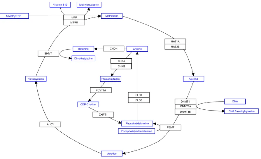
deficiency are associated with teratogenicity, cellular levels must be kept at equilibrium to prevent birth defects. Folate and folic acid have been associated with decreased incidence of neural tube defects. Additionally folate and choline are essential to DNA and RNA repair, synthesis and methylation. Low levels of choline and folate have been associated with DNA errors that could lead to somatic changes in the tumor.
1.5.1 Vitamin A
1.5.1.1 Biologic literature
cranio-neural-crest tissue.142 Excessive vitamin A intake during pregnancy occurs from supplementation.143 Figure 4 summarizes genes and metabolites involved in vitamin A metabolism and transport.
The body does not manufacture retinoids and so they must be acquired through the diet. Vitamin A is taken into the system either in the form animal products as retinyl esters, retinol, or RA or from fruits and vegetables as beta-carotene.144 Dietary retinol can be directly taken up in the intestine. However retinyl ester must first be converted to retinol by retinyl ester hydrolases (REHs) such as carboxyl ester lipase (CEL), and pancreatic lipase-related protein 2
(PNLIPRP2).145,146 Beta-carotene is broken down into retinal by Beta-carotene 15,15'-monooxygenase (BCMO1). When absorbed, all retinoids are converted to retinyl esters by lecithin retinol acyltransferase (LRAT) and is stored in the liver.147
When needed, retinyl esters are hydrolyzed to retinol by REHs in the liver. There is a large family of REHs and the enzyme varies based on location, but in the liver CEL and
carboxylesterase (CES) are mostly responsible.145 The retinol is bound by retinol binding protein (RBP) to be secreted into the bloodstream and made available to all cells including embryonic cells by maternal transfer across the placenta.144 However, research shows that there must be undiscovered placental transfer methods for vitamin A that are not RBP dependent, because homozygous RBP null mutant mice are viable.148 There is evidence that blood retinyl esters can be hydrolyzed by lipoprotein lipase (LPL) in the blood and can be transferred into cells.149 Blood levels of Retinol-RBP are very stable, except in extreme cases of insufficient intake of vitamin A, protein, calories, or zinc.144
retinaldehyde by several alcohol dehydrogenases (ADH) and retinol dehydrogenases (RDH). Retinaldehyde is then oxidized to RA by retinaldehyde dehydrogenases (RALDH).151 To keep a balance of RA in a cell, RA can be degraded to 4-hydroxy-RA or 4-oxo-RA, which are believed to be non-transcriptionally active 152,153 by three cytochrome p450 enzymes.154 Since retinoids are lipid molecules, they must be bound to proteins within cells.155 Several binding proteins have been identified including cellular retinol-binding proteins (CRBP), cellular retinaldehyde–
binding protein (CRalBP) and cellular retinoic acid-binding protein (CRABPI).155 CRBPI has been proposed to facilitate the conversion of retinol to retinyl esters for storage and the oxidation of retinol to retinaldehyde by RDHs.156
RA is the biologically active form and it functions as a ligand for specific nuclear receptors, retinoic acid receptor (RAR) or retinoid X receptor (RXR), which together regulate more than 500 genes.157 All-trans-RA, the most abundant form of RA, binds to RAR, while 9-cis-RA binds to RXR.158 Additionally, RAR binds with RXR to form a heterodimer, suggesting RXR is most likely a scaffold protein to facilitate DNA binding.159 In vivo studies have
demonstrated that binding to RAR is sufficient for rescuing a lethal defect in RA synthesis, while binding to RXR is not.160 These RAR-RXR heterodimers interact with retinoic acid response elements (RARE) in the promoter region of target genes.161
lack Crbp1 have decreased stores of retinyl esters and are sensitive to vitamin A deficiency, but do not have decreased RA synthesis. 165
Mice that are null for Cyp26a1, a gene encoding cytochrome P450 enzyme, have lethal morphogenetic phenotypes. These mice can be phenotypically rescued by disruption of Aldh1a2, suggesting that excess retinoic acid exposure induces these phenotypes.166 Double null mutations in Rar in mice impair survival in utero or shortly after birth and lead to numerous vitamin A deficiency abnormalities.167 Similar results are seen in mice with null mutations in RAR and RXR. These results showed that Rxr-α is the main Rxr involved in developmental signaling.168
1.5.1.2 Epidemiologic literature
Fetal RA level needs to be maintained at a proper concentration. Two studies have found that fetal RA has no correlation with fetal retinol levels, suggesting the variation in fetal RA levels reflects fetal generation and degradation of RA.172,173 Common variants in ALDH1A2 and CRABP2 have been associated with higher cord blood retinoic acid levels in 145 healthy full-term infants.172 A genome wide association study identified common variants near TTR and RBP4 as associated with blood retinol levels in adult males.174 Another GWA study failed to find an association with blood retinol levels, but found that rs6564851, a variant near BMCO1, was associated with higher blood β-carotene levels.175 Similarly, three polymorphisms,
including rs6564851 in BMCO1 were also associated with lower catalytic activity in 28 females.176
Common variants within genes involved in the vitamin A pathway have been associated with neural tube defects. A case-parent triad study of 329 case-parent trios and 281 mother-child or father-child dyads found SNPs within RARA, RARB, and RARG to be negatively associated with meningomyelocele, a severe form of a neural tube defect.177 Another study with 230 case-parent triads and 68 one-case-parent dyads found associations with 3 SNPs in ALDH1A2 and
meningomyelocele.178 Multiple studies have found linkage in the region containing RARA with cleft lip/palate, suggesting these loci may harbor variants.179-181
Adult cancers have also been associated with variants located in the vitamin A pathway. Childhood cancer survivors are at higher risk of adult cancers. However, the reason for this increased risk is unknown since it is unclear if increased risk is due to a general genetic
cancer and ovarian cancer. 184-187 Colorectal cancer, pancreatic cancer, and non-Hodgkin’s lymphoma have been associated with variants in the RXR genes.188-190 These associations could suggest that variation within these genes could be involved in malignant transformation.
1.5.2 Folate
1.5.2.1 Biologic literature
Folate is an essential B vitamin naturally found in foods and is available as folic acid in supplements and food fortification. Food folate has a reduced pteridine ring and a polyglutamate polypeptide that must be hydrolyzed in the intestinal lumen to a monoglutamate form before being absorbed by the intestinal cell and metabolized. Folic acid, which is synthetically produced to fortify foods, contains only a single glutamate and once converted to
tetrahydrofolate (THF) by dihydrofolate reductase (DHFR) is identical to those from food folates.191 Bioavailability of food folate depends on many factors such as the type of food, cooking methods of the food and genetics of the host. Studies have shown that food folate has 30% to 98% of the bioavailability of folic acid.192,193
Folate is necessary in one-carbon metabolism, which is involved in DNA and RNA methylation and DNA synthesis and maintenance.194 Deficiencies in folate while pregnant have been associated with birth defects such as neural tube defects,93,195 low birth weight,196,197 and preterm birth.196 Due to its association with neural tube defects, mandatory folic acid
fortification of cereal products has been in place in the United States since 1997 and in Canada since 1998.93,198
glutamate residuals. These polyglutamated folates cannot be transported out of the cell, so they accumulate in the cell to keep proper cellular folate levels.201
One-carbon metabolism is involved in the biosynthesis of many important
macromolecules such as proteins, lipids, and nucleic acids involved in cells proliferation.16 One-carbon metabolism refers to the metabolic system that uses THF to donate or accept One-carbon units for cellular biosynthetic reactions and occurs in the cytoplasm, mitochondria and nucleus.202 Figure 5 describes the one-carbon pathway in greater detail.
Briefly, during one-carbon metabolism, three major reactions occur in the cytoplasm. 202
1. 10-formyltetrahydrofolate is the one-carbon unit involved in the synthesis of the purine ring by phosphoribosylglycinamide formyltransferase (GART) and 5-aminoimidazole-4-carboxamide ribonucleotide formyltransferase/IMP cyclohydrolase (ATIC).
2. Thymidylate synthetase (TYMS) uses 5,10-methylene tetrahydrofolate as the one-carbon unit for the conversion of deoxyuridine monophosphate (dUMP) to deoxythymidine monophosphate (dTMP).
3. 5-methyltetrahydrofolate is used in for the remethylation of homocysteine to methionine by methyltetrahydrofolate-homocysteine methyltransferase (MTR) and
5-methyltetrahydrofolate-homocysteine methyltransferase reductase (MTRR).
Methionine can be converted to S-adenosylmethionine (AdoMet) by methionine adenosyltransferase, encoded by and MAT1A and MAT2B, which serves as a cofactor for
nucleus. About 10% cellular folate is present in the nucleus and both TYMS and serine hydroxymethyltransferase (SHMT) have been localized in the nucleus.204
The regulation of cellular folate concentration is complex since it is influenced by uptake, polyglutamylation, export, and catabolism. The folate receptor Folbp1 shows localized patterns of expression in the embryo and is highly expressed in the yolk sac, suggesting this receptor is important for maternal-to-fetal transport of folate.205 Additionally, mice that are null for Folbp1 present with the same birth defects as mice with folate deficiencies.206,207 During pregnancy, the need for folate increases due to the growth of the fetus, the placenta, and maternal tissues as well as a requirement for more red blood cells due to uterine enlargement and expansion of blood volume. Although there is an increased need for folate in the mother, newborns have higher red blood cell folate levels compared to maternal levels, 208,209 suggesting the importance of folate to fetal development.
Folate transfer and polyglutamylation are critical to maintain a proper concentration of folate, and disruption of either leads to impaired folate accumulation. Folate monoglutamates can also be transferred to into mitochondria by a specific reduced folate carrier 210,211 and then converted to polyglutamated folates.212 Because of this transfer and conversion, folate concentrations in the cytoplasm are not in equilibrium with folate concentration in the mitochondria.191
are low, dUMP levels tend to accumulate, which leads to increased rates of uracil nucleotide incorporation into DNA and been associated with strand breaks and chromosomal instability.213 Similarly, an insufficient rate of homocysteine remethylation results in an elevated plasma homocysteine, decrease in AdoMet and increase in S-adenosoylhomocysteine (AdoHcy). This leads to a decreased cellular conversion of AdoMet to AdoHcy, which is crucial for cellular methylation and results in decreased levels of 5-methylcytosine, the methylated form of cytosine, in DNA.214,215
39
Figure 5. Folate metabolism and one-carbon pathway within a cell
THF: Tetrahydrofolate; AdoMet: S-Adenosyl methionine; AdoHcy: S-Adenosylhomocysteine ; dTMP: Thymidine monophosphate; dUMP: deoxyuridine
1.5.2.2 Epidemiologic literature
Because of the association of folate with neural tube defects, variants within the one-carbon pathway have been highly studied with respect to birth defects and childhood cancer. Variants within genes involved in the one-carbon pathway have been associated with both adult and childhood cancers as well as certain birth defects.
MTHFR
The most studied gene within the one-carbon metabolism pathway is
methylenetetrahydrofolate reductase (MTHFR), which has two common exonic variants, C677T and A1298C. MTHFR regulation is critical for AdoMet dependent reactions and regulation of homocysteine levels in the cell. The MTHFR reaction is not reversible and commits one-carbon units to methionine biosynthesis.218 Studies have shown the low MTHFR activity may reduce DNA methylation,215 but may enhance synthesis of thymidylate.219
One exonic C677T SNP (rsid: rs1801133) is one of the most common SNPs associated with MTHFR deficiency affecting 5 to 20% of North Americans.220,221 This SNP has been associated with increased plasma homocysteine and decreased plasma and red blood cell folate levels, especially in those with low folate levels.222-225 Another exonic variant A1298C (rsid: rs1801131) has also been associated with decreased enzymatic activity of MTHFR but to a lower extent than the C667T variant.226 Individuals with this polymorphism exhibit increased red blood cell folate levels and homocysteine levels.
neural tube defects. Although there is evidence of between-study heterogeneity, all the studies have a positive trend. One study did not find an independent association with the MTHFR A1298C variant with neural tube defects.227 One meta-analysis of cleft lip/palate found a positive association with maternal C677T, a suggestive association with infant C677T and null associations with A1298C.228 Another meta-analysis found a positive association with infant C677T and cleft lip/palate in Asian populations,229 which was replicated with a newer meta-analysis which found both a maternal and child associations with C677T.230 Additionally, this variant has been associated with increased risk of embryonal central nervous system tumors based on a small study of Thai children.231
Meta-analyses have found the C677T variant to be associated with decreased risk of pediatric acute lymphoblastic leukemia, but results were null for the A1298C variant.232,233 In addition to childhood cancers and birth defects, MTHFR variants have been associated with adult cancers. Although meta-analyses of adult cancers have been largely inconsistent, associations have been found with colon cancer234-236 and ovarian cancer237 among Caucasians, and primary brain tumors 238 among Asians with MTHFR C677T. One meta-analysis pooled all cancer studies together and found that MTHFR C677T was positively associated with cancer in the aggregate, especially in esophageal and stomach cancer and among Asians. 239
Other Genes in one-carbon metabolism
Many other genes within the one-carbon pathway have been associated with blood folate and homocysteine levels. One exonic SNP in reduced folate carrier 1 encoded by gene